Bioimpacts. 13(1):73-84.
doi: 10.34172/bi.2022.23523
Original Article
Inhibitory effects of mixed flavonoid supplements on unraveled DSS-induced ulcerative colitis and arthritis
Siva Prasad Panda 1, *  , Mahamat Sami Adam Mahamat 2, Malikyahia Abdul Rasool 2, DSNBK Prasanth 3, Idris Adam Ismail 2, Moyed Abasher Ahmed Abasher 2, Bikash Ranjan Jena 4
Author information:
1Pharmacology Research Division, Institute of Pharmaceutical Research, GLA University, Mathura, Uttar Pradesh, India
2Pharmacognosy Research Division, College of Pharmacy, Koneru Lakshmaiah Education Foundation, Vaddeswaram, Guntur, India
3Department of Pharmacognosy, KVSR Siddhartha College of Pharmaceutical Sciences, Vijayawada, AP, India
4SIIMS College of Pharmacy, Guntur, India
Abstract
Introduction:
The mixed flavonoid supplement (MFS) [Trimethoxy Flavones (TMF) + epigallocatechin-3-gallate (EGCG)] can be used to suppress inflammatory ulcers as an ethical medicine in Ayurveda. The inflammation of the rectum and anal regions is mostly attributed to nuclear factor kappa beta (NF-κB) signaling. NF-κB stimulates the expression of matrix metalloproteinase (MMP9), inflammatory cytokines tumor necrosis factor (TNF-α), and interleukin-1β (IL-1β). Although much research targeted the NF-κB and MMP9 signaling pathways, a subsequent investigation of target mediators in the inflammatory ulcer healing and NF-κB pathway has not been done.
Methods: The docking studies of compounds TMF and EGCG were performed by applying PyRx and available software to understand ligand binding properties with the target proteins. The synergistic ulcer healing and anti-arthritic effects of MFS were elucidated using dextran sulfate sodium (DSS)-induced colon ulcer in Swiss albino rats. The colon mucosal injury was analyzed by colon ulcer index (CUI) and anorectic tissue microscopy. The IL-1β, tumor necrosis factor (TNF-α), and the pERK, MMP9, and NF-κB expressions in the colon tissue were determined by ELISA and Western blotting. RT-PCR determined the mRNA expression for inflammatory marker enzymes.
Results: The docking studies revealed that EGCG and TMF had a good binding affinity with MMP9 (i.e., -6.8 and -6.0 Kcal/mol) and NF-kB (-9.4 and 8.3 kcal/mol). The high dose MFS better suppressed ulcerative colitis (UC) and associated arthritis with marked low-density pERK, MMP9, and NF-κB proteins. The CUI score and inflammatory mediator levels were suppressed with endogenous antioxidant levels in MFS treated rats.
Conclusion: The MFS effectively unraveled anorectic tissue inflammation and associated arthritis by suppressing NF-κB-mediated MMP9 and cytokines.
Keywords: MFS, Anorectic ulcer, Arthritis, NF-κB, MMP9, Cytokines
Copyright and License Information
© 2023 The Author(s).
This work is published by BioImpacts as an open access article distributed under the terms of the Creative Commons Attribution Non-Commercial License (
http://creativecommons.org/licenses/by-nc/4.0/). Non-commercial uses of the work are permitted, provided the original work is properly cited.
Introduction
The history of Ayurveda was reviewed by Indian scientists in regards to the Indian traditional system of medicine. It was observed that a large population of India depends upon registered Ayurvedic medical practitioners for prevention and getting rid of complications of different diseases.1 Scientists also reviewed the latest incumbent of ayurvedic medicines to treat inflammatory arthritis, ulcerative colitis, and musculoskeletal disorders.2
Ulcerative colitis (UC) means chronic colon inflammation with hallmarks of diarrhea, rectal bleeding, blood with stool, and abdominal pain. An autoimmune disorder is one of the major factors of colitis.3 The UC and other immune-related colitis, Crohn's disease, belong to inflammatory bowel diseases (IBDs). There are other types of UC, including infectious UC, chemical UC, and ischaemic UC.4 The long-term medications used to relieve UC were not enough but causing liver and kidney dysfunction with reduced quality of life. The most common complication of UC is arthritis.5 Therefore, many scientists are in search of good curative both anti-ulcer and anti-arthritic drugs with few or no side effects.
In our previous research, we isolated trimethoxy flavone (TMF) from Tabebuia chrysantha stem extract and revealed the anti-tumor effect of extract and anti-angiogenic effect of TMF due to the suppression of ERK, STAT3, and MMP9 proteins.6,7 Scientists reported the linkage between EGFR and STAT3 with the NF-kB signaling pathway in both inflammation and cancerous growth. Expressions of EGF, MMP-2, MMP9, STAT3, and VEGF were positively correlated with inflammation, tumor size, invasiveness, lymphatic and venous invasion, and metastasis of various carcinomas.8 Polyphenols have a vast interest in the pathogenic alteration of cancer, diabetes, weight reduction, and obesity. Nature synthesized epigallocatechin-3-gallate (EGCG) as prime polyphenols (flavan 3-ols), showing the most potent antiproliferative effects.9 Most of the health promotional strategy of green tea therapy is due to EGCG and related antioxidant activity.10 The mixture of 270 mg of EGCG and 150 mg of caffeine has shown anti-obesity effect.11 Scientists also revealed that the anticancer effect of EGCG is attributed to the dephosphorylation of EGFR.12
Matrix metalloproteinases (MMPs) are a family of endopeptidases that play crucial roles in various pathophysiological processes.13 Osteoarthritis is due to the demethylation of certain CpG sites in the MMP9 promoter, disturbing the synthesis of the MMP9 gene in cartilage tissues.14 Almost all types of arthritis and UC are associated with the significant role of NF-κB signalling.15 Over the last 20 years, the rheumatologist has given attention to anti-inflammatory drugs such as corticosteroids and sulfasalazine for clinical remission of the disease as these can substantially reduce inflammation.16 Despite the current regimens, complete long-term disease remission is not achieved for many patients. The objective of this study was to discover an alternative remedy for UC because the frequency of consumption of conventional drugs by elderly patients leads to potential adverse effects with the development of colon cancer later in life.
Materials and Methods
Raw materials and chemicals
EGCG (Maysar herbal, Faridabad, Haryana), dextran sodium sulfate (DSS) and Mesalazine (Sisco research Lab), TMF (Pharmacology research Lab), and all other chemicals were obtained from Genaxy Scientific, Hyderabad, India.
Docking studies
The MMP enzymes are actively involved in the pathogenesis of inflammation and disease processes such as arthritis and cancer.17 The inflammation of the rectum and anal regions is mostly attributed to NF-κB signalling.18 Citing the above literature findings, the docking studies of compounds TMF and EGCG were performed by applying PyRx and available software to understand ligand binding properties with the target protein. The crystal structure of transcription factor NF-κB P50 homodimer bound to a κB site (1BFT) and MMP9 (6ESM) was derived from the protein data bank.19 The native ligand was also amended to measure the root mean square deviation (RMSD) of the docked pose and X-ray conformable to validate the docking process. Discovery Studio Visualizer was used to analyze and visualize docking implications. All the docked ligands were categorized according to their binding energies and the lowest bonding energy ligands. The structure of all ligands incorporated for docking analysis are represented in Fig. 1.
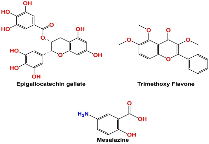
Fig. 1.
Structure of ligands (TMF, EGCG, and Mesalazine).
.
Structure of ligands (TMF, EGCG, and Mesalazine).
Formulation of MFS
Two-dose combinations were selected based on the formula from the literature and our previous research: Low dose A = EGCG 100 µg/mL+ TMF 10 µg/mL, high dose B = EGCG 200 µg/mL + TMF 25 µg/mL.6,20 The powder mixture, low dose A and high dose B, were solubilized separately by pyrogen-free water with DMSO as a solubilizing agent. The LD50 dose value of EGCG was 2000 mg/kg, and the safe dose was 200 mg/kg.21 The formulated mixture was named MFS-LD and MFS-HD. The tribal community of Mangalagiri district, AP, uses the mixture of T. chrysantha stem and tea extract to relieve anorectal inflammation. Our previous research reported that the angiogenesis suppression effect of the TMF was 25 µg/mL.6
Animals
The adult male Wistar albino (WS) rats were acclimatized to laboratory conditions for one week before investigation.
DSS induced inflammatory colitis in WS rats
First, 3% DSS solution (15 g DSS powder in 500 mL sterile water), 5 mL/day, was orally fed to the four groups of rats (Gr-II, Gr III and Gr IV, Gr V; n = 6) for up to 6 days. The treatment schedule is as follows: Gr I, sterile water; Gr-II, DSS; Gr-III, MFS-LD; Gr-IIV, MFS-HD; Gr-V, Mesalazine (0.52 g/kg as a single dose per day).22 Medicines are diluted with distilled water so that all candidates would receive uniform drug components. The body weight, blood in the stool, and stool consistency for all rats (Gr II-IV) were examined during and after treatment to confirm the anti-UC activity of MFS.
Measurement of paw diameter of treated rats
Joint arthritis was evaluated by checking the mean increase in joint diameter for six days via digital vernier caliper. The difference in joint thickness on the 1st and 6th days was calculated for all groups, and the percentage of anti-arthritic effects of MFS-LD and MFS-HD was calculated using the following formula:
Where Vc is Joint diameter in control, and Vt is Joint diameter in treatment groups.
Hematology in DSS-induced UC model
The different blood parameters were analyzed by a blood cell counter (ERBA diagnostic Limited, India). The Rheumatoid factor (RF, IU/mL) was estimated by the available kit (Laila Implex, Vijayawada). The total cholesterol level was measured by the available kit from SPAN diagnostic, India.
Histology of anorectic tissue and liver of rats
After the last dose of MFS, all rats were sacrificed under chloral hydrate anesthesia. The anorectic epithelial tissue section near to anus was removed; a 5-µm longitudinal section was prepared with the help of a spencer micrometer, fixed in formaldehyde solution (3.7%), and stained with hematoxylin-eosin over the slide with by a professional laboratory technician to observe the ulcer characteristics. Similarly, the liver section was prepared and observed by microscope.23
Estimation of ERK, MMP9, NF-κB expression by western blotting
Sample preparation by sonication
Anorectic epithelial tissue was kept in isotonic KCl-0.01M phosphate buffer and centrifuged properly to separate tissue debris. Then 25% tissue homogenate was mixed with 6.0 mL of cold phosphate buffer (pH 7.6) and EDTA to enable thorough lysis of cells and solubilize proteins.
Determination of protein concentration
The protein quantity was estimated in all test sample preparations by applying the Bradford assay (Bradford, 1976). A standard curve was formed to estimate unknown sample concentrations.
Gel electrophoresis and transfer of proteins
The protein samples were passed over 12% SDS–PAGE electrophoresis, allowing separation of proteins by molecular weight. The separated proteins were then transferred overnight to the polyvinylidene difluoride membrane (a western blot) at 48oC.6,7
Blocking and antibodies
The membrane was placed in a dilute albumin protein (bovine serum) solution to mask a non-specific binding site on the membrane. After blocking, the membrane was incubated with primary antibodies. The primary antibodies of the following proteins such as rabbit NF-κB p65 (D14E12, 1/1000 dilution), rabbit MMP-9 (G657, 1/1000 dilution), rabbit pERK1/2 (p44/42, 1/1000 dilution) in 5% BSA (Cell Signaling Technology, Inc.), and monoclonal anti-glyceraldehyde 3-phosphate dehydrogenase (GAPDH) were used for the study. The dilution ratio of GAPDH (sc-166574) was 1/20000. The LI-COR Biosciences software was used to measure bands of all proteins. The GAPDH was procured from Santa Cruz Biotech, Inc., USA.
Estimation of IL-1β and TNFα by ELISA and iNOS, and COX-2 by RT-PCR
The ELISA technique was used to quantify the levels of IL-1β and TNFα in the anorectal tissue. The reader followed the standard detection procedure with an OD value of 450 nm.24 The RT-PCR assay was performed following the same procedure as discussed in our previous research and literature.6,25 For iNOS and COX-2 quantification, 2 μg total RNA was reverse transcribed into cDNA by AMV Reverse Transcriptase. The extracted RNA from colonic mucosa was mixed with primer solution (20 µL) before analysis. The primer sequences are mentioned in Table 1. GAPDH was used as a housekeeping gene.
Table 1.
Sequences of primers
|
Primers
|
|
Sequences
|
| iNOS |
Forward
Reverse |
5′-CACCACCCTCCTTGTTCAAC-3′
5′-CAATCCACAACTCGCTCCAA-3′ |
| COX-2 |
Forward
Reverse |
5′-TGCGATGCTCTT CCGAGCTGTGCT-3′
5′-TCA GGAAGTTCCTTATTTCCTTTC-3′ |
| GAPDH |
Forward
Reverse |
5-GAGTCAACGGATTTGGTCGT-3′
5-GACAAGCTTCCGTTCTCAG-3′ |
Quantitative densitometric analysis
The quantitative densitometric analysis of pERK, STAT3, MMP9, and NF-κB was performed using the quantity one software (Bio-Rad) at the Indian Institute of Chemical Biology, India. Band intensity was analyzed for all proteins of each sample from three independent biological experiments.25
The reduced glutathione (GSH), MPO, SOD, CAT activity
The myeloperoxidase (MPO) of anorectal tissue and GSH, superoxide dismutase (SOD), and catalase (CAT) activity of the liver tissue were determined using respective test kits (GENAXY Scientific, New Delhi).
Results
Docking validation
The MMP9 target structure (PDB-6ESM) was acquired from PDB to dock with the co-crystallized ligand inhibitor. To further confirm the methodology of docking adopted, the RMSD value was calculated. RMSD value of docked and experimentally derived pose of co-crystallized ligand was observed as 0.67532 (Fig. 2). RMSD validation indicates that the technique to estimate the binding affinity of unknown ligands is accurate.
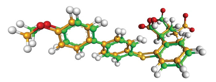
Fig. 2.
Overlay of docked pose and crystallographic pose at the active pocket. Green: Native ligand; Orange: Docked pose.
.
Overlay of docked pose and crystallographic pose at the active pocket. Green: Native ligand; Orange: Docked pose.
To discover a prospective candidate against MMP9 and NF-κB, molecular docking was carried out on two phytoconstituents that are EGCG and TMF. These two phytocompounds were docked towards the target proteins and rated based on their dock performance. For a detailed review, refer to Tables 2 and 3. This table displays the binding energies along with their interactions. Out of these two compounds, EGCG had a good binding score for both the targets, namely, -6.8 and -9.4 with NF-κB and MMP9, respectively (Figs. 3 and 4). Surprisingly, both the compounds EGCG and TMF possess the least binding energies compared to standard with MMP9 and NF-κB.
Table 2.
Binding affinity and interactions of in silico potential molecules against NF-κB (1BFT)
|
Ligands
|
Binding affinity (kcal/mol)
|
Amino acids involved and Distance (A°)
|
|
Hydrogen binding interactions
|
Hydrophobic interactions
|
Electrostatic interactions
|
| Epigallocatechin gallate |
-6.8 |
ARG A:201 (3.03), SER A:203 (3.57), ASP A:210 (3.83), ASN A:200 (4.40). |
ARG A:201 (4.78), ARG A:201 (5.30) |
- |
| Trimethoxy Flavone |
-6.0 |
ARG B:201 (4.01), GLU B: 211 (3.93) |
ARG A:201 (6.06) |
ARG B:253 (7.27) |
| Mesalazine |
-4.8 |
CYS A:197 (3.43), VAL B:244 (4.58), ALA B:242 (4.72), ARG B: 246 (5.05), HIS B:245 (3.56) |
ARG A:198 (6.05), CYS A:197 (5.56), HIS A:245 (4.35) |
- |
Table 3.
Binding affinity and interactions of in silico potential molecules against MMP9 (6ESM) in complex with inhibitor BE4
|
Ligands
|
Binding affinity
(kcal/mol)
|
Amino acids involved and Distance (A°)
|
|
Hydrogen binding interactions
|
Hydrophobic interactions
|
Electrostatic interactions
|
| Epigallocatechin gallate |
-9.4 |
GLY A:186 (3.23, 4.00), TYR A:218 (5.61), MET A:247 (5.77) |
VAL A:223 (5.45), LEU A:188 (3.36) |
HIS A:226 (4.62) |
| Trimethoxy Flavone |
-8.3 |
HIS A:226 (4.91) |
LEU A:187 (5.45), VAL A:223 (5.58), GLY A:186 (5.49) |
HIS A:226 (4.49), HIS A:236 (7.26, 7.52) |
| Mesalazine |
-6.9 |
LEU A:222 (4.38), ARG A:249 (5.24), LEU A:226 (4.88), PRO A:246 (5.80) |
VAL A:223 (5.57) |
HIS A:226 (4.23) |
(2~{S})-2-[2-[4-(4-methoxyphenyl)phenyl]
sulfanylphenyl]pentanedioic
acid (Co-crystallized ligand) |
-10.9 |
ALA A:189 (4.01), ALA A:191 (4.00), HIS A:230 (5.33), GLN A:227 (4.73), HIS A:226 (4.73, 5.55), HIS A:236 (5.40) |
LEU A:187 (4.92), LEU A:188 (4.52), LEU A:222 (4.22), VAL A:223 (5.08), TYR A:248 (4.48), LEU A:243 (4.48, 4.59), ARG A:249 (6.93) |
HIS A:226 (5.23) |
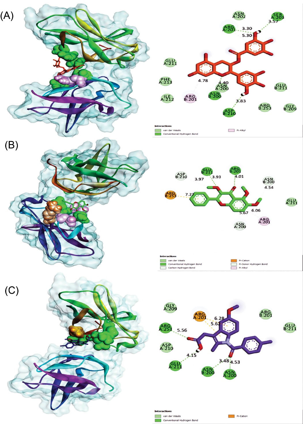
Fig. 3.
Molecular docking (molecular surface view) between NF-κB (1BFT) protein and (A) Epigallocatechin gallate, (B) Trimethoxy Flavone (C) Mesalazine. This molecular docking figure shows compounds at their binding site on the left and the right of the amino acids that interact with the ligand to give resultant binding energy.
.
Molecular docking (molecular surface view) between NF-κB (1BFT) protein and (A) Epigallocatechin gallate, (B) Trimethoxy Flavone (C) Mesalazine. This molecular docking figure shows compounds at their binding site on the left and the right of the amino acids that interact with the ligand to give resultant binding energy.
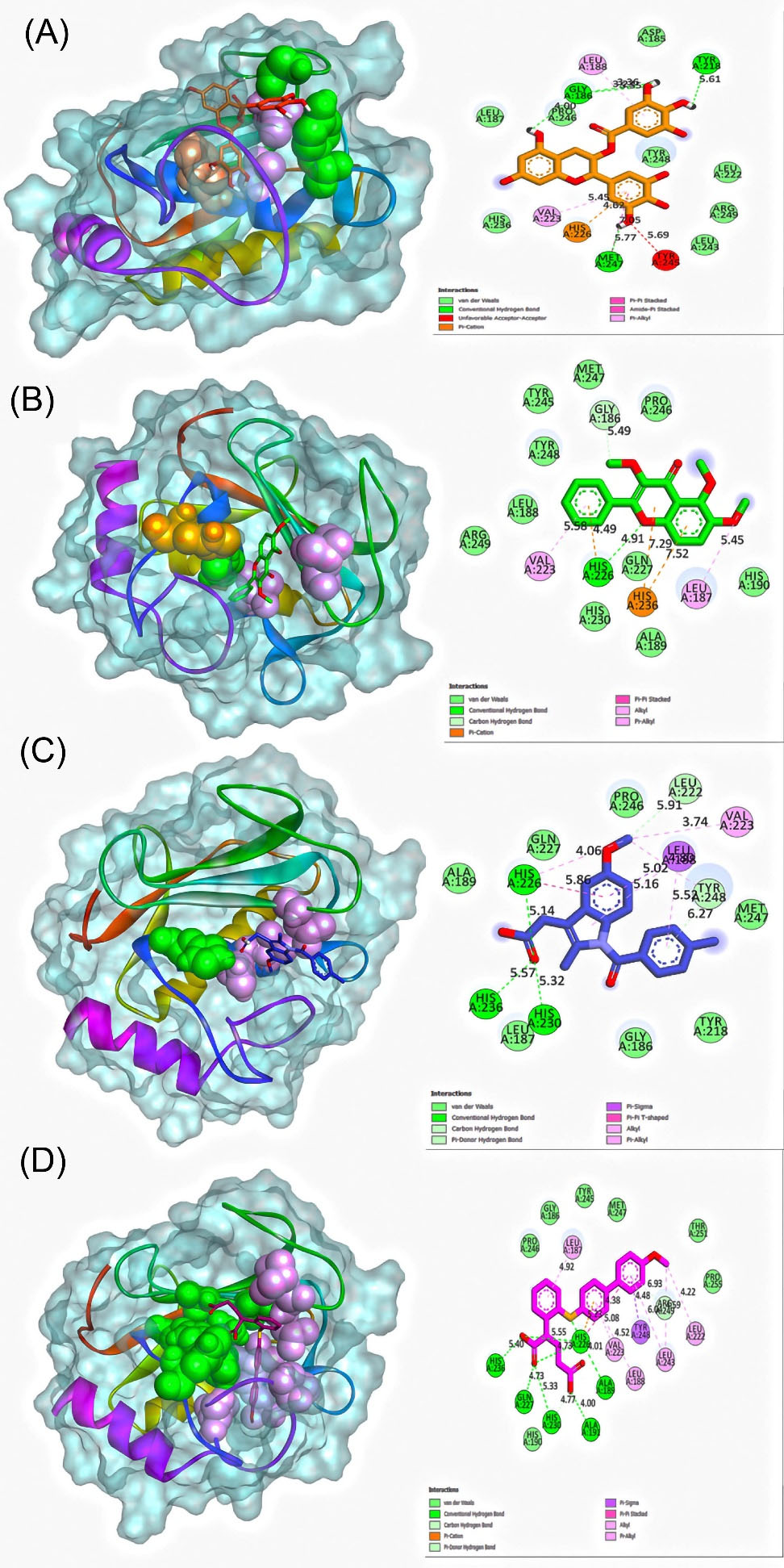
Fig. 4.
Molecular docking (molecular surface view) between MMP9 (6ESM) protein and (A) Epigallocatechin gallate, (B) Trimethoxy Flavone (C) (2~{S})-2-[2-[4-(4-methoxyphenyl)phenyl]sulfanylphenyl]pentanedioic acid (D) Mesalazine. This molecular docking figure shows compounds at their binding site on the left and the right of the amino acids that interact with the ligand to give resultant binding energy.
.
Molecular docking (molecular surface view) between MMP9 (6ESM) protein and (A) Epigallocatechin gallate, (B) Trimethoxy Flavone (C) (2~{S})-2-[2-[4-(4-methoxyphenyl)phenyl]sulfanylphenyl]pentanedioic acid (D) Mesalazine. This molecular docking figure shows compounds at their binding site on the left and the right of the amino acids that interact with the ligand to give resultant binding energy.
Effect of MFS on DSS induced inflammatory colitis (IC) and arthritis in WS rats
On the 6th day, 4 rats were died in the DSS control group because of severe diarrhea and bloody stool. Anorectic lesions like piles or fistula were observed near the anus region of the DSS control group. The MFS-LD and MFS-HD treatment improved body weight along with the nature of stool consistency. No diarrhea, bloody stool, or death cases were noticed in all treated rats (group II-IV) compared to the DSS control group. It is interesting to note that MFS started improving body weight (P < 0.001) and healing anorectal lesions from 2nd day after treatment (Table 4). The MFS and mesalazine treatment restored the paw diameter of normal control rats (Fig. 5; Table 5).
Table 4.
Colon ulcer index
|
Groups
|
Bodyweight (g)
|
Stool nature/observation
|
| Normal control |
24.3 |
Normal stool |
| DSS control |
8.6# |
Diarrhea condition with blood and mucus in the stool. Four rats died on the 7th day from DSS exposure. |
| MFS-LD |
17.15* |
Semisolid stool with mucous but no appearance of a blood clot. |
| MFS-HD |
21.82* |
Soft-hard stool, no mucus, and blood. |
| Mesalazine |
20.64* |
Hard stool consistency. |
Values are mean ± SEM; n=6; #P < 0.05 vs. Normal control; * P < 0.01 vs. DSS control.

Fig. 5.
Footpad image of SW rats: (A) Normal saline control, (B) Development of arthritis as a complication after DSS feed; (C-E) Treatment with MFS-LD, MFS-HD, and Mesalazine. Inflammatory arthritis in the footpad is effectively reduced after six days of treatment with MFS.
.
Footpad image of SW rats: (A) Normal saline control, (B) Development of arthritis as a complication after DSS feed; (C-E) Treatment with MFS-LD, MFS-HD, and Mesalazine. Inflammatory arthritis in the footpad is effectively reduced after six days of treatment with MFS.
Table 5.
Measurement of foot paw diameter
|
Groups
|
Treatment and dose
|
Paw diameter (mm)
|
Percentage inhibition (%)
|
|
2nd day
|
6th day
|
| I |
Normal control |
1.57 ± 0.03 |
3.95 ± 0.02 |
|
| II |
DSS control |
1.42 ± 0.01 |
6.26 ± 0.01 |
|
| III |
MFS-LD |
1.45 ± 0.02* |
2.20 ± 0.02** |
45 |
| IV |
MFS-HD |
1.20 ± 0.01** |
1.18 ± 0.02* |
71 |
| V |
Mesalazine |
1.22 ± 0.01** |
1.18 ± 0.02* |
71 |
Values are mean ± SEM; n=6; ** P < 0.001 vs. Normal control; * P < 0.05 vs. DSS control.
Effect of MFS on hematological parameters in DSS-induced UC model
A severe anemic condition was observed in the DSS control group because of the decrease in Hb value and RBC count. The WBC, Platelet, ESR, total cholesterol, and RF value were increased in the DSS-treated rats. All changes were restored towards a normal condition in the MFS and mesalazine treated groups (Table 6).
Table 6.
Haematological parameters
|
Parameters
|
Normal control
|
DSS control
|
MFS-LD
|
MFS-HD
|
Mesalazine
|
| Hb (g/dL) |
14.50 ± 2.13 |
4.45 ± 3.06# |
10.58 ± 2.52 |
12.52 ± 1.85* |
10.48 ± 3.64* |
| RBC (106/mm3) |
8.50 ± 1.35 |
3.70 ± 1.83 |
7.25 ± 2.16*** |
7.35 ± 0.86 |
7.37 ± 0.52** |
| WBC (103/mm3) |
5.45 ± 2.80 |
12.33 ± 2.59 |
7.47 ± 1.43* |
6.15 ± 0.67** |
6.53± 2.45* |
| Platelets (lakhs/mL) |
2.70 ± 1.65 |
5.30 ± 1.33# |
3.52 ± 1.27** |
3.05 ± 1.17* |
3.23 ± 2.43** |
| ESR (60 min) |
3.45 ± 1.26 |
10.21± 1.36# |
4.73 ± 2.26* |
4.65 ± 1.75* |
4.55 ± 3.77*** |
| RF (IU/mL) |
6.45 ± 0.94 |
19.25 ± 2.24 |
8.35 ± 0.53* |
7.23 ± 0.25* |
7.05 ± 2.76** |
| TC (mg/dL) |
105.27 ± 2.17 |
133.37 ± 0.62# |
110.16 ± 3.72*** |
108.69 ± 2.87** |
105.35 ± 1.80* |
Values are mean ± SEM; n=6; #P <0.001 vs. saline control; *P <0.001, **P <0.01,***P < 0.05 and vs. disease control.
Histology of anorectal and liver tissue of rats
The epithelial tissue of the anus of DSS control rats showed the cell injury, generation, and adhesion of ulcers, infiltration of large numbers of WBCs, platelets, and macrophages to the ulcer region. The above findings were significantly lowered in the MFS and mesalazine-treated rats (P < 0.001) (Fig. 6).
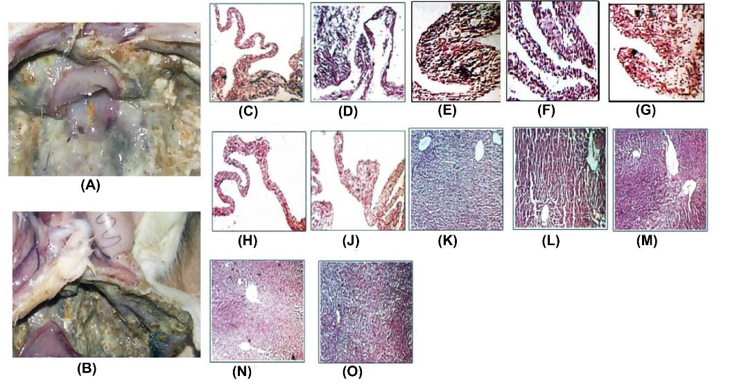
Fig. 6.
Histology of anorectal tissue of rat (original magnification x50): (A) Anus of normal and treated rat (indicated with arrow marks); (B) Anus of colitis rat; (C) Anal epithelium of normal rat; (D-E) Anal epithelial tissue damage of colitis rat observed after DSS feed; (F-G). MFS-LD; h.MFS-HD; (J) mesalazine facilitated wound healing by reducing infiltration of WBCs and macrophages to the damage site; (K) The liver section of normal rat (original magnification x 50); (L) Hepatic tissue necrosis with formation of scar tissue in DSS control rats; (M-O) The treatment with MFS-LD, MFS-HD, and mesalazine normalizes the disturbed hepatic architecture.
.
Histology of anorectal tissue of rat (original magnification x50): (A) Anus of normal and treated rat (indicated with arrow marks); (B) Anus of colitis rat; (C) Anal epithelium of normal rat; (D-E) Anal epithelial tissue damage of colitis rat observed after DSS feed; (F-G). MFS-LD; h.MFS-HD; (J) mesalazine facilitated wound healing by reducing infiltration of WBCs and macrophages to the damage site; (K) The liver section of normal rat (original magnification x 50); (L) Hepatic tissue necrosis with formation of scar tissue in DSS control rats; (M-O) The treatment with MFS-LD, MFS-HD, and mesalazine normalizes the disturbed hepatic architecture.
Report of estimated IL-1β and TNFα by ELISA, and iNOS, and COX-2 by qRT-PCR
In the MFS-HD and Mesalazine treated groups, inflammatory mediators' expressions were significantly lowered compared to the DSS induced group. The decreased level was more obvious to confirm MFS's anti-inflammatory and ulcer healing potential (Fig. 7).
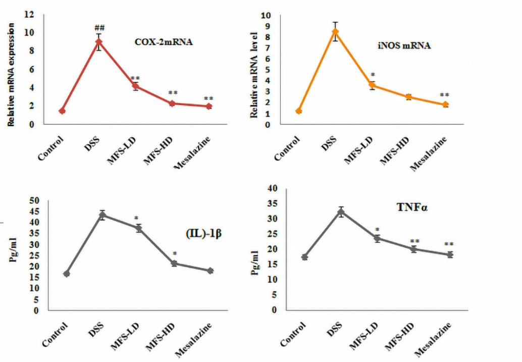
Fig. 7.
Quantification of (IL)-1β and TNFα by ELISA, and expression of iNOS mRNA, and COX-2 mRNA by RT-PCR. Marked values are calculated as mean ± SEM, where n=6; *P <0.001 vs. saline control; and **P <0.01 vs. disease control.
.
Quantification of (IL)-1β and TNFα by ELISA, and expression of iNOS mRNA, and COX-2 mRNA by RT-PCR. Marked values are calculated as mean ± SEM, where n=6; *P <0.001 vs. saline control; and **P <0.01 vs. disease control.
In the MFS-HD and mesalazine treated groups, the inflammatory marker proteins such as p-ERK, MMP9, NF-κB expressions were significantly reduced compared to DSS control groups (Fig. 8).
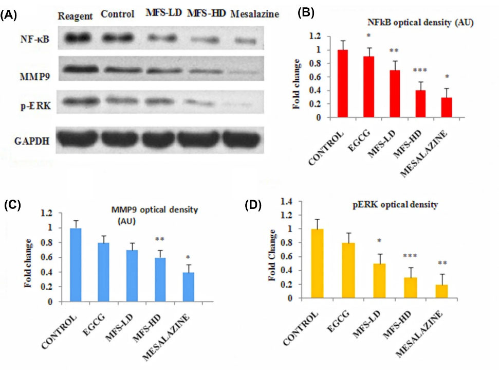
Fig. 8.
(A) Immunoblot images of NF-κB, ERK, and MMP9 signaling with quantification, (B) Quantitative densitometry analysis of NF-κB, (C) Quantitative densitometry analysis of pERK, (D) Quantitative densitometry analysis of MMP9. Marked values are represented as mean ± SEM of three repeated experiments; *P <0.05 vs. saline control; **P <0.001 vs. disease control; and ***P <0.01 vs. disease control.
.
(A) Immunoblot images of NF-κB, ERK, and MMP9 signaling with quantification, (B) Quantitative densitometry analysis of NF-κB, (C) Quantitative densitometry analysis of pERK, (D) Quantitative densitometry analysis of MMP9. Marked values are represented as mean ± SEM of three repeated experiments; *P <0.05 vs. saline control; **P <0.001 vs. disease control; and ***P <0.01 vs. disease control.
Report of GSH, MPO, SOD, and CAT activity
The DSS control group was observed with a lower level of GSH and CAT with increased activity of MPO and SOD. In the MFS-HD and mesalazine treated groups, the GSH, MPO, SOD, and CAT activities were significantly restored to the normal level (Fig. 9).
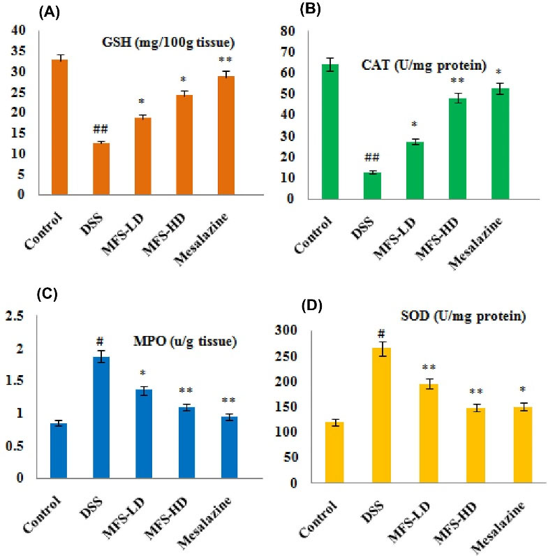
Fig. 9.
GSH and CAT levels in the hepatic tissue of rats in the UC model. Figure 9c-9d: MPO of anorectal tissue and SOD levels in the hepatic tissue of rats in the UC model. Marked values are calculated as mean ± SEM, where n=6; *P < 0.001 vs. saline control; **P < 0.01 vs. disease control; and #P < 0.05 vs. saline control; ##P < 0.01 vs. saline control.
.
GSH and CAT levels in the hepatic tissue of rats in the UC model. Figure 9c-9d: MPO of anorectal tissue and SOD levels in the hepatic tissue of rats in the UC model. Marked values are calculated as mean ± SEM, where n=6; *P < 0.001 vs. saline control; **P < 0.01 vs. disease control; and #P < 0.05 vs. saline control; ##P < 0.01 vs. saline control.
Discussion
IBD is an autoimmune inflammatory disorder where circulated immune cells such as T lymphocytes, B lymphocytes, and macrophages penetrate and reside in the inflammatory loci.22 Arthritis is the most common complication of UC characterized by a rheumatoid factor (RF), TNF, and IL-1β in the blood.26 These cytokines are synthesized and released by B-cells and activated T-cells. The macrophages are activated by cytokines and release inflammatory marker enzyme COX-2, which acts as a catalyst for the synthesis of PGE2 and NOS.27 The IBD affects 0.6% population in western countries, with major determinants as gastrointestinal, cardiovascular disorders, and atherosclerosis.28 To achieve clinical remission, the IBD should be routinely monitored along with adjustment of the treatment regimen.
In recent years scientists have been actively involved in the development of MMP inhibitors. Inflammatory arthritis occurs due to MMP9 production by macrophages in the tissue.29 The overexpression of MMP9 and the production of cytokines are under the control of the transcription factor NF-κB.30 The reduced inflammation and improved motility of postoperative ileum are due to the suppression of MMP9 activity.31 During pathological inflammatory processes, theMMP-2, MMP-3, and MMP9 can both up-and down-regulate IL-1β activity at sites of acute or chronic inflammation.32 The most abundant NF-κB protein dimer is the P50-P65 dimer or NF-κB1/RelA, responsible for an inflammatory response.33 Usually, NF-κB resides in the cytoplasm in the resting stage after combining its Rel homology domain with IK-Bα protein. In the resting stage, the NF-κB cannot translocate to the nucleus and start a nuclear localization sequence function. Hence IK-Bα can be regarded as an NF-κB inhibitor.
EGCG and TMF had a docking score of -6.8 and -6.0 kcal/mol with NF-κB in molecular docking studies. EGCG formed four hydrogen bonds with ARG A:201, SER A:203, ASP A:210, ASN A:200, and one hydrophobic interaction with ARG A:201. TMF interacted with two amino acids ARG B:201 and GLU B: 211, via hydrogen bonding; ARG A:201 by hydrophobic interactions; and ARG B:253 by electrostatic interactions. Unfortunately, the standard mesalazine exhibited low binding energy of -4.8 kcal/mol. Mesalazine formed five hydrogen bonding with amino acids CYS A:197, VAL B:244, ALA B:242, ARG B: 246, HIS B:245 and hydrophobic interactions with ARG A:198, CYS A:197, HIS A:245. The amino acid ARG was involved in hydrogen bonding and hydrophobic interactions with all these three compounds.
The strongest interaction was observed in the EGCG with MMP9 target protein complexes, namely, -9.4 kcal/mol. MMP9 with EGCG complex formed three hydrogen bonds with GLY A:186, TYR A:218, MET A:247; two hydrophobic interactions with VAL A:223 and LEU A:188; one electrostatic interaction with HIS A:226. The receptor-ligand complex of TMF with MMP9 significantly interacted with HIS A:226 by hydrogen bonding; LEU A:187, VAL A:223, GLY A:186 by hydrophobic interactions and HIS A:226 and HIS A:236 via electrostatic interactions with a binding energy of -8.3 kcal/mol. Mesalazine with MMP9 complex formed the four hydrogen bonding with LEU A:222, ARG A:249, LEU A:226, PRO A:246; one hydrophobic interaction with VAL A:223 and one electrostatic interaction with HIS A:226. Based on docking studies, the binding affinities of the compounds concerning MMP9 and NF-κB are as follows:
EGCG > TMF > Mesalazine
During the inflammatory reaction, the pro-inflammatory cytokines such as TNF, ILs, and free radicals of ROS and RNS are involved in the phosphorylation of IK-Bα. The NF-κB p65 became active after detachment of p-IK-Bα from the binding site. The NF-κB translocates to the nucleus and binds to the kappaB site in the promoter region or enhancer region of DNA. Further, the p65 protein of NF-κB will be complex with the promoter of MMP9 to increase MMP9 protein expression.34 The inflammation of the rectum and anal regions is mostly attributed to NF-κB signalling.18 The pro-inflammatory cytokines, oxidative stress, and TNF are responsible for the phosphorylation of the ERK (p-ERK). The p-ERK regulates cell proliferation, dedifferentiation, inflammation, and apoptosis mechanism.35 The free radical scavengers or endogenous antioxidant molecules such as GSH, SOD, and CAT have decreased ERK and IkBα phosphorylation.36 The EGCG had good antioxidant nature because of the phenol ring in its molecular structure.37 The EGCG showed antiproliferative activity by suppressing NF-κB and downregulating MMP-9.38 The TMF and EGCG molecular mixture suppressed the NF-κB signaling in anorectal tissue because of their synergistic antioxidant nature.
The TNF-α and IL-Iβ stimulate cholesterol production in arthritic rats. The MFS significantly normalized different hematological parameters and levels of cholesterol as compared with DSS control and mesalazine treated rats. DSS has a versatile role in medical research to study inflammatory colitis using animal models. The DSS, a branched glucan, damages the inner mucosal layer of the colon by cleavage DNA-histone complex. That results in the infiltration of mucus, inflammatory cells, and mediators to the ulcer area. Administration of MFS alleviated colon mucosal damage, reduced intestinal inflammation, and CUI. The research on UC using DSS is one of the approved strategies to study the anti-inflammatory property of drugs using rats or mice.39 Topical and oral administration of mesalazine effectively controlled chronic UC by dysregulating the immune response against different immunogens such as microbial antigens, dietary antigens, and autoimmune reactions.40
The primary inflammatory reaction is due to the synthesis of histamine, leukotrienes, and interleukins, whereas the late inflammation is due to the synthesis of prostaglandins (PGs).41 The PGs (PGG2 and PGH2) are synthesized from a fatty acid-derived substance, arachidonic acid (AA), by COX-2 enzyme in response to immunological and chemical stimuli. The anti-inflammatory or anti-UC effect of MFS is because of synergistic inhibition of COX-2 and interleukins.
Conclusion
Indians, mostly the peoples of south India, depend upon natural medicine to relieve arthritis and UC-associated anorectal lesions and piles. We formulated an MFS with synergistic antioxidant, anti-ulcer, and anti-inflammatory actions to recall our previous research evidence and traditional use. The beneficial properties of MFS have been established in UC-associated anorectal lesions by improving CUI and arthritis complications. The anti-UC Mechanism of MFS is due to the synergistic suppression of NF-κB signaling, MMP9 gene expression, inflammatory cytokines, and inflammatory marker enzymes.
Research Highlights
What is the current knowledge?
√ The inflammation of the anus and food pad regions is mostly attributed to NF-κB signaling.
What is new here?
√ The MFS unraveled anorectic inflammation and arthritis synergistically by acting on the NF-κB-MMP9-cytokines signaling pathway.
Acknowledgments
Our research works were completed because of support hands from the authority of "KL Deemed to be University, GLA University, Mathura, and Dr. Kartick Pramanik, Ph.D., Post Doc, Assistant professor of Pharmacology, University of Pikeville Kentucky College of Osteopathic Medicine, USA.
Authors’ contribution
SPP, MSAM, IAI, MAR, and MAAA designed the experiment, literature review, in vivo and in vitro analysis, and wrote the manuscript. Docking analysis was done by DSNBK P. MP collected a plant from Guntur, Andhrapradesh, and helped in isolation and animal experiment. SPP supervised the research work.
Funding sources
This study could only be implemented through KL University, Guntur and GLA University, Mathura, Uttar Pradesh.
Ethical statement
All the experimental procedures and protocols used in this study were reviewed and approved by the Institutional Animal Ethical Committee (IAEC) of the SIIMS college of pharmacy (Regd. No.1523/PO/a/11/CPCSEA), India, via approval no. SIIMSCOP/IAEC/PR-5/2019 dated 5/9/2019.
Competing interests
The authors declare no conflict of interest.
References
- Mukherjee P, Wahile A. Integrated approaches towards drug development from Ayurveda and other Indian systems of medicines. J Ethnopharmacol 2006. 103: 25-35. 10.1016/j.jep.2005.09.024.
- Mukherjee PK, Nema NK, Venkatesh P, Debnath PK. A changing scenario for promotion and development of Ayurveda–way forward. J Ethnopharmacol 2012; 143:424-34. doi: 10.1016/j.jep.2012.07.036 [Crossref] [ Google Scholar]
- Sands BE. From symptom to diagnosis: clinical distinctions among various forms of intestinal inflammation. Gastroenterology 2004; 126:1518-32. doi: 10.1053/j.gastro.2004.02.072 [Crossref] [ Google Scholar]
- Guindi M, Riddell RH. Indeterminate colitis. J Clin Pathol 2004; 57:1233-44. doi: 10.1136/jcp.2003.015214 [Crossref] [ Google Scholar]
- Shao J, Liu Z, Wang L, Song Z, Chang H, Han N. Screening of the optimized prescription from Suqingwan in terms of its therapeutic effect on DSS-induced ulcerative colitis by its regulation of inflammatory and oxidative mediators. J Ethnopharmacol 2017; 202:54-62. doi: 10.1016/j.jep.2017.03.006 [Crossref] [ Google Scholar]
- Panda SP, Panigrahy UP, Prasanth D, Gorla US, Guntupalli C, Panda DP. A trimethoxy flavonoid isolated from stem extract of Tabebuia chrysantha suppresses angiogenesis in angiosarcoma. J Pharm Pharmacol 2020; 72:990-9. doi: 10.1111/jphp.13272 [Crossref] [ Google Scholar]
- Panda SP, Panigrahy UP, Panda S, Jena BR. Stem extract of Tabebuia chrysantha induces apoptosis by targeting sEGFR in Ehrlich Ascites Carcinoma. J Ethnopharmacol 2019; 235:219-26. doi: 10.1016/j.jep.2019.02.023 [Crossref] [ Google Scholar]
- Fan Y, Mao R, Yang J. NF-κB, and STAT3 signaling pathways collaboratively link inflammation to cancer. Protein Cell 2013; 4:176-85. doi: 10.1007/s13238-013-2084-3 [Crossref] [ Google Scholar]
- Du G-J, Zhang Z, Wen X-D, Yu C, Calway T, Yuan C-S. Epigallocatechin Gallate (EGCG) is the most effective cancer chemopreventive polyphenol in green tea. Nutrients 2012; 4:1679-91. doi: 10.3390/nu4111679 [Crossref] [ Google Scholar]
- Most J, van Can JG, van Dijk JW, Goossens GH, Jocken J, Hospers JJ. A 3-day EGCG-supplementation reduces interstitial lactate concentration in skeletal muscle of overweight subjects. Sci Rep 2015; 5:17896. doi: 10.1038/srep17896 [Crossref] [ Google Scholar]
- Dulloo AG, Duret C, Rohrer D, Girardier L, Mensi N, Fathi M. Efficacy of a green tea extract rich in catechin polyphenols and caffeine in increasing 24-h energy expenditure and fat oxidation in humans. Am J Clin Nutr 1999; 70:1040-5. doi: 10.1093/ajcn/70.6.1040 [Crossref] [ Google Scholar]
- Hou Z, Sang S, You H, Lee M-J, Hong J, Chin K-V. Mechanism of action of (−)-epigallocatechin-3-gallate: Auto-oxidation–dependent inactivation of epidermal growth factor receptor and direct effects on growth inhibition in human esophageal cancer KYSE 150 cells. Cancer Res 2005; 65:8049-56. doi: 10.1158/0008-5472.CAN-05-0480 [Crossref] [ Google Scholar]
- Sekhon BS. Matrix metalloproteinases–an overview. Res Rep Biol 2010; 1:1-20. doi: 10.2147/RRB.S12043 [Crossref] [ Google Scholar]
- Jackson MT, Moradi B, Smith MM, Jackson CJ, Little CB. Activation of matrix metalloproteinases 2, 9, and 13 by activated protein C in human osteoarthritic cartilage chondrocytes. Arthritis Rheumatol 2014; 66:1525-36. doi: 10.1002/art.38401 [Crossref] [ Google Scholar]
- Brücher BL, Lang F, Jamall IS. NF-κB signaling and crosstalk during carcinogenesis. 4open 2019; 2:13. doi: 10.1051/fopen/2019010 [Crossref] [ Google Scholar]
- Barnett M, Fraser A. Animal models of colitis: lessons learned, and their relevance to the clinic. In: Ulcerative Colitis–Treatments, Special Populations and the Future. IntechOpen; 2011; 161-5. 10.5772/27431.
- Mecham R, Parks W. Matrix metalloproteinases. 1st ed. Elsevier; 1998.. p-362.
- Zaidi D, Wine E. Regulation of Nuclear Factor Kappa-Light-Chain-Enhancer of Activated B Cells (NF-kappa beta) in Inflammatory Bowel Diseases. Front Pediatr 2018; 6:317. doi: 10.1051/10.3389/fped.2018.00317 [Crossref] [ Google Scholar]
- Srinu B, Parameshwar R, Charan GK, Srinivas E, Rao CPK, Chandra JNS. Synthesis, Antitubercular Activity, and Molecular Docking Studies of Novel 2-(4-Chlorobenzylamino)-4-(cyclohexylmethylamino)-pyrimidine-5-carboxamides. Russ J Gen Chem 2019; 89:836-43. doi: 10.1134/S1070363219040327 [Crossref] [ Google Scholar]
- Yuan S, Chen H. Mathematical rules for synergistic, additive, and antagonistic effects of multi-drug combinations and their application in research and development of combinatorial drugs and special medical food combinations. Food Sci Hum Wellness 2019; 8:136-41. doi: 10.1016/j.fshw.2019.01.003 [Crossref] [ Google Scholar]
- Kumar NB, Dickinson SI, Schell MJ, Manley BJ, Poch MA, Pow-Sang J. Green tea extract for prevention of prostate cancer progression in patients on active surveillance. Oncotarget 2018; 9:37798-806. doi: 10.18632/oncotarget.26519 [Crossref] [ Google Scholar]
- Eichele DD, Kharbanda KK. Dextran sodium sulfate colitis murine model: An indispensable tool for advancing our understanding of inflammatory bowel diseases pathogenesis. World J Gastroenterol 2017; 23: 6016-29 10.3748/wjg.v23.i33.6016.
- Boehlert CJ, Rider KA, Flick LM. Biocompatibility evaluation of Ti–15Al–33Nb (at%) and Ti–21Al–29Nb (at%). Mater Trans 2005; 46:1618-26. doi: 10.2320/matertrans.46.1618 [Crossref] [ Google Scholar]
- Ying H, Li Q, Zhao C. Interleukin 1β and tumor necrosis factor α promote hFOB1. 19 cell viability via activating AP1. Am J Transl Res 2016; 8:2411-8. [ Google Scholar]
- Kanojia D, Morshed RA, Zhang L, Miska JM, Qiao J, Kim JW. βIII-Tubulin regulates breast cancer metastases to the brain. Mol Cancer Ther 2015; 14:1152-61. doi: 10.1158/1535-7163 [Crossref] [ Google Scholar]
- Menegatti S, Bianchi E, Rogge L. Anti-TNF Therapy in Spondyloarthritis and Related Diseases, Impact on the Immune System and Prediction of Treatment Responses. Front Immunol 2019; 10:382. doi: 10.3389/fimmu.2019.00382 [Crossref] [ Google Scholar]
- Moita E, Gil-Izquierdo A, Sousa C, Ferreres F, Silva LR, Valentao P. Integrated analysis of COX-2 and iNOS derived inflammatory mediators in LPS-stimulated RAW macrophages pre-exposed to Echium plantagineum L. bee pollen extract. PLoS One 2013; 8:e59131. doi: 10.1371/journal.pone.0059131 [Crossref] [ Google Scholar]
- Yap HY, Tee SZ, Wong MM, Chow SK, Peh SC, Teow SY. Pathogenic role of immune cells in rheumatoid arthritis: Implications in clinical treatment and biomarker development. Cells 2018; 7:161. doi: 10.3390/cells7100161 [Crossref] [ Google Scholar]
- Castano R, Miedinger D, Maghni K, Ghezzo H, Trudeau C, Castellanos L. Matrix metalloproteinase-9 increases in the sputum from allergic occupational asthma patients after specific inhalation challenge. Int Arch Allergy Immunol 2013; 160:161-4. doi: 10.1159/000339737 [Crossref] [ Google Scholar]
- Wang T, Jin X, Liao Y, Sun Q, Luo C, Wang G. Association of Nf-κb and AP-1 with MMP-9 overexpression in 2-chloroethanol exposed rat astrocytes. Cells 2018; 7:96. doi: 10.3390/cells7080096 [Crossref] [ Google Scholar]
- Fana XZ, Zhu HJ, Wu X, Yan J, Xu J, Wang DG. Effects of doxycycline on cx43 distribution and cardiac arrhythmia susceptibility of rats after myocardial infarction. Iran J Pharm Res 2014; 13:613-21. [ Google Scholar]
- Manicone AM, McGuire JK. Matrix metalloproteinases as modulators of inflammation. Semin Cell Dev Biol 2008; 19:34-41. doi: 10.1016/j.semcdb.2007.07.003 [Crossref] [ Google Scholar]
- Giridharan S, Srinivasan M. Mechanisms of NF-κB p65 and strategies for therapeutic manipulation. J Inflamm Res 2018; 11:407-19. doi: 10.2147/JIR.S140188 [Crossref] [ Google Scholar]
- Li Y-F, Xu X-B, Chen X-H, Wei G, He B, Wang J-D. The nuclear factor-κB pathway is involved in matrix metalloproteinase-9 expression in RU486-induced endometrium breakdown in mice. Hum Reprod 2012; 27:2096-106. doi: 10.1093/humrep/des110 [Crossref] [ Google Scholar]
- Plotnikov A, Zehorai E, Procaccia S, Seger R. The MAPK cascades: signaling components, nuclear roles and mechanisms of nuclear translocation. Biochim Biophys Acta 2011; 1813:1619-33. doi: 10.1016/j.bbamcr.2010.12.012 [Crossref] [ Google Scholar]
- Chen B, Liu J, Ho T, Ding X, Mo Y. ERK-mediated NF-κB activation through ASIC1 in response to acidosis. Oncogenesis 2016; 5:e279. doi: 10.1038/oncsis.2016.81 [Crossref] [ Google Scholar]
- Legeay S, Rodier M, Fillon L, Faure S, Clere N. Epigallocatechin Gallate: A Review of Its Beneficial Properties to Prevent Metabolic Syndrome. Nutrients 2015; 7:5443-68. doi: 10.3390/nu7075230 [Crossref] [ Google Scholar]
- Adhami VM, Siddiqui IA, Sarfaraz S, Khwaja SI, Hafeez BB, Ahmad N. Effective prostate cancer chemopreventive intervention with green tea polyphenols in the TRAMP model depends on the stage of the disease. Clin Cancer Res 2009; 15:1947-53. doi: 10.1158/1078-0432.CCR-08-2332 [Crossref] [ Google Scholar]
- Randhawa PK, Singh K, Singh N, Jaggi AS. A review on chemical-induced inflammatory bowel disease models in rodents. Korean J Physiol Pharmacol 2014; 18:279-88. doi: 10.4196/kjpp.2014.18.4.279 [Crossref] [ Google Scholar]
- Karagozian R, Burakoff R. The role of mesalamine in the treatment of ulcerative colitis. Ther Clin Risk Manag 2007; 3:893-903. [ Google Scholar]
- Amdekar S, Roy P, Singh V, Kumar A, Singh R, Sharma P. Anti-inflammatory activity of lactobacillus on carrageenan-induced paw edema in male wistar rats. Int J Inflam 2012; 2012:752015. doi: 10.1155/2012/752015 [Crossref] [ Google Scholar]