Bioimpacts. 15:30973.
doi: 10.34172/bi.30973
Review
Genetic engineering frontiers in cell manipulation-based tissue engineering: A comprehensive review
Amirhossein Mohammadi Conceptualization, Data curation, Investigation, Project administration, Resources, Software, Supervision, Visualization, Writing – original draft, 1 
Mohammad Ali Heydari Methodology, Supervision, Writing – review & editing, 1
Zahra Jamalpoor Conceptualization, Formal analysis, Methodology, Project administration, Supervision, Validation, Writing – review & editing, 1, *
Author information:
1Trauma and Surgery Research Center, Aja University of Medical Sciences, Tehran, Iran
Abstract
Introduction:
A new era of regenerative medicine has been ushered in by the combination of tissue engineering and genetic engineering, offering unprecedented opportunities to address the growing demand for functional tissue replacements. This narrative review explores cutting-edge approaches in cell manipulation-based tissue engineering through the lens of genetic engineering, highlighting the transformative potential of this synergy.
Methods:
We critically examine the application of advanced genetic engineering techniques, including CRISPR-Cas9, TALENs, and synthetic biology, in modifying cellular behaviors and functions for tissue engineering. The review encompasses a diverse range of engineered tissues, from cartilage and bone to cardiac, neural, skin, and vascular constructs, elucidating how genetic manipulation enhances their functionality and physiological relevance. We further investigate the integration of these genetic approaches with emerging technologies such as 3D-bioprinting, microfluidics, and smart biomaterials, which collectively expand the horizons of complex tissue fabrication.
Results:
The review delves into pioneering trends, including in vivo genetic engineering for tissue regeneration and the development of patient-specific engineered tissues, discussing their implications for personalized medicine. We address the field's challenges, including long-term genetic stability, scalability, and off-target effects, while also considering the ethical implications and evolving regulatory landscape of genetically engineered tissues. Emerging technologies in genetic engineering, including base editing and synthetic genetic circuits, have been explored for their potential to create "smart" tissues capable of dynamic environmental responses. The review also highlights the synergistic potential of combining genetic engineering with stem cell technologies to enhance tissue functionality and immunological compatibility.
Conclusion:
This comprehensive review concludes by underscoring the transformative impact of genetic engineering on cell manipulation-based tissue engineering. While significant challenges persist, the rapid advancements in this field herald a future where genetically tailored, functional tissue constructs could revolutionize regenerative medicine, offering new hope for addressing critical unmet medical needs.
Keywords: CRISPR, Bioengineering, Regeneration, Synthetic biology, Personalization, Organoids, Gene editing
Copyright and License Information
© 2025 The Author(s).
This work is published by BioImpacts as an open access article distributed under the terms of the Creative Commons Attribution Non-Commercial License (
http://creativecommons.org/licenses/by-nc/4.0/). Non-commercial uses of the work are permitted, provided the original work is properly cited.
Funding Statement
This research did not receive any specific grant from funding agencies in the public, commercial, or not-for-profit sectors.
Introduction
Tissue engineering has emerged as a promising field in regenerative medicine, aiming to restore, maintain, or enhance tissue function through the development of biological substitutes that can reconstruct or replace damaged tissues and organs.1 This interdisciplinary approach combines principles from engineering, materials science, and life sciences to create functional tissue constructs.2 Concurrently, genetic engineering has revolutionized our ability to manipulate cellular behavior at the molecular level, offering unprecedented opportunities to enhance tissue engineering strategies.3 The integration of these two fields has opened up new avenues for addressing long-standing challenges in regenerative medicine, enabling researchers to engineer more sophisticated and functional tissue constructs.4
Tissue engineering typically involves the intricate interplay of three key components: cells, scaffolds, and bioactive factors.5 Cells serve as the fundamental building blocks, while scaffolds provide essential structural support and guidance for tissue formation. Bioactive factors, such as growth factors and cytokines, play a crucial role in stimulating the cellular processes necessary for tissue development and regeneration.6 The integration of genetic engineering techniques into this paradigm has allowed for precise manipulation of cellular properties, function, and behavior, significantly enhancing the potential of engineered tissues.7
Genetic engineering encompasses a range of techniques that enable the modification of an organism's genetic material. In the context of tissue engineering, these approaches allow researchers to alter gene expression, introduce new genes, or knock out existing ones to enhance cellular functions and tissue formation.8 The most prominent genetic engineering tools currently employed in cell manipulation for tissue engineering include Clustered Regularly Interspaced Short Palindromic Repeats and CRISPR-associated protein 9 (CRISPR-Cas9), Transcription Activator-Like Effector Nucleases (TALENs), and Zinc Finger Nucleases (ZFNs).9,10 These technologies have revolutionized our ability to manipulate cellular genomes with unprecedented precision and efficiency, opening up new possibilities for enhancing cell-based approaches in tissue engineering and regenerative medicine.11
The significance of genetic engineering in cell manipulation for tissue engineering cannot be overstated. By modifying the genetic makeup of cells, researchers can fine-tune their proliferation rates, differentiation potential, and functional properties, leading to the development of superior tissue constructs.12 Genetic engineering approaches in tissue engineering offer several significant advantages, including enhanced cell survival and integration in harsh environments, improved tissue functionality through the expression of specific proteins or factors, controlled differentiation of stem cells into desired cell types, and immunomodulation to reduce the risk of rejection in transplanted tissues.13,14 Furthermore, advanced genetic engineering techniques, such as optogenetics, enable unprecedented spatiotemporal control over cellular processes during tissue formation, allowing for the creation of more complex and physiologically relevant tissue constructs.15
This approach has shown promise in addressing some of the most challenging aspects of tissue engineering, such as vascularization, innervation, and the creation of complex, multi-tissue interfaces.16 Moreover, genetically engineered cells can be designed to respond to external stimuli or environmental cues, enabling the creation of "smart" tissues that can adapt to changing physiological conditions.17 The integration of genetic engineering with other advanced technologies in tissue engineering, such as 3D bioprinting and microfluidics, has further expanded the possibilities for creating highly sophisticated and functional engineered tissues.18
This narrative review aims to provide a comprehensive overview of cell manipulation-based tissue engineering through genetic engineering approaches. The objectives are to examine fundamental principles and techniques, explore applications in various tissue engineering domains, discuss advanced approaches and their integration with other strategies, analyze challenges and ethical considerations, and highlight emerging trends and future perspectives in the field. By addressing these objectives, this review seeks to offer researchers, clinicians, and bioengineers a thorough understanding of the current state and future potential of genetic engineering approaches in cell manipulation-based tissue engineering and regenerative medicine.
Fundamentals of genetic engineering for cell manipulation
Overview of genetic engineering techniques
Genetic engineering techniques have revolutionized the field of cell manipulation for tissue engineering by providing powerful tools to modify cellular genomes with unprecedented precision and efficiency. These techniques allow researchers to alter gene expression, introduce new genes, or knock out existing ones, thereby enhancing cellular functions and tissue formation.11,19 The most prominent genetic engineering tools currently employed in cell manipulation for tissue engineering include CRISPR-Cas9, TALENs, and ZFNs.20 Each of these techniques presents unique advantages and has contributed significantly to advancing the field of tissue engineering.
CRISPR-Cas9
CRISPR-Cas9 has emerged as the most versatile and widely utilized genetic engineering tool due to its simplicity, efficiency, and adaptability.11,21 This system, derived from bacterial adaptive immune mechanisms, consists of two main components: a guide RNA (gRNA) that directs the CRISPR-associated protein 9 (Cas9) endonuclease to a specific DNA sequence, and the Cas9 itself, which cleaves the target DNA.22 The CRISPR-Cas9 system can be easily programmed to target virtually any genomic locus by simply changing the sequence of the gRNA.
In tissue engineering applications, CRISPR-Cas9 has been used to modify cellular properties such as proliferation, differentiation, and functionality. For instance, researchers have employed CRISPR-Cas9 to improve the osteogenic potential of mesenchymal stem cells for bone tissue engineering.23 By targeting the WNT3A gene, they were able to enhance osteogenic differentiation and mineral deposition, leading to improved bone formation both in vitro and in vivo. Similarly, CRISPR-Cas9 has been utilized to modify chondrocytes for cartilage tissue engineering, enhancing their ability to produce extracellular matrix components and resist inflammatory conditions.24
One of the notable applications of CRISPR-Cas9 in tissue engineering is its ability to perform multiplex gene editing, allowing for the simultaneous modification of multiple genes.25 This capability is particularly valuable in addressing complex tissue engineering challenges that require the modulation of multiple cellular pathways. For example, researchers have used multiplex CRISPR-Cas9 editing to enhance the immunomodulatory properties of mesenchymal stem cells, improving their therapeutic potential in various tissue engineering applications.26
CRISPR-Cas9 has also been employed to generate disease models in engineered tissues, facilitating the study of pathological mechanisms and the development of potential therapies.27 By introducing specific mutations associated with genetic disorders, researchers can recapitulate disease phenotypes in engineered tissues, providing valuable insights into disease progression and potential therapeutic interventions.
Recent advancements in CRISPR-Cas9 technology, such as base editing and prime editing, have further expanded its capabilities in tissue engineering applications.28 Base editing allows for the direct conversion of one DNA base to another without inducing double-strand breaks, reducing the risk of unintended mutations. Prime editing provides even greater precision and versatility, enabling the introduction of a wide range of genetic modifications, including insertions, deletions, and all possible base-to-base conversions.29,30
Despite its numerous advantages, the use of CRISPR-Cas9 in tissue engineering still faces some challenges, including off-target effects and delivery efficiency.31 Ongoing research is focused on developing more specific Cas9 variants and optimizing delivery methods to address these limitations. Additionally, ethical considerations and regulatory frameworks surrounding the use of CRISPR-Cas9 in clinical applications of tissue engineering continue to evolve, necessitating careful consideration and adherence to guidelines.20
As CRISPR-Cas9 technology continues to advance, its integration with other tissue engineering approaches, such as 3D bioprinting and organoid development, is opening up new possibilities for creating highly sophisticated and functional engineered tissues.32 The combination of precise genetic manipulation with advanced fabrication techniques promises to revolutionize the field of tissue engineering, bringing us closer to the goal of creating fully functional, patient-specific tissue replacements.33
Fig. 1 is a schematic representation of the CRISPR-Cas9 gene editing system, demonstrating how the Cas9 protein, guided by RNA, targets specific DNA sequences for precise gene insertion, modification, or knockout in tissue engineering applications.
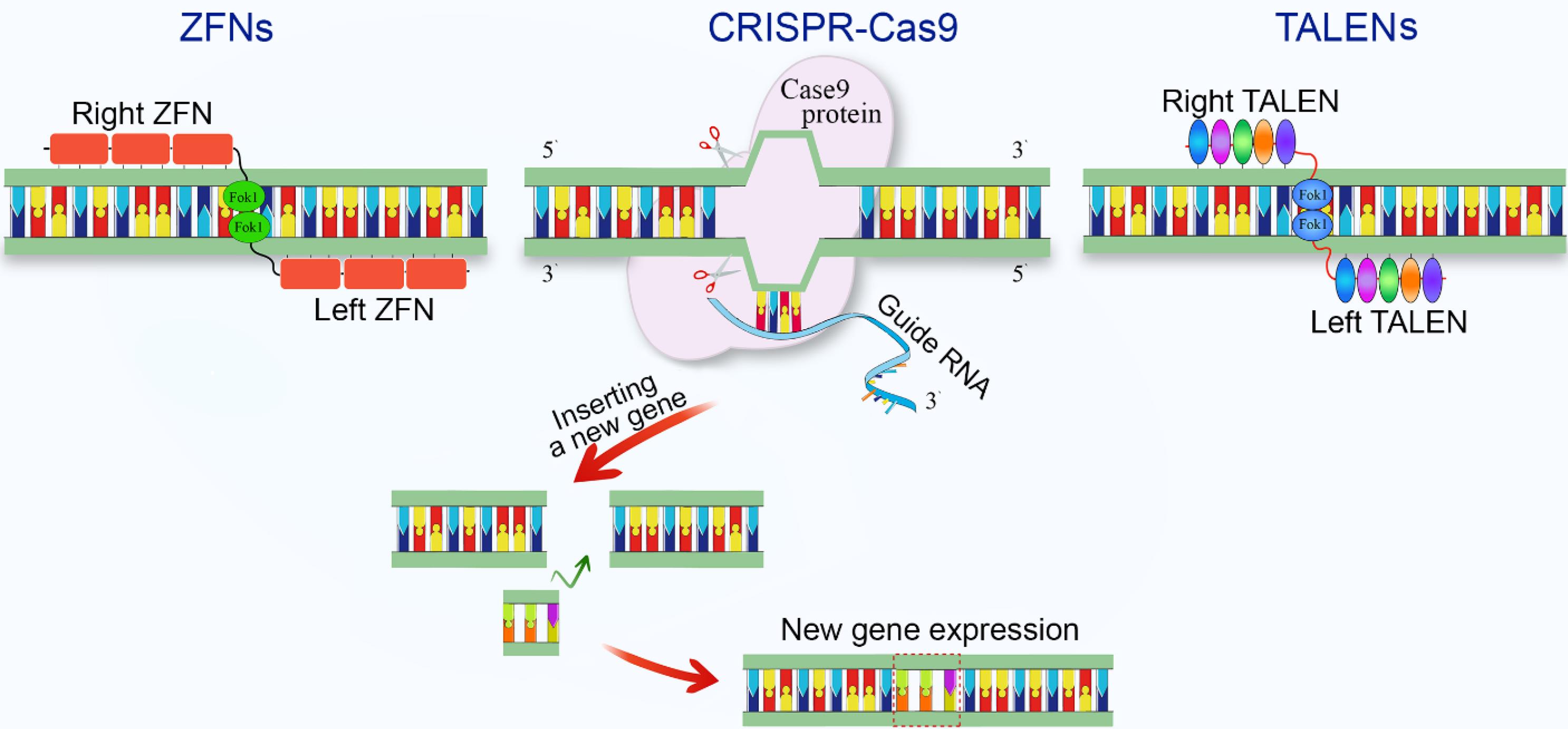
Fig. 1.
Comparison of gene editing techniques and highlighting their mechanisms for targeted DNA modification and gene insertion to enable new gene expression.
.
Comparison of gene editing techniques and highlighting their mechanisms for targeted DNA modification and gene insertion to enable new gene expression.
TALENs
TALENs, another powerful genetic engineering tool widely used in cell manipulation for tissue engineering, consist of a customizable DNA-binding domain fused to a non-specific DNA cleavage domain.34 The DNA-binding domain is composed of repeated modules, each recognizing a single DNA base pair, allowing for highly specific targeting of genomic sequences. This modular structure allows researchers to design TALENs that can bind to virtually any DNA sequence of interest, providing a versatile platform for genetic manipulation in tissue engineering applications.
TALENs have been successfully employed in various tissue engineering applications, including the modification of stem cells for enhanced differentiation and the creation of knockout cell lines for studying gene function in engineered tissues.35 For example, TALENs have been used to modify human pluripotent stem cells to improve their cardiac differentiation efficiency, leading to more robust engineered cardiac tissues.36 In another study, researchers used TALENs to generate knockout cell lines of key extracellular matrix genes in chondrocytes, providing valuable insights into the role of these genes in cartilage tissue engineering.37
One of the advantages of TALENs in tissue engineering is their high specificity and relatively low off-target effects compared to some other genetic engineering techniques.38 This characteristic is particularly important when working with sensitive cell types or when precise genetic modifications are crucial for the desired tissue engineering outcome. Moreover, TALENs have shown high efficiency in generating biallelic modifications, which is often necessary for achieving complete knockout of target genes.39
Despite their advantages, TALENs have limitations in tissue engineering. Their design and assembly are more time-consuming and complex than CRISPR-Cas9, potentially hindering widespread adoption in some tissue engineering applications.40 However, recent advances in TALENs design and assembly protocols have streamlined this process, enhancing accessibility for researchers in the discipline.41
ZFNs
ZFNs were among the first programmable nucleases developed for targeted genome editing and have made significant contributions to cell manipulation in tissue engineering. These engineered proteins consist of a DNA-binding domain composed of zinc finger proteins fused to a DNA cleavage domain.42,43 Each zinc finger recognizes a specific 3-base pair DNA sequence, allowing for the design of ZFNs that target specific genomic loci.
In tissue engineering, ZFNs have been utilized to alter the properties of cells used in tissue constructs, such as improving the immunomodulatory functions of mesenchymal stem cells.44 For instance, researchers have used ZFNs to modify the genome of mesenchymal stem cells to overexpress anti-inflammatory cytokines, enhancing their therapeutic potential in tissue regeneration applications.45 ZFNs have also been employed to create knockout cell lines for studying the role of specific genes in tissue development and regeneration.46
One of the key advantages of ZFNs in tissue engineering is their ability to induce precise genetic modifications with relatively low off-target effects.47,48 This precision is particularly important when working with cells intended for clinical applications, where unintended genetic modifications could have significant consequences. Additionally, ZFNs have shown high efficiency in generating biallelic modifications, which is often necessary for achieving complete knockout of target genes.48,49
However, the complex and time-consuming design and assembly of zinc finger proteins, requiring specialized expertise, have limited their widespread adoption compared to newer techniques like CRISPR-Cas9.50 Furthermore, the range of targetable sequences for ZFNs is more limited compared to TALENs and CRISPR-Cas9, which may restrict their applicability in some tissue engineering scenarios.51,52
Despite these limitations, ZFNs continue to play a role in tissue engineering research, particularly in applications requiring high specificity and a well-established safety profile. As the field of genetic engineering continues to evolve, the integration of ZFNs with other advanced technologies in tissue engineering may lead to new and innovative approaches for cell manipulation and tissue construct development.10,53
Cellular targets for genetic manipulation in tissue engineering
In tissue engineering, various cellular targets have been identified for genetic manipulation to enhance the function, survival, and integration of engineered tissues. These targets play crucial roles in tissue formation and function, and can be broadly categorized into several key areas.
Genes involved in cell proliferation and survival are primary targets for manipulation. By modulating these genes, researchers can enhance cell expansion and longevity in engineered tissues. For instance, overexpression of anti-apoptotic genes like Bcl-2 has been shown to improve the survival of transplanted cells in cardiac tissue engineering.54,55 Similarly, manipulation of cell cycle regulators, such as cyclin-dependent kinases, can promote regulated proliferation in tissue constructs.56 Differentiation factors represent another critical target for genetic manipulation. Altering the expression of key transcription factors or growth factors can guide stem cell differentiation into specific cell types required for various tissues. Overexpression of SOX9 in mesenchymal stem cells, for example, has been used to enhance chondrogenic differentiation for cartilage tissue engineering.57 In another study, genetic manipulation of OCT4 and SOX2 expression in induced pluripotent stem cells was shown to fine-tune their differentiation potential for various tissue engineering applications.58
Extracellular matrix (ECM) production is crucial for the structural integrity and functionality of engineered tissues. Genes encoding ECM proteins or enzymes involved in ECM remodeling are often targeted for modification. For instance, overexpression of elastin in smooth muscle cells has been employed to enhance the mechanical properties of engineered blood vessels.59 Additionally, manipulation of matrix metalloproteinases (MMPs) and their inhibitors can help regulate ECM remodeling in engineered tissues.60
Angiogenic factors are vital targets, especially for large tissue constructs requiring efficient vascularization. Overexpression of angiogenic factors like vascular endothelial growth factor (VEGF), achieved through genetic modification, has been widely used to enhance blood vessel formation in various tissue engineering applications.61 Combinatorial approaches, such as co-expression of VEGF and PDGF-BB, have shown promise in promoting the formation of stable and functional vascular networks in engineered tissues.62
Immunomodulatory molecules are increasingly important targets, particularly for applications involving allogeneic or xenogeneic cell sources. Manipulating cells to express immunomodulatory factors can help reduce immune rejection and promote integration of engineered tissues. For example, genetic modification of mesenchymal stem cells leading to the overexpression of Interleukin 10 (IL-10) has been shown to alter myeloid dendritic cells, reducing immune response and enhancing their immunosuppressive properties.63,64 Similarly, researchers found that genetic modification of mesenchymal stem cells (MSCs) to overexpress angiopoietin 1 (ANGPT1) has been shown to reduce lung inflammation in an ALI/ARDS model. This approach lowered inflammatory markers, including tumor necrosis factor-alpha (TNF-α), interferon-gamma (IFN-γ), interleukin-6 (IL-6), and monocyte chemoattractant protein-1 (MCP-1), demonstrating potential for enhancing MSC-based therapies.65
Lastly, cell adhesion molecules represent another important target for genetic manipulation. Enhancing the expression of these molecules can improve cell-cell and cell-matrix interactions in engineered tissues. Overexpression of N-cadherin in cardiomyocytes, for instance, has been used to enhance electrical coupling in engineered cardiac tissues.66
Gene delivery methods for cell manipulation
Efficient and safe gene delivery is crucial for successful genetic manipulation in tissue engineering. Various methods have been developed and optimized for different cell types and applications, each with its own advantages and limitations.
Viral vectors
Viral vectors, including lentiviruses, adenoviruses, and adeno-associated viruses (AAVs), remain one of the most efficient gene delivery methods due to their high transduction efficiency. Lentiviral vectors are particularly useful for stable, long-term gene expression in both dividing and non-dividing cells.67 AAVs have gained popularity due to their low immunogenicity and ability to transduce a wide range of cell types.68 However, safety concerns regarding insertional mutagenesis and immune responses remain challenges for their clinical application.
Non-viral vectors
In contrast to viral vectors, non-viral vectors offer improved safety profiles but generally lower efficiency. Lipid-based transfection methods, such as lipofection, form complexes with DNA to facilitate cellular uptake. Recent advances in lipid nanoparticle formulations have significantly improved transfection efficiency and reduced cytotoxicity.69 Polymer-based systems, including cationic polymers like poly-ethylene-imine (PEI) and poly-L-lysine (PLL), can condense DNA into nanoparticles for cellular uptake.70
Biodegradable polymers like poly-lactic-co-glycolic acid (PLGA) have shown promise for sustained gene delivery in tissue engineering applications.71
Physical methods
Physical methods for gene delivery have also been explored in tissue engineering. Electroporation uses brief electrical pulses to create temporary pores in cell membranes, allowing DNA entry. It has been effectively used for hard-to-transfect cell types, such as primary neurons and stem cells.72 Another physical method, sonoporation, utilizes ultrasound waves to induce transient pores in cell membrane, which enhance permeability for gene delivery.73
Cell squeezing, also known as microfluidic deformation, represents a promising physical gene delivery method that leverages microfluidic devices to transiently deform cells, facilitating the intracellular delivery of biomolecules, including DNA.74,75 This technique involves passing cells through a narrow constriction or channel within a microfluidic device, inducing a temporary deformation of the cell membrane.76 This deformation creates transient pores or disruptions in the membrane, allowing exogenous molecules, such as DNA, to enter the cell's cytoplasm.77 Unlike other physical methods like electroporation or sonoporation, cell squeezing avoids the use of electrical or ultrasonic forces, potentially reducing cellular stress and toxicity.
Emerging approaches
The use of mRNA instead of DNA for genetic manipulation has gained attention due to its transient nature and reduced risk of genomic integration. Advances in mRNA stabilization and delivery techniques have made this approach increasingly attractive for tissue engineering applications.78 mRNA delivery allows for rapid and transient protein expression, which can be advantageous in certain tissue engineering scenarios where temporary genetic modification is desired.
Nanoparticle-mediated delivery has emerged as a versatile approach for gene delivery in tissue engineering. Various types of nanoparticles, including gold nanoparticles and mesoporous silica nanoparticles, have been developed for this purpose. These systems offer the potential for targeted delivery and controlled release of genetic material.79 Additionally, the use of magnetic nanoparticles for gene delivery, known as magnetofection, has shown promise in enhancing transfection efficiency in various cell types.80
Scaffold-mediated gene delivery represents an innovative approach that combines gene delivery with biomaterial-based tissue engineering strategies. Incorporating genetic material into tissue engineering scaffolds allows for localized and sustained gene delivery. This approach has been particularly useful for promoting tissue regeneration in situ.81 Various techniques, such as layer-by-layer assembly and electrospinning, have been employed to incorporate genetic material into scaffolds for controlled release.82
The choice of gene delivery method depends on various factors, including the target cell type, desired duration of expression, and safety considerations. As the field advances, the development of more efficient and safer gene delivery methods remains a key area of research in genetic manipulation for tissue engineering. The integration of multiple delivery strategies and the development of stimuli-responsive systems are emerging trends that hold promise for enhancing the precision and efficacy of genetic manipulation in tissue engineering applications.83
An overview of the gene delivery methods, including viral, non-viral, physical, and emerging approaches, along with their advantages and applications in tissue engineering, is presented in Fig. 2.
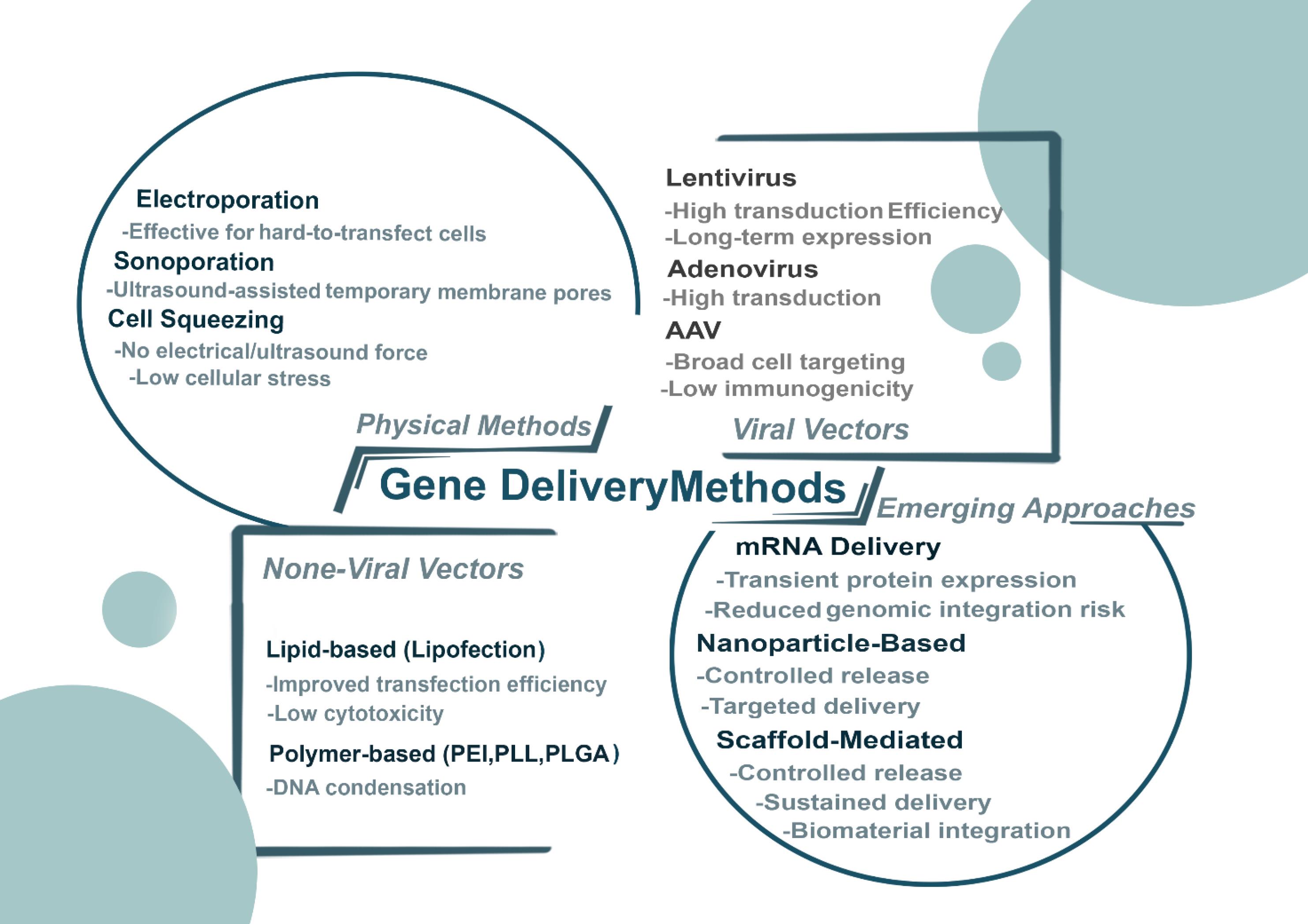
Fig. 2.
Overview of gene delivery methods, including viral, non-viral, physical, and emerging approaches, highlighting their mechanisms, advantages, and applications in gene therapy and biomedical research.
.
Overview of gene delivery methods, including viral, non-viral, physical, and emerging approaches, highlighting their mechanisms, advantages, and applications in gene therapy and biomedical research.
Applications of genetically engineered cells in tissue engineering
Genetic engineering approaches have significantly advanced the field of tissue engineering by enabling precise manipulation of cellular properties and functions. This section explores the applications of genetically engineered cells in various areas of tissue engineering, focusing on cartilage, bone, and cardiac tissue engineering.
The applications of genetically engineered cells in different domains of tissue engineering, such as cartilage, bone, cardiac, vascular, and neural tissues, are summarized in Fig. 3.
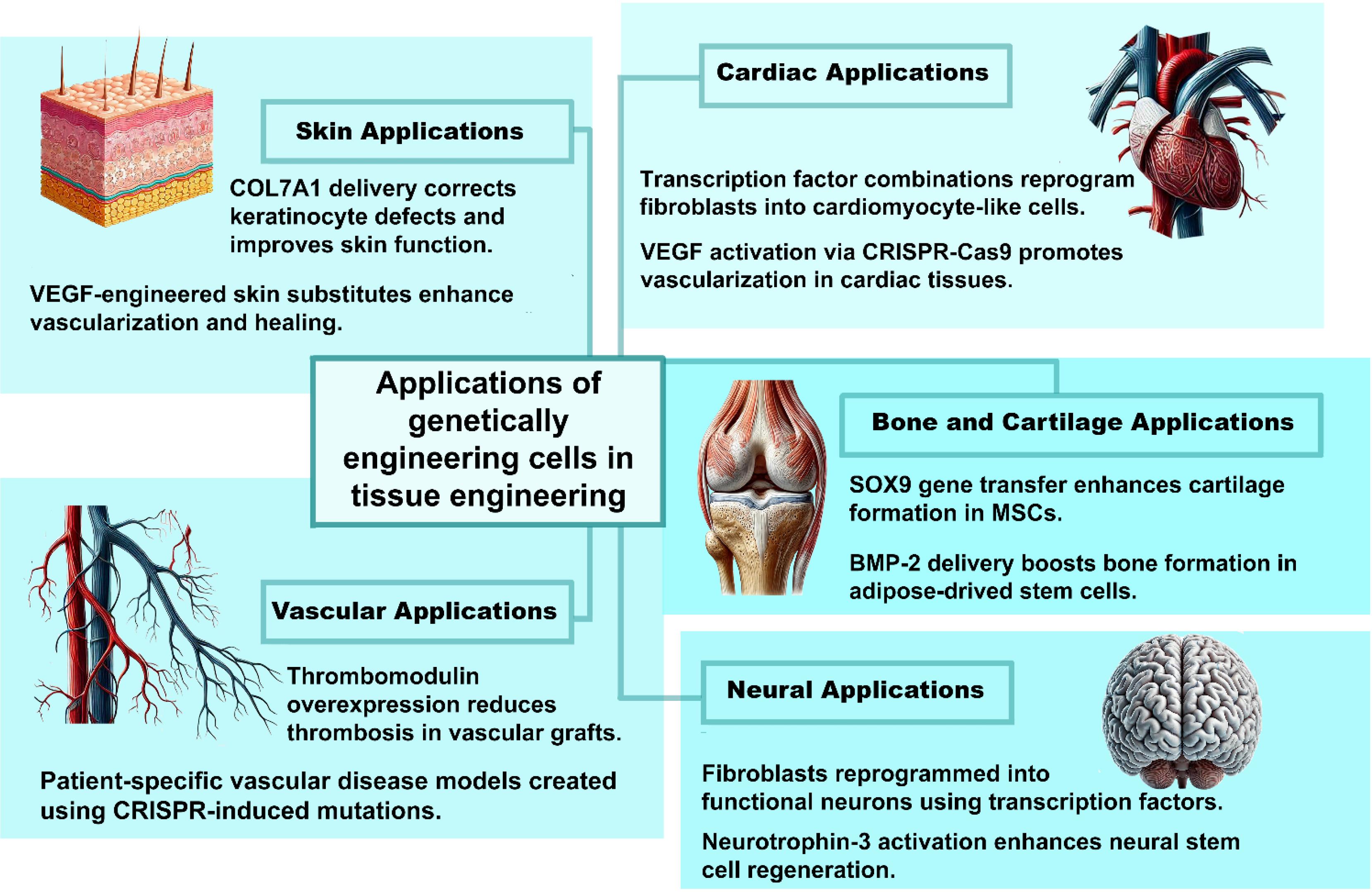
Fig. 3.
Applications of genetically engineered cells in tissue engineering, showcasing techniques like gene knockout, CRISPR-Cas9, and transcription factor reprogramming to improve tissue regeneration in skin, cardiac, vascular, bone, and neural tissues.
.
Applications of genetically engineered cells in tissue engineering, showcasing techniques like gene knockout, CRISPR-Cas9, and transcription factor reprogramming to improve tissue regeneration in skin, cardiac, vascular, bone, and neural tissues.
Cartilage and bone tissue engineering
Cartilage and bone tissue engineering have greatly benefited from genetic engineering approaches, addressing challenges such as limited regenerative capacity and insufficient mechanical properties. Researchers have employed various genetic manipulation techniques to enhance the chondrogenic and osteogenic potential of cells used in these applications.
In cartilage tissue engineering, genetic modification of chondrocytes and MSCs has been extensively studied. One approach involves overexpression of chondrogenic transcription factors such as SOX9, which has been shown to enhance cartilage-specific ECM production.84 For instance, Cao et al used adenoviral-mediated SOX9 gene transfer to enhance chondrogenic differentiation of rabbit bone marrow-derived MSCs, resulting in improved cartilage repair in vivo.85,86
In addition to transcription factor overexpression, CRISPR-Cas9 technology has also been applied to cartilage tissue engineering. As previously discussed,87 this approach demonstrates the potential of genetic engineering in improving cell selection and purity for tissue engineering applications.11,88
In bone tissue engineering, genetic manipulation has been used to enhance osteogenic differentiation and osteogenesis. Overexpression of bone morphogenetic proteins (BMPs), particularly BMP-2 and BMP-7, has been a common strategy to promote osteogenesis.89 For example, Bell et al used a lentiviral vector to deliver BMP-2 to human adipose-derived stem cells, resulting in enhanced bone formation in a critical-sized rat femoral defect model.90-91
Recent advances in genetic engineering have also focused on improving the vascularization of engineered bone tissues. Park et al employed CRISPR-Cas9 to activate the expression of VEGF in MSCs, leading to enhanced angiogenesis and bone regeneration.92
Cardiac tissue engineering
Cardiac tissue engineering aims to develop functional myocardial tissue for repairing damaged hearts or for in vitro disease modeling. Genetic engineering approaches have been instrumental in addressing challenges such as cardiomyocyte maturation, electrical coupling, and tissue vascularization.
One significant application of genetic engineering in cardiac tissue engineering is the direct reprogramming of fibroblasts into cardiomyocytes. This approach offers the potential to convert scar tissue into functional myocardium following a cardiac infarction. For instance, Zhao et al used a combination of transcription factors (Gata4, Mef2c, and Tbx5) delivered via lentiviral vectors to reprogram cardiac fibroblasts into cardiomyocyte-like cells in vivo, improving cardiac function in a mouse model of myocardial infarction.93 Genetic engineering has also been employed to enhance the maturation and functionality of stem cell-derived cardiomyocytes. Luo et al used CRISPR-Cas9 to activate the expression of SERCA2a, a key regulator of calcium handling, in human embryonic stem cell-derived cardiomyocytes. This genetic modification resulted in enhanced calcium reuptake and contractility, addressing a major limitation of stem cell-derived cardiomyocytes for tissue engineering applications.94
Beyond cellular function, genetic engineering has also been used to improve the electrical properties of engineered cardiac tissues. Liao et al used a lentiviral vector to overexpress connexin 43 in neonatal rat cardiomyocytes, enhancing electrical coupling and synchronous contraction in engineered cardiac tissues.95,96 This approach demonstrates the potential of genetic engineering in creating more physiologically relevant tissue constructs. Vascularization of engineered cardiac tissues remains a critical challenge, and genetic engineering approaches have been applied to address this issue. For example, Ye et al used CRISPR-Cas9 to activate the expression of VEGF in human induced pluripotent stem cells-derived (hiPSC) cardiomyocytes, promoting vascularization and improving the survival of engineered cardiac tissues after transplantation in a swine model of myocardial infarction.97,98
The integration of genetic engineering with advanced biomaterials and fabrication techniques has further expanded the possibilities in cardiac tissue engineering. For instance, Gao et al combined CRISPR-Cas9-mediated gene editing with 3D bioprinting to create personalized cardiac tissue patches.99 They engineered hiPSCs to express a genetically encoded voltage indicator, allowing for non-invasive monitoring of electrical activity in the engineered tissues. Within a day, human cardiac muscle patch (hCMP) generated calcium transients and beat synchronously, with contraction/relaxation speeds and transient amplitudes increasing over 7 days. In mice with myocardial infarction, hCMPs improved cardiac function, reduced infarct size/apoptosis, enhanced vascularization, and boosted cell proliferation.100
These applications demonstrate the significant impact of genetic engineering on cardiac tissue engineering. By precisely manipulating cellular properties and cellular functions, genetic engineering approaches have enabled the creation of more functional and physiologically relevant engineered tissues, bringing us closer to addressing critical challenges in regenerative medicine, such as the development of fully functional and transplantable organs.
Neural tissue engineering
Genetic engineering approaches have significantly advanced neural tissue engineering, addressing challenges such as limited neuronal regeneration and impaired functional integration. Researchers have employed various genetic manipulation techniques to enhance neuronal differentiation, axon regeneration, and synaptic plasticity. Direct reprogramming of somatic cells into neurons is a prominent application. Guo et al used a combination of transcription factors (Ascl1, Brn2, and Myt1l) delivered via lentiviral vectors to reprogram human fibroblasts into functional neurons.101,102 This approach offers potential for generating patient-specific neurons for cell replacement therapies or disease modeling.
CRISPR-Cas9 technology has been utilized to enhance the regenerative capacity of neural stem cells. Nori et al employed CRISPR-Cas9 to activate the expression of neurotrophin-3 (NT-3) in hiPSC-derived neural stem cells, resulting in improved neuronal survival and functional recovery in a spinal cord injury model.103,104
Moreover, genetic engineering has been applied to promote axon regeneration in the central nervous system. Axonal damage is an early event in central nervous system disorders, leading to permanent deficits due to poor regeneration. Using an AAV vector to express a dominant-negative form of Unc-51 like autophagy activating kinase 1 (ULK1) (AAV.ULK1.DN), Ribas et al found it enhances axonal regeneration, neuronal survival, and neurite outgrowth. It also promotes axonal protection and neurotransmitter sprouting after spinal cord injury.105
Skin tissue engineering
In skin tissue engineering, genetic engineering approaches have been employed to enhance wound healing, improve skin graft survival, and develop more physiologically relevant skin substitutes.
A significant application is the genetic modification of keratinocytes to enhance their regenerative potential. Hirsch et al used a lentiviral vector to deliver a COL7A1 transgene to keratinocytes from patients with recessive dystrophic epidermolysis bullosa, successfully correcting the genetic defect and improving skin function.106
CRISPR-Cas9 technology has been applied to create genetically modified skin constructs with enhanced properties. Wan et al created a dissolvable microneedle patch loaded with CRISPR-Cas9 gene therapy and glucocorticoids to target the NLRP3 inflammasome. This method allows for precise, localized treatment of conditions like psoriasis and atopic dermatitis by disrupting the NLRP3 gene, thereby improving sensitivity to glucocorticoid therapy. The dual-action approach of CRISPR-mediated gene editing and simultaneous drug delivery shows promise for enhancing the efficacy of treatments for previously uncurable skin disorders.107
Furthermore, genetic engineering has been used to enhance the angiogenic potential of engineered skin substitutes. Supp et al developed genetically modified skin substitutes overexpressing VEGF, leading to improved vascularization and graft take in a porcine wound model.108,109
Vascular tissue engineering
Genetic engineering approaches in vascular tissue engineering have focused on enhancing endothelial cell function, promoting smooth muscle cell differentiation, and improving the mechanical properties of engineered blood vessels.
One key application is the genetic modification of endothelial cells to enhance their anti-thrombogenic properties. Zhang et al employed CRISPR-Cas9 via nanoparticle delivery to robustly edit endothelial cell gene expression in vivo, providing a framework that could be adapted to overexpress thrombomodulin and reduce thrombosis in tissue-engineered vascular grafts.110
Genetic engineering has also been employed to improve the mechanical properties of engineered blood vessels. Rothuizen et al used lentiviral vectors to overexpress elastin in smooth muscle cells, leading to enhanced elasticity and compliance in tissue-engineered vascular grafts.
CRISPR-Cas9 technology has been utilized to create more physiologically relevant models of vascular disease. Li et al used CRISPR-Cas9 to introduce mutations associated with Marfan syndrome in hiPSCs, allowing for the development of patient-specific vascular models for studying disease mechanisms and testing potential therapies.111
The integration of genetic engineering with advanced fabrication techniques has further expanded the possibilities in vascular tissue engineering. For example, Chen et al used single-cell RNA sequencing to profile mouse embryonic ECs, alongside CRISPR-Cas9 loss-of-function in hiPSC-derived arterial and venous ECs.112 This revealed 19 EC subtypes, showing arterial and venous ECs arise from venous-featured capillary precursors, regulated by distinct transcriptional networks, and elucidated EC heterogeneity and arteriovenous differentiation.112
Advanced genetic engineering approaches in tissue engineering
As the field of tissue engineering advances, researchers are exploring increasingly sophisticated genetic engineering approaches to enhance cellular function and tissue performance. This section emphasizes on cutting-edge techniques being applied in tissue engineering.
Epigenetic engineering
Epigenetic engineering involves modifying gene expression patterns without altering the DNA sequence itself. This approach offers the potential for more subtle and reversible control of gene expression over cellular behavior in engineered tissues.113
One significant application of epigenetic engineering in tissue engineering is the modulation of mesenchymal stem cell aging and regenerative capacity through histone modifications, enhancing their potential for therapeutic tissue repair. Haung et al utilized CRISPR-dCas9 fused with histone acetyltransferase p300 to activate NR5A1, GATA4, and DMRT1 in human foreskin fibroblasts, improving their transdifferentiation into testosterone-producing Leydig-like cells compared to dCas9-VP64. This highlights the potential of epigenetic engineering to enhance cell reprogramming for treating male hypogonadism.114
In addition to modulating stem cell differentiation, epigenetic engineering has also been applied to improve the function of mature cells in engineered tissues. Luo et al utilized exosome-delivered CRISPR/dCas9-VP64 to activate pro-quiescence genes in hepatic stellate cells (HSCs), reprogramming them from fibrogenic to quiescent states.115 By leveraging exosomes for targeted delivery, the approach reduced liver fibrosis markers in vitro and in vivo, demonstrating a novel strategy to reverse fibrotic damage and restore liver function without viral vectors or irreversible genome edits.115
Moreover, epigenetic engineering approaches have shown promise in addressing challenges related to cell reprogramming and transdifferentiation. Chakraborty et al used a combination of CRISPR-dCas9-based epigenome editing and traditional transcription factor overexpression to activate the endogenous Myod1 gene in mouse embryonic fibroblasts, effectively reprogramming them into myogenic cells. This approach, which involved fusing two transactivation domains to Cas9, significantly enhanced gene activation, leading to efficient cell phenotype reprogramming.116,117
RNA interference and microRNA-based approaches
RNA interference (RNAi) and microRNA (miRNA)-based approaches offer powerful tools for post-transcriptional regulation of gene expression in engineered tissues.
In cartilage tissue engineering, Legendre et al successfully transfected dedifferentiated chondrocytes with Small interfering RNAs (siRNA) targeting COL1A1, achieving prolonged gene knockdown in mouse chondrocytes within agarose hydrogels and human chondrocytes in collagen sponges cultured with BMP-2.118 This approach demonstrates the potential of RNAi in preserving cell phenotypes in engineered tissues.
Similarly, miRNA-based approaches have also been applied to enhance vascularization in engineered tissues. Devalliere et al developed a scaffold system for controlled delivery of miR-132, promoting angiogenesis and improving the survival of engineered tissues after implantation.119
Furthermore, combinatorial approaches using multiple miRNAs have shown promise in directing cell fate. Paolleti et al used a cocktail of miRNAs to enhance the direct reprogramming of fibroblasts into cardiomyocytes, expanding the possibilities for cardiac tissue engineering.120
Optogenetics and chemogenetics for spatiotemporal control
Optogenetics and chemogenetics provide unprecedented spatiotemporal control over cellular functions in engineered tissues, allowing researchers to modulate tissue behavior with light or specific chemical compounds.
In cardiac tissue engineering, Williams and Entcheva used optogenetic approaches to control the electrical activity of cardiomyocytes in engineered heart tissues, enabling precise pacing and arrhythmia termination.120 This approach allows for creating responsive cardiac tissues for drug testing or therapeutic applications.
Chemogenetic approaches have also been applied in neural tissue engineering. Chen et al used designer receptors exclusively activated by designer drugs (DREADDs) to modulate the activity of transplanted neural progenitor cells in a spinal cord injury model, enhancing functional recovery.88
In addition to DREADDs, the integration of optogenetics with advanced biomaterials has significantly expanded possibilities for controlling engineered tissues. For instance, Hammer et al developed light-responsive hydrogels utilizing light-sensing proteins, enabling dynamic control over matrix properties and cell behavior in 3D tissue constructs.121 Additionally, research on photon upconversion hydrogels has demonstrated the potential for 3D optogenetics, allowing for precise modulation of cellular activities within engineered tissues.122 These advancements highlight the potential of combining optogenetic tools with responsive biomaterials to achieve precise spatiotemporal control in tissue engineering applications.
Synthetic biology and gene circuits for smart tissues
Synthetic biology approaches, including the design of artificial gene circuits, offer the potential to create "smart" engineered tissues capable of sensing and responding to environmental cues.
In this context, Saxena et al developed a synthetic gene circuit that enables engineered pancreatic β-cells to sense glucose levels and produce insulin in a physiologically relevant manner, potentially improving glucose homeostasis in engineered pancreatic tissues for diabetes treatment.123
Beyond glucose sensing, Biomaterial-based delivery of synthetic gene circuits has also proven effective in tissue engineering applications. Shao et al created a hydrogel system for controlled delivery of synthetic transcription factors, enabling spatiotemporal control of gene expression in engineered tissues.124
Moreover, synthetic biology approaches have been applied to enhance the immunomodulatory properties of engineered tissues. Pferdehirt et al designed a synthetic gene circuit that allows mesenchymal stem cells to sense inflammatory signals and respond by producing anti-inflammatory factors, potentially improving the integration and function of engineered tissues in inflammatory environments.125
The integration of these advanced genetic engineering approaches with traditional tissue engineering strategies holds great promise for creating more sophisticated, functional, and responsive engineered tissues. As these technologies continue to evolve, they hold significant potential for addressing long-standing challenges in tissue engineering and regenerative medicine.
Integration of genetic engineering with other tissue engineering strategies
The convergence of genetic engineering with advanced tissue engineering strategies has led to significant advancements in creating complex and functional engineered tissues. This integration providesunprecedented control over tissue architecture, function, and responsiveness to environmental cues.
3D bioprinting with genetically engineered cells
The combination of 3D bioprinting technology and genetically engineered cells offers precise spatial control over tissue architecture and cellular function. This approach allows for the creation of complex tissue structures with enhanced regenerative capabilities, improved vascularization, and tissue-specific functionalities.126 Recent advances have demonstrated the potential of this integrated approach in various applications, including bone regeneration, vascularized tissue constructs, and personalized disease models.127,128
Furthermore, the integration of CRISPR-Cas9 gene editing with 3D bioprinting has further expanded the potential for creating sophisticated tissue models. This combination enables the development of patient-specific disease models and the study of genetic disorders in a physiologically relevant 3D context.129
Microfluidic systems for genetic manipulation and tissue engineering
Microfluidic platforms have revolutionized genetic manipulation and tissue culture by providing precise control over the cellular microenvironment and enabling high-throughput experiments. The integration of genetic engineering with microfluidics has facilitated several advancements, including the optimization of gene editing protocols, the study of gene function in physiologically relevant conditions, and the development of advanced organ-on-a-chip models.130
These systems allow for continuous delivery of genetic manipulation components, real-time monitoring of editing efficiency, and the creation of complex tissue models with genetically engineered cells. The application of microfluidic technologies in combination with genetic engineering has significantly advanced the field of disease modeling and drug screening.131,132
Biomaterial-mediated gene delivery in tissue engineering
The synergy between genetic engineering and advanced biomaterials has led to the development of sophisticated systems for controlled and localized gene delivery in engineered tissues. These approaches enable simultaneous gene editing and tissue regeneration, opening new possibilities for in situ tissue engineering and regenerative medicine applications.133
Biomaterial-mediated gene delivery systems have addressed challenges in transfection efficiency, particularly for hard-to-transfect primary cells. These systems often incorporate stimuli-responsive elements, allowing for spatiotemporal control over gene expression in engineered tissues.133
Building on these advancements, the integration of biomaterial-mediated gene delivery with other tissue engineering strategies, such as 3D bioprinting, has further expanded the possibilities for creating complex, multifunctional tissue constructs with enhanced regenerative potential.134
Organoid development using genetically engineered cells
The combination of genetic engineering with organoid technology has significantly advanced the field of developmental biology, disease modeling, and drug screening. Genetically engineered organoids provide powerful tools for studying complex developmental disorders, modeling genetic diseases, and testing potential therapies in a physiologically relevant 3D context.135
CRISPR-Cas9 technology is widely used in organoid development, enabling the introduction of disease-specific mutations, the creation of reporter lines for real-time monitoring of cellular processes, and the enhancement of organoid functionality.136
The integration of genetic engineering with organoid technology has also facilitated the development of more sophisticated tissue models for drug screening and personalized medicine applications. These advanced organoid systems offer improved predictive power for drug efficacy and toxicity studies.137
In conclusion, the integration of genetic engineering with other advanced tissue engineering strategies has opened new avenues for creating more sophisticated, functional, and physiologically relevant engineered tissues. This synergistic approach is redefining the limits in tissue engineering and regenerative medicine, offering promising solutions for addressing critical challenges in healthcare and biological research.
Challenges and considerations in genetic engineering for tissue engineering
While genetic engineering approaches offer immense potential for advancing tissue engineering,
their safe and effective implementation requires addressing several challenges and considerations. This section discusses key issues related to the application of genetic engineering in tissue engineering.
Despite significant advancements, regulatory hurdles persist. The classification of gene-edited tissues as advanced therapy medicinal products (ATMPs) varies across countries, complicating international clinical trials. Furthermore, regulatory gaps exist around long-term safety and monitoring, especially for in vivo gene editing. Public perception and ethical debates continue to influence the pace of approval and funding.138,139
Off-target effects and safety concerns
One of the primary concerns in genetic engineering for tissue engineering is the potential for off-target effects. These unintended genomic modifications can lead to unforeseen consequences, including altered cellular behavior, oncogenic transformations, or immune responses.140 Recent advancements in gene-editing technologies, such as high-fidelity CRISPR-Cas9 variants, have significantly reduced off-target effects, but complete elimination remains challenging.141
However, ensuring the safety of genetically engineered cells and tissues is paramount for clinical translation. Comprehensive genomic and functional analyses are necessary to detect potential aberrations and assess long-term safety.142 Additionally, the development of inducible or reversible genetic modification systems may provide enhanced control and safety measures for engineered tissues.143
Long-term stability and functionality of genetically engineered tissues
Maintaining the long-term stability and functionality of genetically engineered tissues poses another significant challenge. Epigenetic changes, genetic drift, and in vivo selective pressures can affect introduced gene expression and function over time.144 Strategies to address these issues include the use of stable genomic integration techniques, selection of appropriate promoters, and the development of self-regulating gene circuits.145
Furthermore, ensuring the proper integration and function of engineered tissues within the host environment is crucial. This includes considerations such as vascularization, innervation, and appropriate cellular interactions.146 The development of more sophisticated in vitro models and advanced imaging techniques can help assess and optimize long-term tissue functionality.147
Scalability and reproducibility issues
Translating genetic engineering approaches from small-scale laboratory experiments to clinically relevant tissue engineering applications presents challenges in scalability and reproducibility. Variability in cell sources, genetic modification efficiencies, and tissue culture conditions can impact the consistency of engineered tissues.148
Therefore, addressing these issues requires the development of standardized protocols, quality control measures, and automation technologies. The integration of high-throughput screening methods and machine learning algorithms can help optimize genetic engineering strategies and improve reproducibility.149 Additionally, the development of scalable bioreactor systems and advanced manufacturing techniques is essential for producing clinical-scale quantities of engineered tissues. In this context, the Ambr 250, with its high automation and efficiency, serves as a valuable scale-down model, supporting process optimization and clone selection while minimizing resource utilization.150
Ethical considerations and regulatory landscape
The application of genetic engineering in tissue engineering raises important ethical considerations, particularly regarding human embryonic stem cells, germline modifications, and the creation of chimeric organisms.151 Clear ethical guidelines and public engagement are necessary to address these concerns and ensure responsible research and development in the field.
The regulatory landscape for genetically engineered tissues is complex and evolving. Current regulatory frameworks may not be fully equipped to address the unique challenges posed by these advanced therapies. Harmonization of international regulations, development of specific guidelines for genetically engineered tissues, and close collaboration between researchers, clinicians, and regulatory bodies are crucial for facilitating the clinical translation of these technologies.152
Furthermore, issues of intellectual property, data sharing, and equitable access to genetically engineered tissue therapies require careful consideration to ensure broad societal benefit.153
In conclusion, while genetic engineering offers tremendous potential for advancing tissue engineering, addressing these challenges and considerations is crucial for realizing the full promise of these technologies. Continued research, interdisciplinary collaboration, and ongoing dialogue with stakeholders are essential for overcoming these hurdles and translating genetically engineered tissue therapies into clinical practice. An overview of the gene editing techniques and their applications, advantages, and limitations in tissue engineering can be found in Table 1.
Table 1.
Overview of gene editing and delivery techniques: applications, target tissues, advantages, and limitations
|
Technique
|
Application
|
Tissue
|
Advantages
|
Limitations
|
| CRISPR-Cas9 |
Gene editing, disease modeling, enhanced regeneration, multiplex editing. |
Cartilage, Bone, Cardiac, Neural, Skin, Vascular |
High precision, multiplex editing, versatility, base/prime editing advancements. |
Off-target effects, delivery challenges, ethical concerns, transient vs. stable expression trade-offs. |
| TALENs |
Precise genome modifications, disease modeling, immune cell engineering. |
Engineered iPSC-derived tissues, Cancer, |
High specificity, low off-target effects, suitable for sensitive cell types. |
Complex design/assembly, less efficient than CRISPR, limited adoption due to technical complexity. |
| ZFNs |
Targeted genome editing, immunomodulation. |
Regenerative approaches |
High specificity, established safety profile. |
Limited target range, complex design, largely replaced by CRISPR. |
Lentiviral vectors
gene delivery |
Stable gene delivery, long-term expression. |
Cartilage, Bone, Cardiac, Neural |
High transduction efficiency, stable integration. |
Risk of insertional mutagenesis, immune responses, limited payload capacity. |
| AAV gene delivery |
Axon regeneration, vascular grafts, muscle repair. |
Neural, Vascular, Muscle |
Low immunogenicity, broad tropism, FDA-approved for multiple therapies. |
Limited payload capacity, pre-existing immunity in some patients, transient expression. |
| RNA interference (RNAi) |
Suppressed dedifferentiationAnd Phenotype preservation, anti-fibrotic therapy. |
Cartilage, Liver, Cancer |
Transient effects, reduced genomic integration risk. |
Low delivery efficiency, need for repeated dosing, off-target mRNA silencing. |
| Synthetic gene circuits |
Dynamic environmental sensing (e.g., glucose regulation, inflammation control). |
Pancreatic, Immune-modulated tissues |
Programmable responses, "smart" tissue functionality. |
Complexity, potential immunogenicity, long-term stability concerns. |
| 3D Bioprinting + CRISPR |
Personalized tissue constructs, vascularized models. |
Cardiac, Bone, Vascular |
Spatial control, integration with patient-specific edits. |
Scalability challenges, high cost, regulatory hurdles for clinical translation. |
| Optogenetics |
Spatiotemporal control of tissue activity. |
Neural, Cardiac |
Precise temporal control, non-invasive modulation. |
Requires invasive light delivery, limited depth penetration in tissues. |
| Epigenetic engineering |
Reversible gene expression control, stem cell reprogramming. |
Bone, Liver, Neural |
Reversible modifications, avoids permanent DNA changes. |
Off-target epigenetic effects, transient efficacy, complex delivery requirements. |
Future perspectives
The integration of genetic engineering with tissue engineering continues to evolve rapidly, offering exciting possibilities for addressing unmet medical needs and advancing our understanding of biological systems. Furthermore, the growing pipeline of commercial products and clinical trials underscores the rapid translation of genetic engineering technologies into real-world therapies. This section explores emerging trends and future directions in the field.
Emerging technologies in genetic engineering for tissue engineering
Recent advancements in genetic engineering technologies are expanding the toolkit available for tissue engineering applications. Base editing and prime editing techniques offer more precise and versatile genome modifications with reduced off-target effects compared to traditional CRISPR-Cas9 systems.154 These approaches may enable correcting disease-causing mutations and fine-tuning cellular functions in engineered tissues.
The development of novel delivery methods, such as engineered extracellular vesicles and cell-penetrating peptides, is improving the efficiency and safety of genetic manipulation in various cell types.155 These strategies may overcome current limitations in modifying hard-to-transfect cells and enable more effective in vivo gene editing.
Emerging synthetic biology approaches, including the design of artificial genetic circuits and synthetic genomes, offer unprecedented control over cellular behavior and tissue function.156 These technologies could lead to the creation of "smart" engineered tissues capable of responding dynamically to environmental cues and performing complex biological functions.
Personalized medicine and patient-specific engineered tissues
Recent clinical advances demonstrate that the convergence of genetic engineering, tissue engineering, and personalized medicine is already enabling tailored therapeutic approaches. The ability to generate patient-specific iPSCs and precisely modify their genomes opens new avenues for creating personalized tissue grafts with enhanced functionality and reduced immunogenicity.157
Beyond personalized tissue grafts, advanced organoid technologies combined with genetic engineering are enabling the development of patient-specific disease models for drug screening and personalized treatment strategies.158 These miniature tissue constructs can recapitulate complex genetic disorders and provide valuable insights into individual disease mechanisms.
The integration of genetic engineering with 3D bioprinting technologies facilitates the creation of personalized tissue constructs with precise spatial control over cellular composition and function.159 This approach holds promise for developing custom-designed tissue replacements tailored to individual patient needs.
In vivo genetic engineering for tissue regeneration
Direct in vivo genetic engineering for tissue regeneration represents an exciting frontier in the field. Advancements in targeted delivery systems and tissue-specific gene editing approaches are enabling localized genetic modifications in native tissues.160 This strategy may allow for the stimulation of endogenous repair mechanisms and the reprogramming of resident cells to promote tissue regeneration.
The development of switchable gene circuits and chemically inducible systems offers the potential for temporal control over genetic modifications in vivo.161 These approaches may enable the fine-tuning of regenerative processes and provide enhanced safety measures for in vivo genetic engineering applications.
Combining in vivo genetic engineering with biomaterial-based delivery systems is showing promise for controlled and sustained genetic manipulation in target tissues.162 This integrated approach may enhance the efficacy and durability of genetic interventions for tissue regeneration.
Combining genetic engineering with stem cell technologies
The synergy between genetic engineering and stem cell technologies continues to drive innovation in tissue engineering. CRISPR-Cas9 screening approaches are facilitating the identification of key regulators of stem cell fate and differentiation, providing new targets for optimizing tissue-specific cell derivation.163
Genetic engineering strategies are being employed to enhance the functionality and survival of stem cell-derived tissues. For example, the introduction of anti-apoptotic genes or factors promoting vascularization can improve the engraftment and long-term viability of transplanted tissues.164
Furthermore, the development of genetically engineered "universal" stem cell lines, designed to evade immune recognition, holds promise for creating off-the-shelf tissue products suitable for allogeneic transplantation.165 This approach may address challenges related to scalability and accessibility of engineered tissues.
In conclusion, genetic engineering in tissue engineering has a bright future, with emerging technologies and interdisciplinary approaches opening new possibilities for creating more sophisticated, functional, and personalized engineered tissues. As the field continues to advance, addressing ethical considerations, refining regulatory frameworks, and ensuring equitable access to these technologies will be crucial for realizing their full potential in regenerative medicine and beyond. This potential is evident in the fascinating studies and commercial products that have already emerged in the field Recent developments in clinical applications underscore the translational potential of genetic engineering. For example, CRISPR-based therapies such as CASGEVY (exa-cel) for sickle cell disease, Edit-101 for inherited retinal diseases, and NTLA-2001 for transthyretin amyloidosis have entered clinical trials. These therapies represent early real-world validation of the technologies discussed in this review. A summary of selected examples is included in Table 2.
Table 2.
Current commercial products and clinical trials of gene editing and therapy technologies
|
Technique
|
Product
|
Company/ Developer
|
Description
|
Application
|
Status
|
Ref
|
| CRISPR-Cas9 |
CTX131: Anti-CD70 allogenic CAR-T |
CRISPR therapeutics |
Designed to enhance CAR-T potency and reduce CAR-T exhaustion |
Treatment of solid tumors and hematological malignancies |
Clinical trials (Phase 1/2) |
166
|
|
|
CTX320: Lp(a) for cardiovascular disease |
CRISPR therapeutics |
In vivo gene-editing therapy using lipid nanoparticle (LNP) delivery of cas9 mRNA and gRNA to the liver to reduce expression of Lp(a) |
Reduce plasma Lp(a) levels in humans |
Clinical trials |
167,168
|
|
|
CASGEVY (exa-cel) |
CRISPR therapeutics & vertex |
Autologous, ex vivo CRISPR/Cas9 gene-editing therapy to produce fetal hemoglobin in red blood cells. |
Treatment of sickle cell disease and beta-thalassemia. |
FDA-approved (2023) |
169
|
|
|
Edit-101 |
Editas Medicine |
In vivo CRISPR therapy for retinal degeneration (LCA10) |
Repairs Retinal Tissue Via Gene Editing |
Clinical trials (Phase 1/2) |
170,171
|
|
|
Nexiguran Ziclumeran (NTLA-2001) |
Intellia therapeutics |
In vivo CRISPR therapy targeting the gene encoding transthyretin (TTR), reducing serum TTR levels. |
Treatment for transthyretin amyloidosis. |
Clinical trials (Phase 1) |
172,173
|
| TALENs |
UCART19 |
Cellectis |
TALEN-edited allogeneic CAR-T cells for leukemia, modifying immune cells for tissue repair. |
Leukemia treatment, immune tissue repair. |
Clinical trials (Phase 1) |
174,175
|
|
|
Cemacabtagene Ansegedleucel (Allo-501a) |
Allogene Therapeutics
& Cellectis |
Anti-CD19 allogeneic CAR-T product for large B-cell lymphoma (LBCL). |
Treatment for early diagnosed LBCL. |
Clinical trials (Phase 1/2) |
176
|
|
|
CYT001 |
Cytlimic & nec corp |
A novel cancer vaccine using AI-designed TALEN-edited T-cells targeting solid tumors. |
Enhances immune response against cancer cells |
Clinical trials (Phase 1) |
177
|
| ZFNs |
SB-318 |
Sangamo Therapeutics |
ZFN Therapy For MPS I Via Liver Gene Editing. |
Liver tissue function improvement. |
Clinical trials (Phase 1/2) |
178
|
|
|
SB-FIX |
Sangamo therapeutics |
ZFN therapy for hemophilia B, editing hepatocytes DNA under the control of the albumin promoter. |
Blood-related tissue repair. |
Clinical trials (Phase 1/2) |
179
|
|
|
SB-728 |
University of pennsylvania |
ZFNs used to genetically modify autologous T-cells by removing CCR5 protein to prevent HIV entry. |
Enhances immune function and prolongs CD4 + T-cell survival. |
Clinical trials (Phase 1)
completed |
180
|
|
|
Isaralgagene civaparvovec
(ST-920) |
Sangamo Therapeutics |
A liver-targeted ZFN-mediated and AAV2/8-mediated vector carrying the cDNA for human α-Gal |
Improves kidney/heart tissue function |
Clinical trials (Phase 1/2) |
181-183
|
Lentiviral vectors
gene delivery |
AVXS-101 (Zolgensma) |
Novartis Pharmaceuticals |
Lentiviral vector delivering the SMN gene to treat SMA by replacing the missing SMN1 gene. |
Treatment of spinal muscular atrophy (SMA). |
FDA-approved (2019)
Clinical trials (Phase 3 long-term follow-up) |
184,185
|
|
|
CTL019 (kymriahtm) |
Novartis Pharmaceuticals |
Lentiviral vectors engineering T-cells for cancer, particularly B-cell acute lymphoblastic leukemia (ALL). |
Immune cell modification for B-cell ALL. |
FDA-approved (2017) |
186-188
|
|
|
Axicabtagene Ciloleucel (YESCARTA) |
Kite pharma |
Lentiviral vector-based CAR-T therapy modifies patient T-cells to create CAR-T cells, activating T-cell proliferation, cytokine release, and apoptosis of CD19-expressing cancer cells in large B-cell lymphoma. |
Enhances immune tissue mechanisms and cancer treatment. |
FDA-approved (2017) |
189,190
|
| AAV gene delivery |
Luxturna |
Spark Therapeutics |
AAV2 vector delivering RPE65 gene for retinal dystrophy, replacing the mutated gene. |
Retinal tissue function restoration. |
FDA-approved (2017) |
191,192
|
|
|
ELEVIDYS (SRP-9001) |
Sarepta therapeutics |
AAV therapy delivering a gene encoding micro-dystrophin for Duchenne muscular dystrophy, restoring dystrophin expression. |
Treatment of duchenne muscular dystrophy |
FDA-approved (2023) |
193,194
|
|
|
Hemgenix |
CSL Behring |
AAV5 vector delivering the FIX gene to treat hemophilia B, promoting long-term clotting factor production. |
Blood tissue regeneration for hemophilia B. |
FDA-approved (2022),Ongoing observational study (2023-2043) |
195,196
|
| RNA interference (RNAi) |
Onpattro (patisiran) |
Alnylam pharmaceuticals |
siRNA therapy targeting transthyretin (TTR) to silence the mutated TTR gene, reducing amyloid deposits that cause organ damage in amyloidosis patients. |
Promotes liver tissue health by using lipid nanoparticles to deliver siRNA directly to the liver. |
FDA-approved (2018) |
197,198
|
|
|
Leqvio (inclisiran) |
Novartis |
Long-acting siRNA targeting and reducing PCSK9 production in the liver for hypercholesterolemia. |
Vascular tissue maintenance and LDL cholesterol reduction. |
FDA-approved (2021) |
199,200
|
|
|
Givosiran |
Alnylam Pharmaceuticals |
siRNA therapy for acute hepatic porphyria, silencing ALAS1 expression to decrease porphyrin precursors. |
Enhances liver tissue function, reducing attack frequency. |
FDA-approved (2019) |
201,202
|
| Synthetic gene circuits |
Synnotch receptor |
Okada Lab, University of California |
Synthetic receptor system for programmable cell behavior, enhancing specificity and tumor cell killing. |
Tumor-specific CAR-T for glioblastoma in preclinical models. |
Clinical trials (Phase 1) |
203,204
|
|
|
Logic-gated CAR-T
(SENTI-202) |
Senti biosciences |
Targeting CD33 and/or FLT3 in hematological malignancies with an inhibitory CAR (iCAR) for tissue protection. |
Enhances tissue-specific immune. |
Clinical trials (Phase 1) |
205,206
|
| 3D Bioprinting + CRISPR |
Organovo tissue models |
Organovo |
Bioprinted tissues with CRISPR-modified cells (e.g., liver). |
Mimics native tissue structure, making it ideal for toxicology and disease modeling. |
Commercially available |
207
|
|
|
3d Bioprinted LNO (Lymph Node Organoids) |
Prellis Biologics |
Via two-photon holography bioprinter enable to generate lymph node organoids, which recapture the complex biology of immune responses |
In vitro High-resolution bioprinting with gene-edited cells |
Research and development |
208
|
|
|
Full-stack
Tissue therapeutic
Platform |
Aspect biosystems |
a platform designed to develop implantable tissues, offering transformative potential for treating severe metabolic and endocrine disorders. |
Supports tissue model development |
Research/Commercial tools |
209
|
| Optogenetics |
Neurolux optogenetics kits |
Neurolux |
Implantable optogenetic devices for neural tissue stimulation |
Neural tissue regulation. |
Commercially available |
210,211
|
|
|
Channelrhodopsin tools |
Thorlabs |
Optogenetic tools for precise neural tissue manipulation. |
Enables precise neural tissue manipulation |
Commercially available |
212
|
| Epigenetic engineering |
Tempo platforms |
Tune therapeutics |
Epigenetic editing platform for chronic diseases, enabling gene silencing and modulation. |
Chronic disease treatment via gene modulation. |
Pre-clinical |
213,214
|
|
|
PCSK9 epigenetic editor platform |
Chroma medicine |
Epigenetic editing platform for silencing/activating PCSK9 genes to maintain reduced LDL-C levels. |
Long-term LDL-C regulation and cardiovascular health. |
Pre-clinical |
215,216
|
Conclusion
The integration of genetic engineering approaches with cell manipulation-based tissue engineering has ushered in a new era of possibilities for regenerative medicine and personalized therapies. This narrative review has explored the diverse applications, cutting-edge techniques, and future perspectives of genetic engineering in tissue engineering, highlighting its transformative potential in addressing critical challenges in the field.
Throughout this review, we have seen how genetic engineering techniques, particularly CRISPR-Cas9 and its variants, have revolutionized our ability to precisely manipulate cellular functions and behaviors. These advancements have enabled the creation of more sophisticated, functional, and physiologically relevant engineered tissues across various applications, including cartilage, bone, cardiac, neural, skin, and vascular tissue engineering.
The integration of genetic engineering with other advanced technologies, such as 3D bioprinting, microfluidics, and biomaterial-mediated gene delivery, has further expanded the possibilities for creating complex tissue constructs with enhanced functionality. These synergistic approaches offer unprecedented control over tissue architecture, cellular composition, and spatiotemporal gene expression, paving the way for more accurate disease models and improved therapeutic outcomes.
Emerging technologies in genetic engineering, such as base editing and prime editing, hold promise for even more precise and versatile genome modifications with reduced off-target effects. The development of synthetic biology approaches and artificial genetic circuits offers the potential to create "smart" engineered tissues capable of responding dynamically to environmental cues and performing complex biological functions.
The convergence of genetic engineering, tissue engineering, and personalized medicine is opening new avenues for tailored therapeutic approaches. Patient-specific engineered tissues, derived from genetically modified induced pluripotent stem cells, have the potential to revolutionize transplantation medicine by providing immunocompatible grafts with enhanced functionality. Moreover, the application of genetic engineering in organoid development is facilitating the creation of more accurate disease models for drug screening and personalized treatment strategies.
Despite these remarkable advancements, several challenges remain to be addressed. Off-target effects, long-term stability of genetic modifications, scalability, and reproducibility issues continue to be areas of active research. The development of more sophisticated in vitro models, advanced imaging techniques, and standardized protocols will be crucial for overcoming these hurdles and translating genetically engineered tissue therapies into clinical practice.
The ethical implications and regulatory landscape surrounding genetically engineered cells for tissue engineering applications require careful consideration. As the field progresses, it is essential to maintain an open dialogue between researchers, clinicians, policymakers, and the public to ensure responsible development and implementation of these technologies.
Looking to the future, in vivo genetic engineering for tissue regeneration represents an exciting frontier, offering the potential for localized and controlled genetic modifications to stimulate endogenous repair mechanisms. The combination of genetic engineering with stem cell technologies continues to drive innovation, promising the development of off-the-shelf tissue products and improved engraftment strategies.
In conclusion, genetic engineering approaches have become indispensable tools in cell manipulation-based tissue engineering, offering unprecedented control over cellular function and tissue development. As these technologies continue to evolve and integrate with other advanced approaches, they hold immense potential for addressing unmet medical needs, advancing our understanding of biological systems, and revolutionizing regenerative medicine. While challenges remain, the rapid pace of innovation in this field suggests a future where personalized, genetically engineered tissue therapies become a reality, fundamentally transforming the landscape of healthcare and tissue engineering.
Review Highlights
What is the current knowledge?
-
Genetic engineering has revolutionized tissue engineering by enhancing cellular functions and tissue development.
-
CRISPR-Cas9 is widely used to modify genes and study diseases in engineered tissues.
-
Off-target effects and the stability of genetic modifications remain critical concerns in gene editing for tissue engineering.
What is new here?
-
Integration of genetic engineering with advanced tissue engineering methods is enhancing tissue functionality and complexity.
-
Advances in gene delivery methods, including biomaterial-based systems, enable controlled and localized gene expression in tissues.
-
The convergence of synthetic biology and tissue engineering is enabling the development of "smart" tissues that can dynamically respond to environmental signals.
Competing Interests
The authors declare that there are no conflicts of interest or competing interests related to this work.
Data Availability Statement
No new data or code were generated during the study. The article is based on a review of previously published literature, and all data supporting the findings are available in the referenced sources.
Declaration of AI Use in Scientific Writing
The authors used ChatGPT AI to improve the readability, academic tone, and language quality of the text. No original data, analysis, or substantive content was generated by AI.
Ethical Approval
As this article is a review of existing literature and does not involve human or animal subjects, ethical approval was not required.
References
- Han F, Wang J, Ding L, Hu Y, Li W, Yuan Z. Tissue engineering and regenerative medicine: achievements, future, and sustainability in Asia. Front Bioeng Biotechnol 2020; 8:83. doi: 10.3389/fbioe.2020.00083 [Crossref] [ Google Scholar]
- Kasoju N, Sunilkumar A. Convergence of tissue engineering and sustainable development goals. Biotechnol Sustain Mater 2024; 1:20. doi: 10.1186/s44316-024-00021-y [Crossref] [ Google Scholar]
- Kolanu ND. CRISPR-Cas9 gene editing: curing genetic diseases by inherited epigenetic modifications. Glob Med Genet 2024; 11:113-22. doi: 10.1055/s-0044-1785234 [Crossref] [ Google Scholar]
- Sharami SH, Vojoudi E, Zarei-Behjani Z, Hasanzadi A, Shoarishoar SS, Vaez A. From challenges to solutions: the promise of regenerative medicine and tissue engineering in restoring female reproductive health. Heliyon 2025; 11:e43484. doi: 10.1016/j.heliyon.2025.e43484 [Crossref] [ Google Scholar]
- Ullah S, Zainol I. Fabrication and applications of biofunctional collagen biomaterials in tissue engineering. Int J Biol Macromol 2025; 298:139952. doi: 10.1016/j.ijbiomac.2025.139952 [Crossref] [ Google Scholar]
- Aiman AR, Vigneswari S, Amran NA, Murugaiyah V, Amirul AA, Ramakrishna S. Advancing regenerative medicine through the development of scaffold, cell biology, biomaterials and strategies of smart material. Regen Eng Transl Med 2022; 8:298-320. doi: 10.1007/s40883-021-00227-w [Crossref] [ Google Scholar]
- Hussen BM, Taheri M, Yashooa RK, Abdullah GH, Abdullah SR, Kheder RK. Revolutionizing medicine: recent developments and future prospects in stem-cell therapy. Int J Surg 2024; 110:8002-24. doi: 10.1097/js9.0000000000002109 [Crossref] [ Google Scholar]
- Gupta A, Kang K, Pathania R, Saxton L, Saucedo B, Malik A, et al. Harnessing genetic engineering to drive economic bioproduct production in algae. Front Bioeng Biotechnol 2024. 12: 1350722. doi: 10.3389/fbioe.2024.1350722.
- Alamillo JM, López CM, Martínez Rivas FJ, Torralbo F, Bulut M, Alseekh S. Clustered regularly interspaced short palindromic repeats/CRISPR-associated protein and hairy roots: a perfect match for gene functional analysis and crop improvement. Curr Opin Biotechnol 2023; 79:102876. doi: 10.1016/j.copbio.2022.102876 [Crossref] [ Google Scholar]
- Aljabali AA, El-Tanani M, Tambuwala MM. Principles of CRISPR-Cas9 technology: advancements in genome editing and emerging trends in drug delivery. J Drug Deliv Sci Technol 2024; 92:105338. doi: 10.1016/j.jddst.2024.105338 [Crossref] [ Google Scholar]
- Ansori AN, Antonius Y, Susilo RJ, Hayaza S, Kharisma VD, Parikesit AA. Application of CRISPR-Cas9 genome editing technology in various fields: A review. Narra J 2023; 3:e184. doi: 10.52225/narra.v3i2.184 [Crossref] [ Google Scholar]
- Bao Y, Hu Y, Hao M, Zhang Q, Yang G, Jiang Z. Genetically modified cell membrane proteins in tissue engineering and regenerative medicine. Biofabrication 2025; 17:032004. doi: 10.1088/1758-5090/add625 [Crossref] [ Google Scholar]
- Bektas CK, Luo J, Conley B, Le KN, Lee KB. 3D bioprinting approaches for enhancing stem cell-based neural tissue regeneration. Acta Biomater 2025; 193:20-48. doi: 10.1016/j.actbio.2025.01.006 [Crossref] [ Google Scholar]
- Iqbal MZ, Riaz M, Biedermann T, Klar AS. Breathing new life into tissue engineering: exploring cutting-edge vascularization strategies for skin substitutes. Angiogenesis 2024; 27:587-621. doi: 10.1007/s10456-024-09928-6 [Crossref] [ Google Scholar]
- Zhu L, Wang Y, Wu X, Wu G, Zhang G, Liu C. Protein design accelerates the development and application of optogenetic tools. Comput Struct Biotechnol J 2025; 27:717-32. doi: 10.1016/j.csbj.2025.02.014 [Crossref] [ Google Scholar]
- Norouzi S, Saveh Shemshaki N, Norouzi E, Latifi M, Azimi B, Danti S. Recent advances in biomaterials for tissue-engineered constructs: Essential factors and engineering techniques. Mater Today Chem 2024; 37:102016. doi: 10.1016/j.mtchem.2024.102016 [Crossref] [ Google Scholar]
- Bril M, Fredrich S, Kurniawan NA. Stimuli-responsive materials: a smart way to study dynamic cell responses. Smart Mater Med 2022; 3:257-73. doi: 10.1016/j.smaim.2022.01.010 [Crossref] [ Google Scholar]
- De Spirito M, Palmieri V, Perini G, Papi M. Bridging the gap: integrating 3D bioprinting and microfluidics for advanced multi-organ models in biomedical research. Bioengineering (Basel) 2024; 11:664. doi: 10.3390/bioengineering11070664 [Crossref] [ Google Scholar]
- Garg P, Singhal G, Pareek S, Kulkarni P, Horne D, Nath A. Unveiling the potential of gene editing techniques in revolutionizing cancer treatment: a comprehensive overview. Biochim Biophys Acta Rev Cancer 2025; 1880:189233. doi: 10.1016/j.bbcan.2024.189233 [Crossref] [ Google Scholar]
- Chehelgerdi M, Chehelgerdi M, Khorramian-Ghahfarokhi M, Shafieizadeh M, Mahmoudi E, Eskandari F. Comprehensive review of CRISPR-based gene editing: mechanisms, challenges, and applications in cancer therapy. Mol Cancer 2024; 23:9. doi: 10.1186/s12943-023-01925-5 [Crossref] [ Google Scholar]
- Verdezoto-Prado J, Chicaiza-Ortiz C, Mejía-Pérez AB, Freire-Torres C, Viteri-Yánez M, Deng L. Advances in environmental biotechnology with CRISPR/Cas9: bibliometric review and cutting-edge applications. Discov Appl Sci 2025; 7:167. doi: 10.1007/s42452-025-06609-x [Crossref] [ Google Scholar]
- Asmamaw M, Zawdie B. Mechanism and applications of CRISPR/Cas-9-mediated genome editing. Biologics 2021; 15:353-61. doi: 10.2147/btt.S326422 [Crossref] [ Google Scholar]
- Park SY, Lee JK, Lee SH, Kim DS, Jung JW, Kim JH. Multifunctional vitamin D-incorporated PLGA scaffold with BMP/VEGF-overexpressed tonsil-derived MSC via CRISPR/Cas9 for bone tissue regeneration. Mater Today Bio 2024; 28:101254. doi: 10.1016/j.mtbio.2024.101254 [Crossref] [ Google Scholar]
- Li C, Du Y, Zhang T, Wang H, Hou Z, Zhang Y. "Genetic scissors" CRISPR/Cas9 genome editing cutting-edge biocarrier technology for bone and cartilage repair. Bioact Mater 2023; 22:254-73. doi: 10.1016/j.bioactmat.2022.09.026 [Crossref] [ Google Scholar]
- Chen F, Chen L, Yan Z, Xu J, Feng L, He N. Recent advances of CRISPR-based genome editing for enhancing staple crops. Front Plant Sci 2024; 15:1478398. doi: 10.3389/fpls.2024.1478398 [Crossref] [ Google Scholar]
- Dubey AK, Mostafavi E. Biomaterials-mediated CRISPR/Cas9 delivery: recent challenges and opportunities in gene therapy. Front Chem 2023; 11:1259435. doi: 10.3389/fchem.2023.1259435 [Crossref] [ Google Scholar]
- Abdelnour SA, Xie L, Hassanin AA, Zuo E, Lu Y. The potential of CRISPR/Cas9 gene editing as a treatment strategy for inherited diseases. Front Cell Dev Biol 2021; 9:699597. doi: 10.3389/fcell.2021.699597 [Crossref] [ Google Scholar]
- Li ZH, Wang J, Xu JP, Wang J, Yang X. Recent advances in CRISPR-based genome editing technology and its applications in cardiovascular research. Mil Med Res 2023; 10:12. doi: 10.1186/s40779-023-00447-x [Crossref] [ Google Scholar]
- Li M, Lin Y, Cheng Q, Wei T. Prime editing: a revolutionary technology for precise treatment of genetic disorders. Cell Prolif 2025; 58:e13808. doi: 10.1111/cpr.13808 [Crossref] [ Google Scholar]
- Chen PJ, Liu DR. Prime editing for precise and highly versatile genome manipulation. Nat Rev Genet 2023; 24:161-77. doi: 10.1038/s41576-022-00541-1 [Crossref] [ Google Scholar]
- Liu W, Li L, Jiang J, Wu M, Lin P. Applications and challenges of CRISPR-Cas gene-editing to disease treatment in clinics. Precis Clin Med 2021; 4:179-91. doi: 10.1093/pcmedi/pbab014 [Crossref] [ Google Scholar]
- Huang S, Wu Y, Zhao H, Song K, Liu Y, Mao J, et al. Advancements in bone organoids: perspectives on construction methodologies and application strategies. J Adv Res 2025. doi: 10.1016/j.jare.2025.06.011.
- Harley WS, Li CC, Toombs J, O'Connell CD, Taylor HK, Heath DE. Advances in biofabrication techniques towards functional bioprinted heterogeneous engineered tissues: a comprehensive review. Bioprinting 2021; 23:e00147. doi: 10.1016/j.bprint.2021.e00147 [Crossref] [ Google Scholar]
- Li H, Yang Y, Hong W, Huang M, Wu M, Zhao X. Applications of genome editing technology in the targeted therapy of human diseases: mechanisms, advances and prospects. Signal Transduct Target Ther 2020; 5:1. doi: 10.1038/s41392-019-0089-y [Crossref] [ Google Scholar]
- Hazrati A, Malekpour K, Soudi S, Hashemi SM. CRISPR/Cas9-engineered mesenchymal stromal/stem cells and their extracellular vesicles: a new approach to overcoming cell therapy limitations. Biomed Pharmacother 2022; 156:113943. doi: 10.1016/j.biopha.2022.113943 [Crossref] [ Google Scholar]
- Saeed S, Khan SU, Khan WU, Abdel-Maksoud MA, Mubarak AS, Aufy M. Genome editing technology: a new frontier for the treatment and prevention of cardiovascular diseases. Curr Probl Cardiol 2023; 48:101692. doi: 10.1016/j.cpcardiol.2023.101692 [Crossref] [ Google Scholar]
- Li C, Du Y, Zhang T, Wang H, Hou Z, Zhang Y. "Genetic scissors" CRISPR/Cas9 genome editing cutting-edge biocarrier technology for bone and cartilage repair. Bioact Mater 2023; 22:254-73. doi: 10.1016/j.bioactmat.2022.09.026 [Crossref] [ Google Scholar]
- Khan SH. Genome-editing technologies: concept, pros, and cons of various genome-editing techniques and bioethical concerns for clinical application. Mol Ther Nucleic Acids 2019; 16:326-34. doi: 10.1016/j.omtn.2019.02.027 [Crossref] [ Google Scholar]
- Mikkelsen NS, Bak RO. Enrichment strategies to enhance genome editing. J Biomed Sci 2023; 30:51. doi: 10.1186/s12929-023-00943-1 [Crossref] [ Google Scholar]
- Azeez SS, Hamad RS, Hamad BK, Shekha MS, Bergsten P. Advances in CRISPR-Cas technology and its applications: revolutionising precision medicine. Front Genome Ed 2024; 6:1509924. doi: 10.3389/fgeed.2024.1509924 [Crossref] [ Google Scholar]
- Jones CH, Androsavich JR, So N, Jenkins MP, MacCormack D, Prigodich A. Breaking the mold with RNA-a "RNAissance" of life science. NPJ Genom Med 2024; 9:2. doi: 10.1038/s41525-023-00387-4 [Crossref] [ Google Scholar]
- Saha SK, Saikot FK, Rahman MS, Jamal M, Rahman SM, Islam SM. Programmable molecular scissors: applications of a new tool for genome editing in biotech. Mol Ther Nucleic Acids 2019; 14:212-38. doi: 10.1016/j.omtn.2018.11.016 [Crossref] [ Google Scholar]
- Bhuyan SJ, Kumar M, Ramrao Devde P, Rai AC, Mishra AK, Singh PK. Progress in gene editing tools, implications and success in plants: a review. Front Genome Ed 2023; 5:1272678. doi: 10.3389/fgeed.2023.1272678 [Crossref] [ Google Scholar]
- Zhu X, Ma D, Yang B, An Q, Zhao J, Gao X. Research progress of engineered mesenchymal stem cells and their derived exosomes and their application in autoimmune/inflammatory diseases. Stem Cell Res Ther 2023; 14:71. doi: 10.1186/s13287-023-03295-7 [Crossref] [ Google Scholar]
- Shimizu Y, Ntege EH, Inoue Y, Matsuura N, Sunami H, Sowa Y. Optimizing mesenchymal stem cell extracellular vesicles for chronic wound healing: bioengineering, standardization, and safety. Regen Ther 2024; 26:260-74. doi: 10.1016/j.reth.2024.06.001 [Crossref] [ Google Scholar]
- Zhang D, Zhang Z, Unver T, Zhang B. CRISPR/Cas: a powerful tool for gene function study and crop improvement. J Adv Res 2021; 29:207-21. doi: 10.1016/j.jare.2020.10.003 [Crossref] [ Google Scholar]
- Zheng Y, Li Y, Zhou K, Li T, VanDusen NJ, Hua Y. Precise genome-editing in human diseases: mechanisms, strategies and applications. Signal Transduct Target Ther 2024; 9:47. doi: 10.1038/s41392-024-01750-2 [Crossref] [ Google Scholar]
- Liao H, Wu J, VanDusen NJ, Li Y, Zheng Y. CRISPR-Cas9-mediated homology-directed repair for precise gene editing. Mol Ther Nucleic Acids 2024; 35:102344. doi: 10.1016/j.omtn.2024.102344 [Crossref] [ Google Scholar]
- Xu Y, Li Z. CRISPR-Cas systems: overview, innovations and applications in human disease research and gene therapy. Comput Struct Biotechnol J 2020; 18:2401-15. doi: 10.1016/j.csbj.2020.08.031 [Crossref] [ Google Scholar]
- Cavazza A, Molina-Estévez FJ, Reyes ÁP, Ronco V, Naseem A, Malenšek Š. Advanced delivery systems for gene editing: a comprehensive review from the GenE-HumDi COST Action Working Group. Mol Ther Nucleic Acids 2025; 36:102457. doi: 10.1016/j.omtn.2025.102457 [Crossref] [ Google Scholar]
- Guo C, Ma X, Gao F, Guo Y. Off-target effects in CRISPR/Cas9 gene editing. Front Bioeng Biotechnol 2023; 11:1143157. doi: 10.3389/fbioe.2023.1143157 [Crossref] [ Google Scholar]
- González Castro N, Bjelic J, Malhotra G, Huang C, Alsaffar SH. Comparison of the feasibility, efficiency, and safety of genome editing technologies. Int J Mol Sci 2021; 22:10355. doi: 10.3390/ijms221910355 [Crossref] [ Google Scholar]
- Asiamah JY, Mahdi SH, Tamang KR, Carson CB, Koirala P, Reed EA. Genome editing in grain legumes for food security. Front Genome Ed 2025; 7:1572292. doi: 10.3389/fgeed.2025.1572292 [Crossref] [ Google Scholar]
- Sindhi K, Pingili RB, Beldar V, Bhattacharya S, Rahaman J, Mukherjee D. The role of biomaterials-based scaffolds in advancing skin tissue construct. J Tissue Viability 2025; 34:100858. doi: 10.1016/j.jtv.2025.100858 [Crossref] [ Google Scholar]
- Kaloni D, Diepstraten ST, Strasser A, Kelly GL. BCL-2 protein family: attractive targets for cancer therapy. Apoptosis 2023; 28:20-38. doi: 10.1007/s10495-022-01780-7 [Crossref] [ Google Scholar]
- Hegde M, Singh AK, Kannan S, Kolkundkar U, Seetharam RN. Therapeutic applications of engineered mesenchymal stromal cells for enhanced angiogenesis in cardiac and cerebral ischemia. Stem Cell Rev Rep 2024; 20:2138-54. doi: 10.1007/s12015-024-10787-3 [Crossref] [ Google Scholar]
- Huang Y, Wang Z. Therapeutic potential of SOX family transcription factors in osteoarthritis. Ann Med 2025; 57:2457520. doi: 10.1080/07853890.2025.2457520 [Crossref] [ Google Scholar]
- Matiukhova M, Ryapolova A, Andriianov V, Reshetnikov V, Zhuravleva S, Ivanov R. A comprehensive analysis of induced pluripotent stem cell (iPSC) production and applications. Front Cell Dev Biol 2025; 13:1593207. doi: 10.3389/fcell.2025.1593207 [Crossref] [ Google Scholar]
- Yuan Z, Li Y, Zhang S, Wang X, Dou H, Yu X. Extracellular matrix remodeling in tumor progression and immune escape: from mechanisms to treatments. Mol Cancer 2023; 22:48. doi: 10.1186/s12943-023-01744-8 [Crossref] [ Google Scholar]
- Cabral-Pacheco GA, Garza-Veloz I, Castruita-De la Rosa C, Ramirez-Acuña JM, Perez-Romero BA, Guerrero-Rodriguez JF. The roles of matrix metalloproteinases and their inhibitors in human diseases. Int J Mol Sci 2020; 21:9739. doi: 10.3390/ijms21249739 [Crossref] [ Google Scholar]
- Ghalehbandi S, Yuzugulen J, Pranjol MZ, Pourgholami MH. The role of VEGF in cancer-induced angiogenesis and research progress of drugs targeting VEGF. Eur J Pharmacol 2023; 949:175586. doi: 10.1016/j.ejphar.2023.175586 [Crossref] [ Google Scholar]
- Alasvand N, Behnamghader A, Milan PB, Simorgh S, Mobasheri A, Mozafari M. Tissue-engineered small-diameter vascular grafts containing novel copper-doped bioactive glass biomaterials to promote angiogenic activity and endothelial regeneration. Mater Today Bio 2023; 20:100647. doi: 10.1016/j.mtbio.2023.100647 [Crossref] [ Google Scholar]
- Li Y, Jin M, Guo D, Shen S, Lu K, Pan R. Unveiling the immunogenicity of allogeneic mesenchymal stromal cells: challenges and strategies for enhanced therapeutic efficacy. Biomed Pharmacother 2024; 180:117537. doi: 10.1016/j.biopha.2024.117537 [Crossref] [ Google Scholar]
- Ceccarelli S, Pontecorvi P, Anastasiadou E, Napoli C, Marchese C. Immunomodulatory effect of adipose-derived stem cells: the cutting edge of clinical application. Front Cell Dev Biol 2020; 8:236. doi: 10.3389/fcell.2020.00236 [Crossref] [ Google Scholar]
- Ma W, Tang S, Yao P, Zhou T, Niu Q, Liu P. Advances in acute respiratory distress syndrome: focusing on heterogeneity, pathophysiology, and therapeutic strategies. Signal Transduct Target Ther 2025; 10:75. doi: 10.1038/s41392-025-02127-9 [Crossref] [ Google Scholar]
- Tsai YW, Tseng YS, Wu YS, Song WL, You MY, Hsu YC. N-cadherin promotes cardiac regeneration by potentiating pro-mitotic β-catenin signaling in cardiomyocytes. Nat Commun 2025; 16:896. doi: 10.1038/s41467-025-56216-y [Crossref] [ Google Scholar]
- Wang JH, Gessler DJ, Zhan W, Gallagher TL, Gao G. Adeno-associated virus as a delivery vector for gene therapy of human diseases. Signal Transduct Target Ther 2024; 9:78. doi: 10.1038/s41392-024-01780-w [Crossref] [ Google Scholar]
- Shirley JL, de Jong YP, Terhorst C, Herzog RW. Immune responses to viral gene therapy vectors. Mol Ther 2020; 28:709-22. doi: 10.1016/j.ymthe.2020.01.001 [Crossref] [ Google Scholar]
- Wang C, Pan C, Yong H, Wang F, Bo T, Zhao Y. Emerging non-viral vectors for gene delivery. J Nanobiotechnology 2023; 21:272. doi: 10.1186/s12951-023-02044-5 [Crossref] [ Google Scholar]
- Zakeri A, Jadidi Kouhbanani MA, Beheshtkhoo N, Beigi V, Mousavi SM, Hashemi SA. Polyethylenimine-based nanocarriers in co-delivery of drug and gene: a developing horizon. Nano Rev Exp 2018; 9:1488497. doi: 10.1080/20022727.2018.1488497 [Crossref] [ Google Scholar]
- Joshi G, Sharma V, Saxena R, Yadav KS. Polylactic coglycolic acid (PLGA)-based green materials for drug delivery. In: Ahmed S, ed. Applications of Advanced Green Materials. Woodhead Publishing; 2021. p. 425-40. doi: 10.1016/b978-0-12-820484-9.00017-9.
- Meacham JM, Durvasula K, Degertekin FL, Fedorov AG. Physical methods for intracellular delivery: practical aspects from laboratory use to industrial-scale processing. J Lab Autom 2014; 19:1-18. doi: 10.1177/2211068213494388 [Crossref] [ Google Scholar]
- Liu F, Su R, Jiang X, Wang S, Mu W, Chang L. Advanced micro/nano-electroporation for gene therapy: recent advances and future outlook. Nanoscale 2024; 16:10500-21. doi: 10.1039/d4nr01408a [Crossref] [ Google Scholar]
- Kang G, Carlson DW, Kang TH, Lee S, Haward SJ, Choi I. Intracellular nanomaterial delivery via spiral hydroporation. ACS Nano 2020; 14:3048-58. doi: 10.1021/acsnano.9b07930 [Crossref] [ Google Scholar]
- Sharei A, Cho N, Mao S, Jackson E, Poceviciute R, Adamo A, et al. Cell squeezing as a robust, microfluidic intracellular delivery platform. J Vis Exp 2013; e50980. doi: 10.3791/50980.
- An L, Ji F, Zhao E, Liu Y, Liu Y. Measuring cell deformation by microfluidics. Front Bioeng Biotechnol 2023; 11:1214544. doi: 10.3389/fbioe.2023.1214544 [Crossref] [ Google Scholar]
- Mao Z, Shi B, Wu J, Gao X. Mechanically mediated cargo delivery to cells using microfluidic devices. Biomicrofluidics 2024; 18:061302. doi: 10.1063/5.0240667 [Crossref] [ Google Scholar]
- Ouranidis A, Vavilis T, Mandala E, Davidopoulou C, Stamoula E, Markopoulou CK. mRNA therapeutic modalities design, formulation and manufacturing under pharma 40 principles. Biomedicines 2021; 10:50. doi: 10.3390/biomedicines10010050 [Crossref] [ Google Scholar]
- Huang H, Liu R, Yang J, Dai J, Fan S, Pi J. Gold nanoparticles: construction for drug delivery and application in cancer immunotherapy. Pharmaceutics 2023; 15:1868. doi: 10.3390/pharmaceutics15071868 [Crossref] [ Google Scholar]
- Azadpour B, Aharipour N, Paryab A, Omid H, Abdollahi S, Madaah Hosseini H. Magnetically-assisted viral transduction (magnetofection) medical applications: an update. Biomater Adv 2023; 154:213657. doi: 10.1016/j.bioadv.2023.213657 [Crossref] [ Google Scholar]
- Madry H, Venkatesan JK, Carballo-Pedrares N, Rey-Rico A, Cucchiarini M. Scaffold-mediated gene delivery for osteochondral repair. Pharmaceutics 2020; 12:930. doi: 10.3390/pharmaceutics12100930 [Crossref] [ Google Scholar]
- Liu X, Zhou C, Xie Q, Xia L, Liu L, Bao W. Recent advances in layer-by-layer assembly scaffolds for co-delivery of bioactive molecules for bone regeneration: an updated review. J Transl Med 2024; 22:1001. doi: 10.1186/s12967-024-05809-0 [Crossref] [ Google Scholar]
- Quadir SS, Choudhary D, Singh S, Choudhary D, Chen MH, Joshi G. Genetic frontiers: exploring the latest strategies in gene delivery. J Drug Deliv Sci Technol 2024; 101:106316. doi: 10.1016/j.jddst.2024.106316 [Crossref] [ Google Scholar]
- Mamachan M, Sharun K, Banu SA, Muthu S, Pawde AM, Abualigah L. Mesenchymal stem cells for cartilage regeneration: insights into molecular mechanism and therapeutic strategies. Tissue Cell 2024; 88:102380. doi: 10.1016/j.tice.2024.102380 [Crossref] [ Google Scholar]
- Weissenberger M, Weissenberger MH, Gilbert F, Groll J, Evans CH, Steinert AF. Reduced hypertrophy in vitro after chondrogenic differentiation of adult human mesenchymal stem cells following adenoviral SOX9 gene delivery. BMC Musculoskelet Disord 2020; 21:109. doi: 10.1186/s12891-020-3137-4 [Crossref] [ Google Scholar]
- Xu X, Xu L, Xia J, Wen C, Liang Y, Zhang Y. Harnessing knee joint resident mesenchymal stem cells in cartilage tissue engineering. Acta Biomater 2023; 168:372-87. doi: 10.1016/j.actbio.2023.07.024 [Crossref] [ Google Scholar]
- Freitas GP, Lopes HB, Souza AT, Gomes MP, Quiles GK, Gordon J. Mesenchymal stem cells overexpressing BMP-9 by CRISPR-Cas9 present high in vitro osteogenic potential and enhance in vivo bone formation. Gene Ther 2021; 28:748-59. doi: 10.1038/s41434-021-00248-8 [Crossref] [ Google Scholar]
- Chen Y, Luo X, Kang R, Cui K, Ou J, Zhang X. Current therapies for osteoarthritis and prospects of CRISPR-based genome, epigenome, and RNA editing in osteoarthritis treatment. J Genet Genomics 2024; 51:159-83. doi: 10.1016/j.jgg.2023.07.007 [Crossref] [ Google Scholar]
- Bharadwaz A, Jayasuriya AC. Osteogenic differentiation cues of the bone morphogenetic protein-9 (BMP-9) and its recent advances in bone tissue regeneration. Mater Sci Eng C Mater Biol Appl 2021; 120:111748. doi: 10.1016/j.msec.2020.111748 [Crossref] [ Google Scholar]
- Zhu L, Liu Y, Wang A, Zhu Z, Li Y, Zhu C. Application of BMP in bone tissue engineering. Front Bioeng Biotechnol 2022; 10:810880. doi: 10.3389/fbioe.2022.810880 [Crossref] [ Google Scholar]
- Bell JA, Mayfield CK, Collon K, Chang S, Gallo MC, Lechtholz-Zey E. In vivo effects of cell seeding technique in an ex vivo regional gene therapy model for bone regeneration. J Biomed Mater Res A 2024; 112:1688-98. doi: 10.1002/jbm.a.37718 [Crossref] [ Google Scholar]
- Liu H, Chen H, Han Q, Sun B, Liu Y, Zhang A. Recent advancement in vascularized tissue-engineered bone based on materials design and modification. Mater Today Bio 2023; 23:100858. doi: 10.1016/j.mtbio.2023.100858 [Crossref] [ Google Scholar]
- Yamada Y, Sadahiro T, Ieda M. Development of direct cardiac reprogramming for clinical applications. J Mol Cell Cardiol 2023; 178:1-8. doi: 10.1016/j.yjmcc.2023.03.002 [Crossref] [ Google Scholar]
- Hong Y, Zhao Y, Li H, Yang Y, Chen M, Wang X. Engineering the maturation of stem cell-derived cardiomyocytes. Front Bioeng Biotechnol 2023; 11:1155052. doi: 10.3389/fbioe.2023.1155052 [Crossref] [ Google Scholar]
- Simon-Chica A, Wülfers EM, Kohl P. Nonmyocytes as electrophysiological contributors to cardiac excitation and conduction. Am J Physiol Heart Circ Physiol 2023; 325:H475-91. doi: 10.1152/ajpheart.00184.2023 [Crossref] [ Google Scholar]
- Liao SY, Liu Y, Siu CW, Zhang Y, Lai WH, Au KW. Proarrhythmic risk of embryonic stem cell-derived cardiomyocyte transplantation in infarcted myocardium. Heart Rhythm 2010; 7:1852-9. doi: 10.1016/j.hrthm.2010.09.006 [Crossref] [ Google Scholar]
- Kim M, Hwang DG, Jang J. Bioprinting approaches in cardiac tissue engineering to reproduce blood-pumping heart function. iScience 2025; 28:111664. doi: 10.1016/j.isci.2024.111664 [Crossref] [ Google Scholar]
- Ye L, Chang YH, Xiong Q, Zhang P, Zhang L, Somasundaram P. Cardiac repair in a porcine model of acute myocardial infarction with human induced pluripotent stem cell-derived cardiovascular cells. Cell Stem Cell 2014; 15:750-61. doi: 10.1016/j.stem.2014.11.009 [Crossref] [ Google Scholar]
- Gao L, Kupfer ME, Jung JP, Yang L, Zhang P, Da Sie Y. Myocardial tissue engineering with cells derived from human-induced pluripotent stem cells and a native-like, high-resolution, 3-dimensionally printed scaffold. Circ Res 2017; 120:1318-25. doi: 10.1161/circresaha.116.310277 [Crossref] [ Google Scholar]
- Kieda J, Shakeri A, Landau S, Wang EY, Zhao Y, Lai BF. Advances in cardiac tissue engineering and heart-on-a-chip. J Biomed Mater Res A 2024; 112:492-511. doi: 10.1002/jbm.a.37633 [Crossref] [ Google Scholar]
- Hammam IA, Winters R, Hong Z. Advancements in the application of biomaterials in neural tissue engineering: a review. Biomed Eng Adv 2024; 8:100132. doi: 10.1016/j.bea.2024.100132 [Crossref] [ Google Scholar]
- Guo Z, Zhang L, Wu Z, Chen Y, Wang F, Chen G. In vivo direct reprogramming of reactive glial cells into functional neurons after brain injury and in an Alzheimer's disease model. Cell Stem Cell 2014; 14:188-202. doi: 10.1016/j.stem.2013.12.001 [Crossref] [ Google Scholar]
- Imai R, Tamura R, Yo M, Sato M, Fukumura M, Takahara K. Neuroprotective effects of genome-edited human iPS cell-derived neural stem/progenitor cells on traumatic brain injury. Stem Cells 2023; 41:603-16. doi: 10.1093/stmcls/sxad028 [Crossref] [ Google Scholar]
- Nori S, Okada Y, Nishimura S, Sasaki T, Itakura G, Kobayashi Y. Long-term safety issues of iPSC-based cell therapy in a spinal cord injury model: oncogenic transformation with epithelial-mesenchymal transition. Stem Cell Reports 2015; 4:360-73. doi: 10.1016/j.stemcr.2015.01.006 [Crossref] [ Google Scholar]
- Ribas VT, Vahsen BF, Tatenhorst L, Estrada V, Dambeck V, Almeida RA. AAV-mediated inhibition of ULK1 promotes axonal regeneration in the central nervous system in vitro and in vivo. Cell Death Dis 2021; 12:213. doi: 10.1038/s41419-021-03503-3 [Crossref] [ Google Scholar]
- Hirsch T, Rothoeft T, Teig N, Bauer JW, Pellegrini G, De Rosa L. Regeneration of the entire human epidermis using transgenic stem cells. Nature 2017; 551:327-32. doi: 10.1038/nature24487 [Crossref] [ Google Scholar]
- Wan T, Pan Q, Ping Y. Microneedle-assisted genome editing: a transdermal strategy of targeting NLRP3 by CRISPR-Cas9 for synergistic therapy of inflammatory skin disorders. Sci Adv 2021; 7:eabe2888. doi: 10.1126/sciadv.abe2888 [Crossref] [ Google Scholar]
- Zhang M, Xing J, Zhong Y, Zhang T, Liu X, Xing D. Advanced function, design and application of skin substitutes for skin regeneration. Mater Today Bio 2024; 24:100918. doi: 10.1016/j.mtbio.2023.100918 [Crossref] [ Google Scholar]
- Supp DM, Hahn JM, McFarland KL, Combs KA, Lee KS, Inceoglu B. Soluble epoxide hydrolase inhibition and epoxyeicosatrienoic acid treatment improve vascularization of engineered skin substitutes. Plast Reconstr Surg Glob Open 2016; 4:e1151. doi: 10.1097/gox.0000000000001151 [Crossref] [ Google Scholar]
- Zhang X, Jin H, Huang X, Chaurasiya B, Dong D, Shanley TP. Robust genome editing in adult vascular endothelium by nanoparticle delivery of CRISPR-Cas9 plasmid DNA. Cell Rep 2022; 38:110196. doi: 10.1016/j.celrep.2021.110196 [Crossref] [ Google Scholar]
- Li T, Ma B, Yang H, Zhu G, Shu C, Luo M. Generation of a CRISPR/Cas9-corrected-hiPSC (NCCDFWi001-A-1) from a Marfan syndrome patient hiPSC with a heterozygous c2613A > C variant in the fibrillin 1 (FBN1) gene. Stem Cell Res 2021; 56:102543. doi: 10.1016/j.scr.2021.102543 [Crossref] [ Google Scholar]
- Chen J, Zhang X, DeLaughter DM, Trembley MA, Saifee S, Xiao F. Molecular and spatial signatures of mouse embryonic endothelial cells at single-cell resolution. Circ Res 2024; 134:529-46. doi: 10.1161/circresaha.123.323956 [Crossref] [ Google Scholar]
- Jurkowska RZ. Role of epigenetic mechanisms in the pathogenesis of chronic respiratory diseases and response to inhaled exposures: from basic concepts to clinical applications. Pharmacol Ther 2024; 264:108732. doi: 10.1016/j.pharmthera.2024.108732 [Crossref] [ Google Scholar]
- Huang H, Zhang W, Zhang J, Zhao A, Jiang H. Epigenome editing based on CRISPR/dCas9(p300) facilitates transdifferentiation of human fibroblasts into Leydig-like cells. Exp Cell Res 2023; 425:113551. doi: 10.1016/j.yexcr.2023.113551 [Crossref] [ Google Scholar]
- Luo N, Li J, Chen Y, Xu Y, Wei Y, Lu J. Hepatic stellate cell reprogramming via exosome-mediated CRISPR/dCas9-VP64 delivery. Drug Deliv 2021; 28:10-8. doi: 10.1080/10717544.2020.1850917 [Crossref] [ Google Scholar]
- Chakraborty S, Ji H, Kabadi AM, Gersbach CA, Christoforou N, Leong KW. A CRISPR/Cas9-based system for reprogramming cell lineage specification. Stem Cell Reports 2014; 3:940-7. doi: 10.1016/j.stemcr.2014.09.013 [Crossref] [ Google Scholar]
- Shakirova KM, Ovchinnikova VY, Dashinimaev EB. Cell reprogramming with CRISPR/Cas9 based transcriptional regulation systems. Front Bioeng Biotechnol 2020; 8:882. doi: 10.3389/fbioe.2020.00882 [Crossref] [ Google Scholar]
- Legendre F, Ollitrault D, Gomez-Leduc T, Bouyoucef M, Hervieu M, Gruchy N. Enhanced chondrogenesis of bone marrow-derived stem cells by using a combinatory cell therapy strategy with BMP-2/TGF-β1, hypoxia, and COL1A1/HtrA1 siRNAs. Sci Rep 2017; 7:3406. doi: 10.1038/s41598-017-03579-y [Crossref] [ Google Scholar]
- Devalliere J, Chang WG, Andrejecsk JW, Abrahimi P, Cheng CJ, Jane-wit D. Sustained delivery of proangiogenic microRNA-132 by nanoparticle transfection improves endothelial cell transplantation. FASEB J 2014; 28:908-22. doi: 10.1096/fj.13-238527 [Crossref] [ Google Scholar]
- Williams JC, Entcheva E. Optogenetic versus electrical stimulation of human cardiomyocytes: modeling insights. Biophys J 2015; 108:1934-45. doi: 10.1016/j.bpj.2015.03.032 [Crossref] [ Google Scholar]
- Hammer JA, Ruta A, West JL. Using tools from optogenetics to create light-responsive biomaterials: LOVTRAP-PEG hydrogels for dynamic peptide immobilization. Ann Biomed Eng 2020; 48:1885-94. doi: 10.1007/s10439-019-02407-w [Crossref] [ Google Scholar]
- Oh TJ, Fan H, Skeeters SS, Zhang K. Steering molecular activity with optogenetics: recent advances and perspectives. Adv Biol (Weinh) 2021; 5:e2000180. doi: 10.1002/adbi.202000180 [Crossref] [ Google Scholar]
- Saxena P, Heng BC, Bai P, Folcher M, Zulewski H, Fussenegger M. A programmable synthetic lineage-control network that differentiates human IPSCs into glucose-sensitive insulin-secreting beta-like cells. Nat Commun 2016; 7:11247. doi: 10.1038/ncomms11247 [Crossref] [ Google Scholar]
- Shao Y, Sang J, Fu J. On human pluripotent stem cell control: the rise of 3D bioengineering and mechanobiology. Biomaterials 2015; 52:26-43. doi: 10.1016/j.biomaterials.2015.01.078 [Crossref] [ Google Scholar]
- Pferdehirt L, Ross AK, Brunger JM, Guilak F. A synthetic gene circuit for self-regulating delivery of biologic drugs in engineered tissues. Tissue Eng Part A 2019; 25:809-20. doi: 10.1089/ten.TEA.2019.0027 [Crossref] [ Google Scholar]
- Huang MS, Christakopoulos F, Roth JG, Heilshorn SC. Organoid bioprinting: from cells to functional tissues. Nat Rev Bioeng 2025; 3:126-42. doi: 10.1038/s44222-024-00268-0 [Crossref] [ Google Scholar]
- Kang HW, Lee SJ, Ko IK, Kengla C, Yoo JJ, Atala A. A 3D bioprinting system to produce human-scale tissue constructs with structural integrity. Nat Biotechnol 2016; 34:312-9. doi: 10.1038/nbt.3413 [Crossref] [ Google Scholar]
- Jain P, Kathuria H, Dubey N. Advances in 3D bioprinting of tissues/organs for regenerative medicine and in-vitro models. Biomaterials 2022; 287:121639. doi: 10.1016/j.biomaterials.2022.121639 [Crossref] [ Google Scholar]
- Salaris F, Rosa A. Construction of 3D in vitro models by bioprinting human pluripotent stem cells: challenges and opportunities. Brain Res 2019; 1723:146393. doi: 10.1016/j.brainres.2019.146393 [Crossref] [ Google Scholar]
- Wang H, Ning X, Zhao F, Zhao H, Li D. Human organoids-on-chips for biomedical research and applications. Theranostics 2024; 14:788-818. doi: 10.7150/thno.90492 [Crossref] [ Google Scholar]
- Li Z, Hui J, Yang P, Mao H. Microfluidic organ-on-a-chip system for disease modeling and drug development. Biosensors (Basel) 2022; 12:370. doi: 10.3390/bios12060370 [Crossref] [ Google Scholar]
- Dornhof J, Kieninger J, Muralidharan H, Maurer J, Urban GA, Weltin A. Microfluidic organ-on-chip system for multi-analyte monitoring of metabolites in 3D cell cultures. Lab Chip 2022; 22:225-39. doi: 10.1039/d1lc00689d [Crossref] [ Google Scholar]
- Chernonosova V, Khlebnikova M, Popova V, Starostina E, Kiseleva E, Chelobanov B. Electrospun scaffolds enriched with nanoparticle-associated DNA: general properties, DNA release and cell transfection. Polymers (Basel) 2023; 15:3202. doi: 10.3390/polym15153202 [Crossref] [ Google Scholar]
- Khanna A, Ayan B, Undieh AA, Yang YP, Huang NF. Advances in three-dimensional bioprinted stem cell-based tissue engineering for cardiovascular regeneration. J Mol Cell Cardiol 2022; 169:13-27. doi: 10.1016/j.yjmcc.2022.04.017 [Crossref] [ Google Scholar]
- Corrò C, Novellasdemunt L, Li VS. A brief history of organoids. Am J Physiol Cell Physiol 2020; 319:C151-65. doi: 10.1152/ajpcell.00120.2020 [Crossref] [ Google Scholar]
- Takahashi T. Organoids for drug discovery and personalized medicine. Annu Rev Pharmacol Toxicol 2019; 59:447-62. doi: 10.1146/annurev-pharmtox-010818-021108 [Crossref] [ Google Scholar]
- Hu W, Lazar MA. Modelling metabolic diseases and drug response using stem cells and organoids. Nat Rev Endocrinol 2022; 18:744-59. doi: 10.1038/s41574-022-00733-z [Crossref] [ Google Scholar]
- Fischer P, Reiss T, Mahlich J, Gicquel E, Aichinger H, Pullmann L. Unlocking the value of innovative medicines: insights from the advanced therapy medicinal products (ATMP) innovation systems in Germany and Sweden. Health Policy Technol 2023; 12:100744. doi: 10.1016/j.hlpt.2023.100744 [Crossref] [ Google Scholar]
- Salazar-Fontana LI. A regulatory risk-based approach to ATMP/CGT development: integrating scientific challenges with current regulatory expectations. Front Med (Lausanne) 2022; 9:855100. doi: 10.3389/fmed.2022.855100 [Crossref] [ Google Scholar]
- Park H, Kang YK, Shim G. CRISPR/Cas9-mediated customizing strategies for adoptive T-cell therapy. Pharmaceutics 2024; 16:346. doi: 10.3390/pharmaceutics16030346 [Crossref] [ Google Scholar]
- Merlin JPJ, Abrahamse H. Optimizing CRISPR/Cas9 precision: mitigating off-target effects for safe integration with photodynamic and stem cell therapies in cancer treatment. Biomed Pharmacother 2024; 180:117516. doi: 10.1016/j.biopha.2024.117516 [Crossref] [ Google Scholar]
- Kosicki M, Tomberg K, Bradley A. Repair of double-strand breaks induced by CRISPR-Cas9 leads to large deletions and complex rearrangements. Nat Biotechnol 2018; 36:765-71. doi: 10.1038/nbt.4192 [Crossref] [ Google Scholar]
- Zhuo C, Zhang J, Lee JH, Jiao J, Cheng D, Liu L. Spatiotemporal control of CRISPR/Cas9 gene editing. Signal Transduct Target Ther 2021; 6:238. doi: 10.1038/s41392-021-00645-w [Crossref] [ Google Scholar]
- Issa JP. Aging and epigenetic drift: a vicious cycle. J Clin Invest 2014; 124:24-9. doi: 10.1172/jci69735 [Crossref] [ Google Scholar]
- Carneiro DC, Rocha VP, Damasceno PK, Barbosa JD, Soares MB. Therapeutic applications of synthetic gene/genetic circuits: a patent review. Front Bioeng Biotechnol 2024; 12:1425529. doi: 10.3389/fbioe.2024.1425529 [Crossref] [ Google Scholar]
- Das S, Gordián-Vélez WJ, Ledebur HC, Mourkioti F, Rompolas P, Chen HI. Innervation: the missing link for biofabricated tissues and organs. NPJ Regen Med 2020; 5:11. doi: 10.1038/s41536-020-0096-1 [Crossref] [ Google Scholar]
- Nam SY, Ricles LM, Suggs LJ, Emelianov SY. Imaging strategies for tissue engineering applications. Tissue Eng Part B Rev 2015; 21:88-102. doi: 10.1089/ten.TEB.2014.0180 [Crossref] [ Google Scholar]
- Moy AB, Kamath A, Ternes S, Kamath J. The challenges to advancing induced pluripotent stem cell-dependent cell replacement therapy. Med Res Arch 2023; 11:4784. doi: 10.18103/mra.v11i11.4784 [Crossref] [ Google Scholar]
- Lawson CE, Martí JM, Radivojevic T, Jonnalagadda SV, Gentz R, Hillson NJ. Machine learning for metabolic engineering: a review. Metab Eng 2021; 63:34-60. doi: 10.1016/j.ymben.2020.10.005 [Crossref] [ Google Scholar]
- Xu P, Clark C, Ryder T, Sparks C, Zhou J, Wang M. Characterization of TAP Ambr 250 disposable bioreactors, as a reliable scale-down model for biologics process development. Biotechnol Prog 2017; 33:478-89. doi: 10.1002/btpr.2417 [Crossref] [ Google Scholar]
- Hyun I, Wilkerson A, Johnston J. Embryology policy: revisit the 14-day rule. Nature 2016; 533:169-71. doi: 10.1038/533169a [Crossref] [ Google Scholar]
- de Kanter AJ, Jongsma KR, Verhaar MC, Bredenoord AL. The ethical implications of tissue engineering for regenerative purposes: a systematic review. Tissue Eng Part B Rev 2023; 29:167-87. doi: 10.1089/ten.TEB.2022.0033 [Crossref] [ Google Scholar]
- Hernández-Melchor D, López-Bayghen E, Padilla-Viveros A. The patent landscape in the field of stem cell therapy: closing the gap between research and clinic. F1000Res 2022; 11:997. doi: 10.12688/f1000research.123799.4 [Crossref] [ Google Scholar]
- Anzalone AV, Randolph PB, Davis JR, Sousa AA, Koblan LW, Levy JM. Search-and-replace genome editing without double-strand breaks or donor DNA. Nature 2019; 576:149-57. doi: 10.1038/s41586-019-1711-4 [Crossref] [ Google Scholar]
- Liang Y, Duan L, Lu J, Xia J. Engineering exosomes for targeted drug delivery. Theranostics 2021; 11:3183-95. doi: 10.7150/thno.52570 [Crossref] [ Google Scholar]
- Xie M, Fussenegger M. Designing cell function: assembly of synthetic gene circuits for cell biology applications. Nat Rev Mol Cell Biol 2018; 19:507-25. doi: 10.1038/s41580-018-0024-z [Crossref] [ Google Scholar]
- Armstrong JP, Stevens MM. Emerging technologies for tissue engineering: from gene editing to personalized medicine. Tissue Eng Part A 2019; 25:688-92. doi: 10.1089/ten.TEA.2019.0026 [Crossref] [ Google Scholar]
- Kim J, Koo BK, Knoblich JA. Human organoids: model systems for human biology and medicine. Nat Rev Mol Cell Biol 2020; 21:571-84. doi: 10.1038/s41580-020-0259-3 [Crossref] [ Google Scholar]
- Skylar-Scott MA, Uzel SG, Nam LL, Ahrens JH, Truby RL, Damaraju S. Biomanufacturing of organ-specific tissues with high cellular density and embedded vascular channels. Sci Adv 2019; 5:eaaw2459. doi: 10.1126/sciadv.aaw2459 [Crossref] [ Google Scholar]
- Gaharwar AK, Singh I, Khademhosseini A. Engineered biomaterials for in situ tissue regeneration. Nat Rev Mater 2020; 5:686-705. doi: 10.1038/s41578-020-0209-x [Crossref] [ Google Scholar]
- Teixeira AP, Fussenegger M. Synthetic gene circuits for regulation of next-generation cell-based therapeutics. Adv Sci (Weinh) 2024; 11:e2309088. doi: 10.1002/advs.202309088 [Crossref] [ Google Scholar]
- Yu Y, Gao Y, He L, Fang B, Ge W, Yang P. Biomaterial-based gene therapy. MedComm (2020) 2023; 4:e259. doi: 10.1002/mco2.259 [Crossref] [ Google Scholar]
- Mair B, Aregger M, Tong AH, Chan KS, Moffat J. A method to map gene essentiality of human pluripotent stem cells by genome-scale CRISPR screens with inducible Cas9. Methods Mol Biol 2022; 2377:1-27. doi: 10.1007/978-1-0716-1720-5_1 [Crossref] [ Google Scholar]
- Devetzi M, Goulielmaki M, Khoury N, Spandidos DA, Sotiropoulou G, Christodoulou I. Genetically-modified stem cells in treatment of human diseases: tissue kallikrein (KLK1)-based targeted therapy (review). Int J Mol Med 2018; 41:1177-86. doi: 10.3892/ijmm.2018.3361 [Crossref] [ Google Scholar]
- Zhao W, Lei A, Tian L, Wang X, Correia C, Weiskittel T. Strategies for genetically engineering hypoimmunogenic universal pluripotent stem cells. iScience 2020; 23:101162. doi: 10.1016/j.isci.2020.101162 [Crossref] [ Google Scholar]
- CRISPR Therapeutics. A Safety and Efficacy Study Evaluating CTX131 in Adult Subjects with Relapsed/Refractory Hematologic Malignancies. Report No: NCT06492304. 2025. Available from: https://clinicaltrials.gov/study/NCT06492304.
- Morrow PK, D'Souza S, Wood T, Gowda V, Lee N, Lei Zhang M. CTX320: an investigational in vivo CRISPR-based therapy efficiently and durably reduces lipoprotein (a) levels in non-human primates after a single dose. Circulation 2023; 148:A17013. doi: 10.1161/circ.148.suppl_1.17013 [Crossref] [ Google Scholar]
- CRISPR Therapeutics. Pipeline. Available from: https://crisprtx.com/pipeline.
- Singh A, Irfan H, Fatima E, Nazir Z, Verma A, Akilimali A. Revolutionary breakthrough: FDA approves CASGEVY, the first CRISPR/Cas9 gene therapy for sickle cell disease. Ann Med Surg (Lond) 2024; 86:4555-9. doi: 10.1097/ms9.0000000000002146 [Crossref] [ Google Scholar]
- Editas Medicine Inc. Single Ascending Dose Study in Participants with LCA10. Report No: NCT03872479. 2022. Available from: https://www.clinicaltrials.gov/study/NCT03872479.
- Harvey JP, Sladen PE, Yu-Wai-Man P, Cheetham ME. Induced pluripotent stem cells for inherited optic neuropathies-disease modeling and therapeutic development. J Neuroophthalmol 2022; 42:35-44. doi: 10.1097/wno.0000000000001375 [Crossref] [ Google Scholar]
- Fontana M, Solomon SD, Kachadourian J, Walsh L, Rocha R, Lebwohl D. CRISPR-Cas9 gene editing with Nexiguran ziclumeran for ATTR cardiomyopathy. N Engl J Med 2024; 391:2231-41. doi: 10.1056/NEJMoa2412309 [Crossref] [ Google Scholar]
- Intellia Therapeutics. A Phase 3 Study of NTLA-2001 in ATTRv-PN. Report No: NCT06672237. 2025. Available from: https://clinicaltrials.gov/study/NCT06672237.
- Cellectis. Servier and Pfizer Announce Results of UCART19 First-in-Human Trials to Be Presented at the 44th EBMT (European Society for Blood and Marrow Transplantation) Annual Meeting. 2018. Available from: https://www.cellectis.com/en/press/servier-and-pfizer-announce-results-of-ucart19-first-in-human-trials-to-be-presented-at-the-44th-ebmt-european-society-for-blood-and-marrow-transplantation-annual-meeting.
- Benjamin R, Graham C, Yallop D, Jozwik A, Mirci-Danicar OC, Lucchini G. Genome-edited, donor-derived allogeneic anti-CD19 chimeric antigen receptor T cells in paediatric and adult B-cell acute lymphoblastic leukaemia: results of two phase 1 studies. Lancet 2020; 396:1885-94. doi: 10.1016/s0140-6736(20)32334-5 [Crossref] [ Google Scholar]
- Locke FL, Munoz JL, Tees MT, Lekakis LJ, de Vos S, Nath R. Allogeneic chimeric antigen receptor T-cell products cemacabtagene ansegedleucel/ALLO-501 in relapsed/refractory large B-cell lymphoma: phase I experience from the ALPHA2/ALPHA clinical studies. J Clin Oncol 2025; 43:1695-705. doi: 10.1200/jco-24-01933 [Crossref] [ Google Scholar]
- Ogasawara S, Kanzaki H, Koroki K, Kanayama K, Maruta S, Kobayashi K. Phase I study of a new concept cancer vaccine composed artificial intelligence (AI)-designed shared-antigen peptides plus combined synergistically activating antigen-specific CTL reaction (CYT001) in patients with advanced hepatocellular carcinoma (CRESCENT 1). J Clin Oncol 2020; 38:TPS595. doi: 10.1200/JCO.2020.38.4_suppl.TPS595 [Crossref] [ Google Scholar]
- Sangamo Therapeutics. Ascending Dose Study of Genome Editing by the Zinc Finger Nuclease (ZFN) Therapeutic SB-318 in Subjects with MPS I. 2016. Available from: https://clinicaltrials.gov/study/NCT02702115.
- Sangamo Therapeutics. Long Term Follow-up (LTFU) of Subjects Who Received SB-318, SB-913, or SB-FIX (LTFU). 2020. Available from: https://clinicaltrials.gov/study/NCT04628871.
- University of Pennsylvania. Autologous T-Cells Genetically Modified at the CCR5 Gene by Zinc Finger Nucleases SB-728 for HIV (Zinc-Finger). 2009. Available from: https://clinicaltrials.gov/study/NCT00842634.
- Sangamo Therapeutics. Dose-Ranging Study of ST-920, an AAV2/6 Human Alpha Galactosidase A Gene Therapy in Subjects with Fabry Disease (STAAR). 2019. Available from: https://clinicaltrials.gov/study/NCT04046224.
- Pagant S, Huston MW, Moreira L, Gan L, St Martin S, Sproul S. ZFN-mediated in vivo gene editing in hepatocytes leads to supraphysiologic α-Gal A activity and effective substrate reduction in Fabry mice. Mol Ther 2021; 29:3230-42. doi: 10.1016/j.ymthe.2021.03.018 [Crossref] [ Google Scholar]
- Yasuda M, Huston MW, Pagant S, Gan L, St Martin S, Sproul S. AAV2/6 gene therapy in a murine model of Fabry disease results in supraphysiological enzyme activity and effective substrate reduction. Mol Ther Methods Clin Dev 2020; 18:607-19. doi: 10.1016/j.omtm.2020.07.002 [Crossref] [ Google Scholar]
- A Long-term Safety Study in Brazilian Patients with a Confirmed Diagnosis of Spinal Muscular Atrophy (SMA) Treated with Onasemnogene Abeparvovec (Zolgensma®) - ARISER Study. 2023. Available from: https://clinicaltrials.gov/study/NCT06019637.
- Ogbonmide T, Rathore R, Rangrej SB, Hutchinson S, Lewis M, Ojilere S. Gene therapy for spinal muscular atrophy (SMA): a review of current challenges and safety considerations for onasemnogene abeparvovec (Zolgensma). Cureus 2023; 15:e36197. doi: 10.7759/cureus.36197 [Crossref] [ Google Scholar]
- Newman H, Li Y, Liu H, Myers RM, Tam V, DiNofia A. Impact of poverty and neighborhood opportunity on outcomes for children treated with CD19-directed CAR T-cell therapy. Blood 2023; 141:609-19. doi: 10.1182/blood.2022017866 [Crossref] [ Google Scholar]
- Novartis Pharmaceuticals. Study of Efficacy and Safety of CTL019 in Pediatric ALL Patients (ENSIGN). 2014. Available from: https://clinicaltrials.gov/study/NCT02228096.
- Novartis. Novartis Receives First Ever FDA Approval for a CAR-T Cell Therapy, Kymriah (TM) (CTL019), for Children and Young Adults with B-Cell All that is Refractory or Has Relapsed at Least Twice. Available from: https://www.novartis.com/news/media-releases/novartis-receives-first-ever-fda-approval-car-t-cell-therapy-kymriahtm-ctl019-children-and-young-adults-b-cell-all-refractory-or-has-relapsed-least-twice.
- University Health Network, Toronto. Optimizing lymphoDepletion to Improve Outcomes in Patients Receiving Cell Therapy with Yescarta (ODIN). 2023. Available from: https://clinicaltrials.gov/study/NCT05950802.
- AlDallal SM. Yescarta: a new era for non-Hodgkin lymphoma patients. Cureus 2020; 12:e11504. doi: 10.7759/cureus.11504 [Crossref] [ Google Scholar]
- Novartis Pharmaceuticals. Study of Efficacy and Safety of Voretigene Neparvovec in Japanese Patients with Biallelic RPE65 Mutation-associated Retinal Dystrophy. 2020. Available from: https://clinicaltrials.gov/study/NCT04516369.
- Keeler AM, Flotte TR. Recombinant adeno-associated virus gene therapy in light of Luxturna (and Zolgensma and Glybera): where are we, and how did we get here?. Annu Rev Virol 2019; 6:601-21. doi: 10.1146/annurev-virology-092818-015530 [Crossref] [ Google Scholar]
- Hoy SM. Delandistrogene moxeparvovec: first approval. Drugs 2023; 83:1323-9. doi: 10.1007/s40265-023-01929-x [Crossref] [ Google Scholar]
- Sarepta Therapeutics. A Gene Transfer Therapy to Evaluate the Safety and Efficacy of Delandistrogene Moxeparvovec (SRP-9001) Following Therapeutic Plasma Exchange (Plasmapheresis) in Participants with Duchenne Muscular Dystrophy (DMD) and Pre-existing Antibodies to AAVrh74 (HORIZON). 2024. Available from: https://clinicaltrials.gov/study/NCT06597656.
- CSL Behring. An Observational Cohort Study to Characterize the Effectiveness and Safety of HEMGENIX® in Patients with Hemophilia B (IX-TEND 4001). 2023. Available from: https://clinicaltrials.gov/study/NCT06008938.
- Pipe SW, Leebeek FWG, Recht M, Key NS, Castaman G, Miesbach W. Gene therapy with etranacogene dezaparvovec for hemophilia B. N Engl J Med 2023; 388:706-18. doi: 10.1056/NEJMoa2211644 [Crossref] [ Google Scholar]
- Hu B, Zhong L, Weng Y, Peng L, Huang Y, Zhao Y. Therapeutic siRNA: state of the art. Signal Transduct Target Ther 2020; 5:101. doi: 10.1038/s41392-020-0207-x [Crossref] [ Google Scholar]
- Alnylam Pharmaceuticals. APOLLO: The Study of an Investigational Drug, Patisiran (ALN-TTR02), for the Treatment of Transthyretin (TTR)-Mediated Amyloidosis. 2013. Available from: https://clinicaltrials.gov/study/NCT01960348.
- Anonymous. CADTH Reimbursement Reviews and Recommendations. Inclisiran (Leqvio): CADTH Reimbursement Review: Therapeutic area: Primary hypercholesterolemia. Ottawa (ON): Canadian Agency for Drugs and Technologies in Health; 2022.
- Novartis. A Real-world Study of Inclisiran Adherence, Treatment Patterns, Patient Characteristics, and Effectiveness in ASCVD Patients with Hypercholesterolemia, ASCVD-risk Equivalent Patients with Hypercholesterolemia and Familial Hypercholesterolemia. 2024. Available from: https://clinicaltrials.gov/study/NCT06507852.
- Ricci A, Ventura P. Givosiran for the treatment of acute hepatic porphyria. Expert Rev Clin Pharmacol 2022; 15:383-93. doi: 10.1080/17512433.2022.2075848 [Crossref] [ Google Scholar]
- Alnylam Pharmaceuticals. Expanded Access Protocol of Givosiran for Patients with Acute Hepatic Porphyria. 2019. Available from: https://clinicaltrials.gov/study/NCT04056481.
- Choe JH, Watchmaker PB, Simic MS, Gilbert RD, Li AW, Krasnow NA. SynNotch-CAR T cells overcome challenges of specificity, heterogeneity, and persistence in treating glioblastoma. Sci Transl Med 2021; 13:eabe7378. doi: 10.1126/scitranslmed.abe7378 [Crossref] [ Google Scholar]
- Hideho Okada. Anti-EGFRvIII synNotch Receptor Induced Anti-EphA2/IL-13Ralpha2 CAR (E-SYNC) T Cells. 2023. Available from: https://clinicaltrials.gov/study/NCT06186401.
- Senti Biosciences. SENTI-202: Off-the-shelf Logic Gated CAR NK Cell Therapy in Adults with CD33 and/or FLT3 Blood Cancers Including AML/MDS. 2024. Available from: https://clinicaltrials.gov/study/NCT06325748.
- Zhu I, Liu R, Garcia JM, Hyrenius-Wittsten A, Piraner DI, Alavi J, et al. Modular design of synthetic receptors for programmed gene regulation in cell therapies. Cell 2022; 185: 1431-43.e16. doi: 10.1016/j.cell.2022.03.023.
- Neff EP. Printing cures: Organovo advances with 3D-printed liver tissue. Lab Anim (NY) 2017; 46:57. doi: 10.1038/laban.1203 [Crossref] [ Google Scholar]
- Prellis Biologics. Prellis Biologics: Antibody Discovery with AI & Immune Diversity. 2022. Available from: https://prellisbio.com/.
- Aspect Biosystems. Anonymous. Full-Stack Tissue Therapeutic Platform. Available from: https://www.aspectbiosystems.com/technology.
- Anonymous. NeuroLux – Discovery Tools for Neuroscience. 2024.
- Zhang Y, Castro DC, Han Y, Wu Y, Guo H, Weng Z. Battery-free, lightweight, injectable microsystem for in vivo wireless pharmacology and optogenetics. Proc Natl Acad Sci U S A 2019; 116:21427-37. doi: 10.1073/pnas.1909850116 [Crossref] [ Google Scholar]
- Thorlabs. Thorlabs - Photonics Products & Solutions. Available from: https://www.thorlabs.com.
- Tune Therapeutics. TEMPO Platform. 2022. Available from: https://tunetx.com/the-science/tempo-platform/.
- McCutcheon SR, Swartz AM, Brown MC, Barrera A, McRoberts Amador C, Siklenka K. Transcriptional and epigenetic regulators of human CD8( + ) T cell function identified through orthogonal CRISPR screens. Nat Genet 2023; 55:2211-23. doi: 10.1038/s41588-023-01554-0 [Crossref] [ Google Scholar]
- Cappelluti MA, Mollica Poeta V, Valsoni S, Quarato P, Merlin S, Merelli I. Durable and efficient gene silencing in vivo by hit-and-run epigenome editing. Nature 2024; 627:416-23. doi: 10.1038/s41586-024-07087-8 [Crossref] [ Google Scholar]
- Tremblay F, Kelly K, Shah S, El Sebae G, Ko CW, Clarkson S, et al. An epigenetic editor targeting human PCSK9 efficiently and durably lowers Low Density Lipoprotein Cholesterol (LDL-C) in non-human primates. Eur Heart J 2024; 45: ehae666.3666. doi: 10.1093/eurheartj/ehae666.3666.