Bioimpacts. 15:32495.
doi: 10.34172/bi.32495
Review
Exosomes-derived mesenchymal stem cells in corneal regeneration: Properties and challenges
Ning Yao Conceptualization, Writing – original draft, 1 
Qian Zhang Writing – original draft, 1
Rongli Zhao Writing – original draft, 1
Xuemei Yang Writing – original draft, 1
Misbah Ullah Khan Writing – original draft, 2
Yan Dai Conceptualization, Supervision, Writing – original draft, Writing – review & editing, 1, *
Author information:
1Department of Ophthalmology, Lanzhou Petrochemical General Hospital (Fourth Affiliated Hospital of Gansu University of Chinese Medicine), Lanzhou, 730060, China
2Centre for Nanosciences, University of Okara, Okara 56130 Pakistan
Abstract
The field of mesenchymal stem cell (MSC) therapy has grown rapidly over the last ten years. MSCs' regenerative, and immunomodulatory properties have led to extensive research into them as therapeutic agents for the cell-based management of chronic ocular diseases. However, poor biocompatibility, penetration, and transport to the target ocular tissues restrict the use of MSC-based treatment. The involvement of exosomes in MSC biological functions has been clarified by a growing body of research, which also indicates that MSC-derived extracellular vesicles (EVs) have similar anti-inflammatory, anti-apoptotic, tissue-repairing, neuroprotective, and immunomodulatory qualities as MSCs. Recent developments in exosomes produced from MSCs may help address the difficulties MSC therapy faces. MSC-derived exosomes' nanoscale size enables them to quickly cross biological barriers and enter immune-privileged organs. This enables the effective delivery of therapeutic factors, like trophic and immunomodulatory agents, to ocular tissues that are normally difficult for both conventional therapy and MSC transplantation to target. Furthermore, mesenchymal stem cell transplantation hazards are reduced when EVs are used. The properties of EVs produced by MSCs and their biological roles in corneal regeneration are the main topics of this review of the literature.
Keywords: Cataracts, Corneal regeneration, Extracellular vesicles, Mesenchymal stem cells-derived exosomes, Regenerative therapy
Copyright and License Information
© 2025 The Author(s).
This work is published by BioImpacts as an open access article distributed under the terms of the Creative Commons Attribution Non-Commercial License (
http://creativecommons.org/licenses/by-nc/4.0/). Non-commercial uses of the work are permitted, provided the original work is properly cited.
Funding Statement
No funding source was required.
Introduction
One condition that has been found to impair clear vision is cataracts. Numerous factors, such as, aging, eye trauma, inflammation, and other associated conditions, contribute to this illness. Surgery, which involves removing the natural lens of the eye and replacing it with an artificial one, is the most effective way to treat cataracts.1,2 Problems with the development of posterior capsule opacification (PCO) may arise following surgery. On the surface of the lens capsule, there are faint lines or folds that obstruct light transmission due to the development and multiplication of remaining epithelial cells.3 As a result, it is critical for PCO and cataract prevention.
Methods for treating cataracts have been studied. Therapeutic techniques based on cells and tissues are novel approaches to this goal. In 2007, O'Connor et al demonstrated that after 43 days of vitreous body induction, lens epithelial fragments coupled with their apical surfaces facing one another could proliferate and differentiate. These structures exhibit lens-like characteristics, as demonstrated by electron microscopy, conventional light, and immunohistochemistry.3 Therefore, choosing the right stem cells for treatment is a crucial decision.4 In this instance, the chosen stem cells need to be capable of differentiating into lens fiber cells. Mesenchymal stem cell (MSC) therapy has attracted a lot of attention lately due to its possible therapeutic use in the management of eye conditions. MSC-based therapy has a number of drawbacks despite its strong immunomodulatory, reparatory, and regenerative qualities.5,6 More precisely, the usefulness of MSC-based therapies is limited by inadequate biocompatibility, penetration, and delivery to the target ocular tissues.2,7 Researchers have turned their attention to a novel feature of MSCs—their exosomes—in an attempt to get around these difficulties.
Like their parent MSCs, exosomes are nanoscale vesicles with anti-inflammatory, anti-apoptotic, tissue-repairing, neuroprotective, and immunomodulatory qualities. The potential benefits of employing MSC-derived exosomes (MSC-exosomes) as a drug-delivery method can be fully realized because, based on their ability to breach the blood-brain barrier, they may be able to better infiltrate barriers like the blood-retinal barrier (Fig. 1).8 Furthermore, their payload is shielded from deterioration, which raises its bioavailability in ocular tissue.9
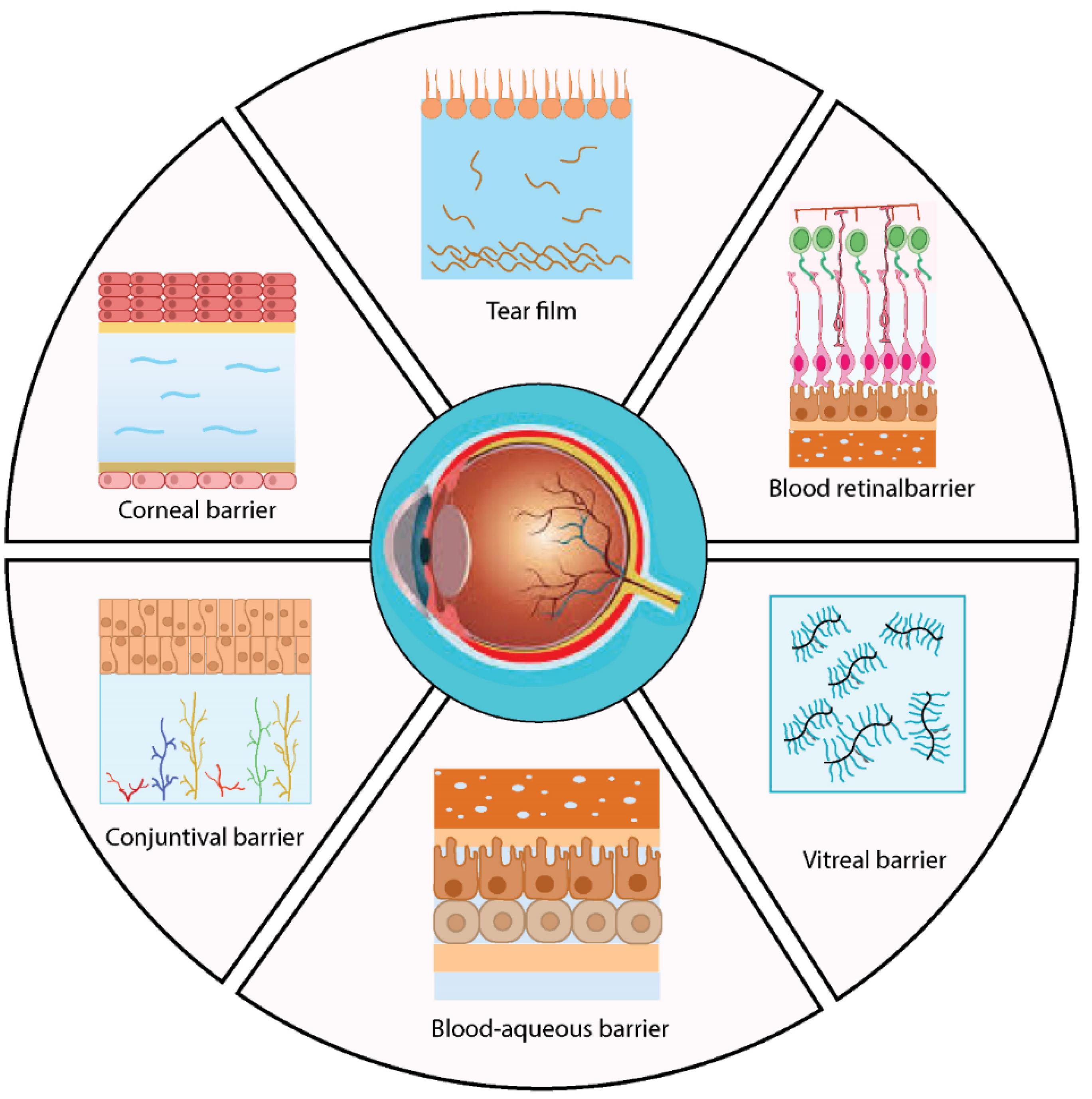
Fig. 1.
The eye's biological boundaries. Lacrimal fluid production continuously removes the three layers of the tear film barrier—lipid, aqueous, and mucin—from the surface of the eye, allowing for quick medication clearance. The corneal barrier restricts the entry of external substances into the eye by acting as a mechanical and chemical barrier. The surface of the corneal epithelium has tight connections that stop hydrophilic and macromolecular molecules from diffusing. Although intravitreal injection provides a direct route to the retina and vitreous, it may hinder the diffusion of bigger, positively charged medications to the choroid through the retinal pigment epithelium (RPE) barrier. A major obstacle to topical and systemic drug administration in the anterior and posterior chambers of the eye is the blood-ocular barrier (BOB). It includes the blood-retinal barrier (BRB) as well as the blood-aqueous barrier (BAB). The iris, ciliary muscle, pigmented and non-pigmented epithelial cells, endothelial cells, and tight junctions that limit drug molecule access make up the BAB, which is connected to the anterior chamber.
.
The eye's biological boundaries. Lacrimal fluid production continuously removes the three layers of the tear film barrier—lipid, aqueous, and mucin—from the surface of the eye, allowing for quick medication clearance. The corneal barrier restricts the entry of external substances into the eye by acting as a mechanical and chemical barrier. The surface of the corneal epithelium has tight connections that stop hydrophilic and macromolecular molecules from diffusing. Although intravitreal injection provides a direct route to the retina and vitreous, it may hinder the diffusion of bigger, positively charged medications to the choroid through the retinal pigment epithelium (RPE) barrier. A major obstacle to topical and systemic drug administration in the anterior and posterior chambers of the eye is the blood-ocular barrier (BOB). It includes the blood-retinal barrier (BRB) as well as the blood-aqueous barrier (BAB). The iris, ciliary muscle, pigmented and non-pigmented epithelial cells, endothelial cells, and tight junctions that limit drug molecule access make up the BAB, which is connected to the anterior chamber.
Although MSC transplantation has enormous potential in the field of regenerative medicine, a new study indicates that treatment with MSC-exosomes may offer a number of advantages over conventional MSC-based therapies. By using exosomes in a different way, practitioners can avoid some of the hazards that come with MSC-centered therapy. Avoiding these hazards is essential for maximizing therapy results. These risks include intravenous MSC injection-induced occlusion of small veins, unwanted differentiation, and allogeneic immunological rejection.10
Here, we highlight new developments in the research of exosomes produced from MSCs in corneal regeneration and cataract therapy. We describe the properties and biological roles of MSC-exosomes and investigate how they might be used to treat a range of conditions affecting cornea. We also look at the potential of exosome-based treatments in a clinical setting and the issues that need to be resolved in preclinical research (both in vitro and animal-based) to ensure a seamless transfer to clinical trials.
Extracellular vesicles
The secreted nanoscale vesicles known as extracellular vesicles (EVs) can be found in milk, serum, urine, plasma, and cerebrospinal fluid.11 EVs are useful for research on tissue repair and regeneration because they are low-antigenic cellular substructures that include a large number of proteins, nucleic acids, growth factors, and other physiologically active components.12 Furthermore, because of their structural features, EVs can be employed to transport nucleic acids or medications. Exosomes, micro vesicles (MVs), and apoptotic bodies are the three EV subtypes that are grouped based on their size and biogenesis. Exosomes are EVs with a diameter of 30–150 nm that exit multivesicular endosome pathways. Exosomes are produced constitutively and in response to stimulation during the exocytosis of multivesicular bodies (MVBs), which is a crucial stage in endolysosomal trafficking.13 Exosomes have the ability to carry HIV particles, oncogenic receptors, miRNA, and mRNA horizontally.
The phospholipid bilayer-encased MVs are about the same size as bacteria, with a diameter of 100–1000 nm. Although the molecular makeup of MVs is unknown, matrix metalloproteinases (MMPs), glycoproteins, and integrins seem to be significant depending on the kind of cell. Cancer cells create MVs, also referred to as Oncosomes. Oncosomes may boost organotropic metastatic propagation, facilitate invasion, and offer proteolytic activity by altering the genetic and metabolic capabilities of their target cells.14
Apoptotic bodies, the most varied kind of EVs, are created during apoptosis and come in a variety of shapes. Within the platelet size spectrum, their diameter ranges from 1 to 4 μm.15 Apoptosis will undoubtedly remove damaged, old, diseased, or abnormal cells from healthy tissue. The intentional breakdown of a cell into apoptotic bodies, which are made of cellular waste, is known as apoptosis.
An overview of MSCs-derived EVs
Paracrine signaling is believed to be crucial to the function of MSCs, which are now the focus of regenerative medicine research. MSC-EVs contribute to the process of tissue healing and regeneration by travelling to the appropriate areas, engrafting, and then differentiating into mature, functioning cells.16 By acting directly or indirectly on target cells, the proteins, mRNAs, miRNAs, long non-coding RNAs, and lipid components found in MSC-EVs activate pertinent signaling pathways. As a result, target cells experience regenerative and reparative effects from these pathways.
A more recent theory states that MSCs use different ways to repair tissue and influence nearby cells by promoting cell viability, proliferation, and differentiation, reducing fibrosis and apoptosis, promoting extracellular matrix (ECM) remodeling, and occasionally modifying the responses of the local immune system to prevent inflammation. The secretion of EVs, intercellular connections made possible by tunnelling nanotubes, and the synthesis and release of certain trophic factors, cytokines, chemokines, and hormones are some of these other strategies that support paracrine signaling between MSCs and their surrounding cells.17
Exosomes: Characteristics and biogenesis
Exosomes are often defined as a nanoscale subset of EVs with a payload encased in a lipid bilayer, with a diameter ranging from 30 to 150 nm. A diverse collection of proteins, amino acids, metabolites, lipids, and nucleic acids (including, but not limited to, DNA, miRNA, mRNA, and lncRNA) make up the exosome cargo, which is indicative of the cell of origin.18 The endosomal limiting membrane invaginates and buds, creating MVBs that contain intraluminal vesicles, which starts exosome biogenesis. The endosomal sorting complex required for transport (ESCRT) and ESCRT-independent pathways work together to sort the exosome payload.19 Through the interaction of Rab GTPases with SNARE proteins, MVBs fuse with the plasma membrane to form cup-shaped exosomes that can be released into the extracellular environment. Exosomes can remain in the ocular structure for a long time after release because the lipid bilayer shields the intracellular payload from enzymatic destruction, preserving biological potency and integrity. When target cells internalize exosomes and release their protected cargo, they function as mediators of intercellular communication, making them ideal for long-distance transport in biological fluids. Internalization methods include phagocytosis, micropinocytosis, receptor-mediated endocytosis, and direct fusion with the target membrane. Gene expression and cellular function are altered as a result of interaction with the target cell and the release of the contained payload into the intracellular environment. All cell types can manufacture exosomes, but MSCs are significantly more capable of producing and secreting them than cells from mesodermal lineages (Fig. 2).20
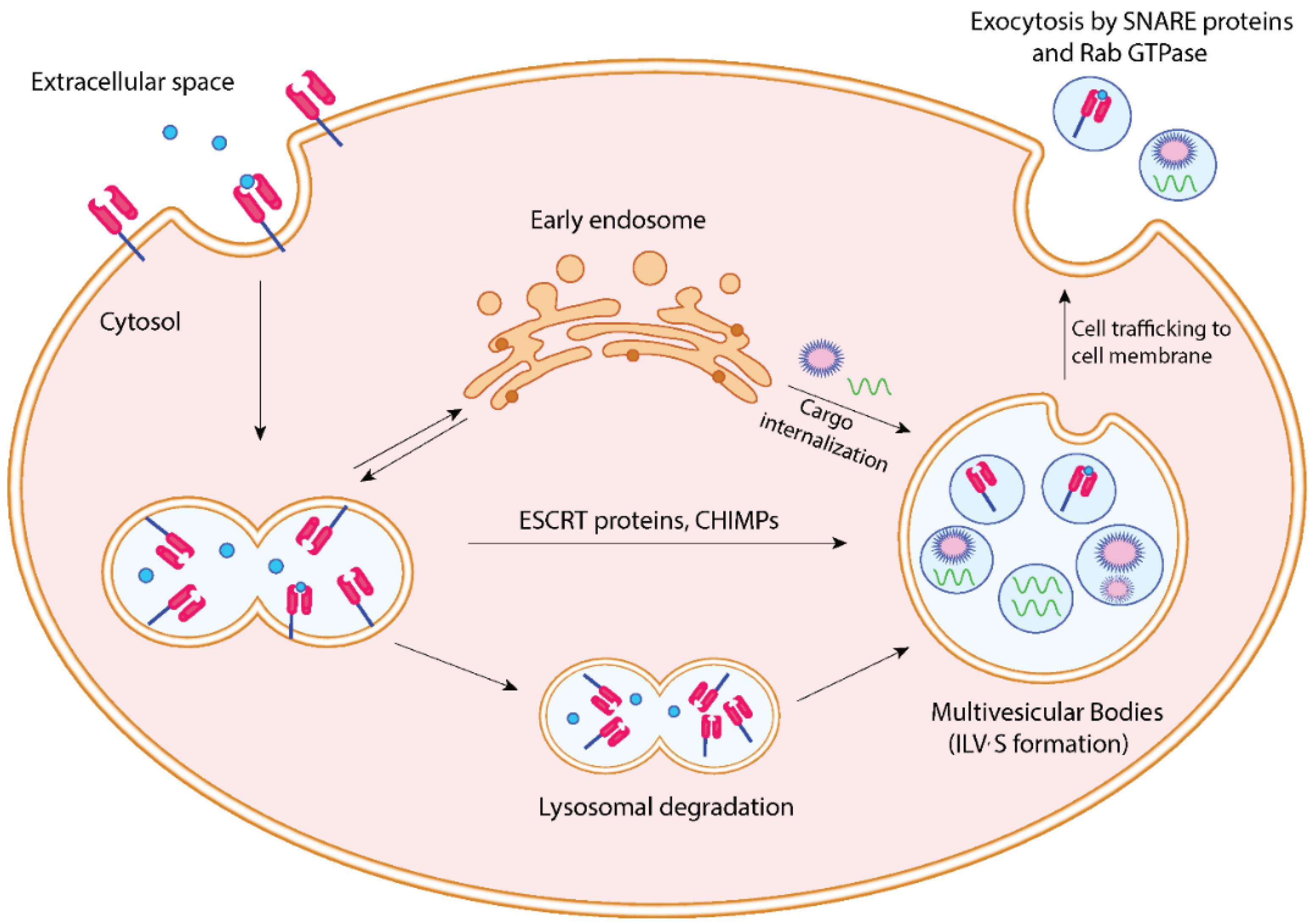
Fig. 2.
Exosome biogenesis characteristics. Early endosomes are formed in the cytoplasm when lipids and components of the cell membrane are endocytosed. This is the first step in the production of exosomes. After being sorted by the proteins ESCRT and CHMP, these endosomes form multivesicular bodies that contain intraluminal vesicles. Following their trafficking to the cell membrane, these multivesicular structures release exosomes into the extracellular environment via the exocytic pathway.
.
Exosome biogenesis characteristics. Early endosomes are formed in the cytoplasm when lipids and components of the cell membrane are endocytosed. This is the first step in the production of exosomes. After being sorted by the proteins ESCRT and CHMP, these endosomes form multivesicular bodies that contain intraluminal vesicles. Following their trafficking to the cell membrane, these multivesicular structures release exosomes into the extracellular environment via the exocytic pathway.
Exosomes' function in cellular communication
MSC-exosomes can trigger different responses in a variety of target cell types because of the distinct composition of the contained cargo. With more than 4000 different types found in exosome cargo, there is mounting evidence that miRNAs play a significant role as mediators of intercellular communication. MSC-exosomes produced from bone marrow (BM), adipose, and umbilical cord cells all have a distinct complement of miRNA types, which are indicative of the donor cell's identity and condition.21
MSC-exosomes have a great deal of demonstrated and potential therapeutic utility because of their anti-inflammatory, pro-regenerative, pro-angiogenic, immunomodulatory, and immunosuppressive qualities. Despite being an immune-privileged region in comparison to other organs, ocular tissue can sustain substantial damage from immune-mediated illnesses that impact the anterior and posterior segments.21,22 By regulating the hyperactive immune response that is characteristic of these conditions, MSC-exosomes have demonstrated effectiveness in treating a range of immune-mediated ocular illnesses, including autoimmune uveitis, corneal allograft rejection, and Sjögren's syndrome dry eye.23 Interestingly, the fundamental mechanism is the same for all of these conditions and will be covered in more detail in the next part.24
MSC-exosome benefits for ophthalmology
MSC-exosome-based therapies avoid the inflammatory response linked to cell-based therapies and many of the hazards associated with MSC transplantation, many of which stem from undesirable cell differentiation.24 Partial and total vision loss may result from serious and irreversible downstream effects of such differentiation and inflammation.25 Other issues associated with MSC transplantation include malignant transformation, proliferative vitreous retinopathy that results in retinal detachment, vitreous opacification, vitreous hemorrhage, retinal artery and vein occlusion, and allogeneic immunological rejection.24,25 The use of MSC-exosomes can greatly minimize these possible side effects related to MSC transplantation. Since the majority of the therapeutic effects of MSC transplantation come from the generation of soluble paracrine chemicals rather than from direct cell replacement, using MSC-exosomes offers a means to achieve equal efficacy with a more favorable safety profile.26
Other advantageous features of MSC-exosomes over MSC transplantation are as follows (Fig. 3):
-
Because MSC-exosomes can express several surface molecules, they can operate on particular tissues and cells in a selective manner.27 They have the ability to engage with recipient cells and deliver their cargo, resulting in favorable modifications to cellular function and gene expression.20
-
MSC-exosomes can last in the ocular structure for an extended period of time due to the protection provided by the lipid bilayer. In particular, the bilayer shields the contained cargo from early enzymatic breakdown and offers stability and structural stiffness.
-
Because of their inherent presence in bodily fluids and the bilipid membrane they inherited from their parent cells, they are extremely biocompatible.28 For example, exosome-based therapies are less likely to cause embolization, graft-versus-host rejection, and teratoma formation than MSC therapy.29
-
Because of their small size and lipid bilayer membrane, they may be able to pass through the biological barriers of the eye, including the tear film, corneal stroma, vitreous, and blood-retinal and blood-aqueous barriers. It is crucial to remember that there are not any publications that particularly discuss exosomes' capacity to pass through the cornea, tear film, or other ocular barriers at the moment. However, some studies suggest that exosomes can pass through the blood-brain barrier (BBB).30 Additionally, research in different bodily regions has shown that exosomes can get past difficult obstacles.31 Given these data, it is possible to speculate that exosomes could be used as a delivery system to get past ocular obstacles such as the corneal and tear films. Nevertheless, more investigation is required to confirm this theory. More options for administration routes and the capacity to distribute more bioactive molecules to the intended location would result from this.
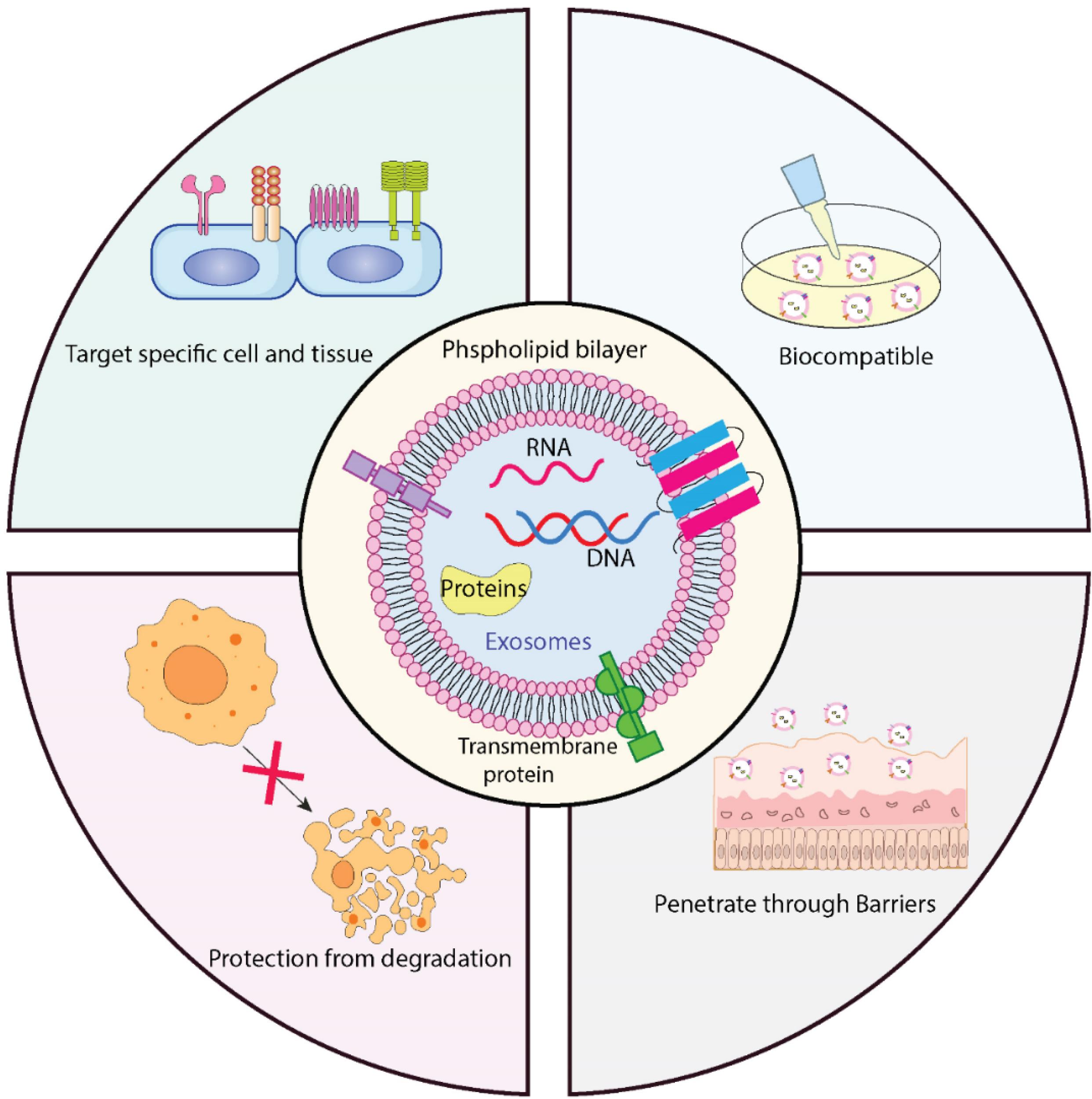
Fig. 3.
Advantageous properties of MSC-exosomes.
.
Advantageous properties of MSC-exosomes.
As previously mentioned, MSC-exosomes' effectiveness and safety characteristics make them a viable new therapeutic agent for the treatment of a range of eye conditions. They exhibit great potential, particularly in the treatment of eye illnesses that are resistant to existing treatments, although more research is definitely needed.
MSC-exosomes for corneal regeneration
The process of corneal regeneration is dynamic and intricate, typically including ECM remodeling, cellular proliferation, and inflammation. After corneal damage, dead or injured cells release mediators and cytokines that draw immune cells to the area and cause surviving keratocytes to divide and become fibroblasts, which move in the direction of the injury. In addition to secreting ECM and ECM remodeling enzymes like collagenase and MMPs, these fibroblasts can also differentiate into myofibroblasts, which help in wound contraction and closure.32 Prolonged or excessive myofibroblast activation brought on by persistent epithelial abnormalities results in corneal opacification, scar tissue development, and incorrect collagen fiber deposition. Corneal scarring and opacity may also be caused by excessive inflammation and abnormal angiogenesis.33 Successful corneal regeneration depends on maintaining corneal transparency, and therapeutic treatment of corneal wounds must encourage healing while reducing inflammation, neovascularization, and disorders of the collagen-rich ECM.
Because exosomes have been demonstrated to support tissue regeneration and reduce inflammation, they have garnered attention in the treatment of corneal diseases.34 Recent research has shown that MSC-exosomes accelerate re-epithelialization by promoting corneal epithelial cell migration and proliferation in vitro.35 In vivo animal studies have effectively applied these findings, showing that corneal wound healing is markedly enhanced in eyes treated with MSC-exosomes.36 Since miR-21 suppression resulted in a partial reduction of wound healing effects through its modulation of the PTEN/PI3K/Akt pathway, it was suggested that the presence of miR-21 within the MSC-exosomes contributed to the exosomes' ability to cure corneal wounds.37 Furthermore, downregulation of RNA levels of proinflammatory cytokines such as interleukin (IL)-1β, IL-8, TNF-α, and NF-κB, as well as of the pro-apoptotic protein Caspase-8, suggests that MSC-exosomes decrease inflammation and apoptosis after corneal injury.38 The downregulation of the production of the pro-angiogenic factor vascular endothelial growth factor (VEGF) and the angiogenesis-associated matrix metalloproteinases MMP-2 and MMP-9 suggests that these MSC-exosomes may also have anti-angiogenic qualities.39 The therapeutic advantages of MSC-exosomes on corneal stromal stem cells (CSSCs) have also been clarified by studies. The progenitor cells of keratocytes found in the limbal stroma CSSCs, have the therapeutic capacity to restructure disrupted ECM, potentially leading to the restoration of transparency.40 It has been demonstrated that adipose-derived MSC-exosomes stimulate the proliferation of CSSCs and block their apoptosis in a dose-dependent manner, indicating that they may be used in ECM remodeling to lessen corneal opacity.39
It has been shown that autophagy, which is the process by which cells self-destruct in order to preserve homeostasis by breaking down and recycling pieces, might contribute to corneal injury.41 The effect of MSC-exosomes coupled with Rapamycin (50 nM), an autophagy activator (AA), on corneal regeneration was assessed in a study by Ma et al.36 It was shown that AA had positive effects on migration and proliferation in corneal epithelial cells that were comparable to those of human umbilical MSC-exosome treatment, and that the combined effect of exosome-AA treatment was stronger than that of either exosomes or AA alone. The mouse eyes treated with Exo-AA had the lowest percentage of apoptotic cells, while the group treated with a combination of exosomes and an autophagy inhibitor (AI) had the greatest percentage. The Exo-AA group also showed a decrease in haze grade and the expression of proinflammatory genes TNF-α, IL-1β, IL-6, and CXCL-2. Therefore, autophagy activators may be a promising addition to treatments based on exosomes.
The use of bioengineered hydrogels as scaffolds for corneal restoration that have been altered to release MSC-exosomes continuously has been investigated in a number of animal studies. When thermosensitive chitosan-based hydrogels (CHI) of sustained-release induced pluripotent stem cell-derived MSC (iPSC-MSC)-exosomes were applied to a rat corneal anterior lamellar injury model, the corneal transparency increased and collagen expression decreased compared to untreated eyes. Further examination of the overexpressed miRNA in iPSC-MSC-exosomes revealed that miR-432-5p modulates the translocation-associated membrane protein 2 (TRAM-2) to downregulate collagen manufacturing in CSSCs.42 The regulated release of exosomes rich in miRNA-24-3p from adipocyte-derived MSCs was achieved by developing a thermosensitive hydrogel of modified hyaluronic acid with di (ethylene glycol) monomethyl ether methacrylate (DEGMA). In a study employing an alkali burn model in rabbits, it was discovered that this hydrogel produced a transparent and consistent coating on the ocular surface and could be removed by blinking. The usage of miRNA 24-3p has been shown to improve corneal epithelial defect healing, reduce corneal stromal fibrosis, and reduce macrophage activation. It is believed to promote corneal epithelial cell migration and corneal repair.43 An alternative to the traditional penetrating keratoplasty, which is linked to a number of problems including corneal transplant rejection, wound leak, and endophthalmitis, may be possible with current research into treatments that combine biosynthetic hydrogels with MSC-exosome delivery.
Overcoming challenges in the clinical translation of MSC-exosomes
Even though MSC-exosome therapies show great promise, there are still a number of unresolved issues, including low yield capacity, unclear treatment mechanisms of action, non-uniformity in MSC-exosome isolation and purification, unstandardized large-scale production protocols, and performance characteristics that are still lacking, such as clinical sensitivity and specificity or sample stability.44 However, the issue of exosome product variability and the absence of established quality assessment standards appear to be major barriers to clinical translation. In the end, heterogeneity lowers reproducibility in both in vitro and in vivo settings by impeding the quality and management of MSC-exosome products. Criteria for distinguishing MSCs derived from different targets have been devised by the International Society for Cell and Gene Therapy.45
Overcoming the barrier of MSC-exosome heterogeneity
Parental and exosome heterogeneity, as previously mentioned, impairs the quality and control of MSC-exosome products, decreasing their reproducibility in both in vitro and in vivo settings.12 It goes without saying that this problem will yield a variety of outcomes. Furthermore, published research indicates that the therapeutic effects will vary depending on the parental source. For example, BMSC-MSC-exosomes are four times more angiogenic than adipose tissue-derived stem cell (ADSC)-derived exosomes; endometrial-derived MSC-exosomes are noticeably superior to both in this respect. However, it is known that MSC-exomes generated from ADSCs produce higher VEGF and hepatocyte growth factor (HGF), two cardioprotective factors. Accordingly, exosomes produced from bone marrow-derived mesenchymal stem cells (BMSCs) and ADSCs can cause M2 polarization of macrophages; the former can do so by increasing CD206 expression by 3.2 times, while the latter can only do so by 1.5 times. In order to identify the best sourcing and extraction procedure for patients, further study is required to examine the complex impacts of various parental sources and their corresponding exosomes.
Exosome product extraction from MSCs produced from human pluripotent stem cells (hPSCs) is one solution proposed by Kou et al.12 The theory that hPSCs would address source heterogeneity and, in turn, exosome product heterogeneity lends theoretical support to this proposal. The effectiveness of this approach has been studied in clinical trials looking into its use in patients with refractory GVHD. Similar benefits were seen in reducing rejection after transplanting abdominal organs. Passage counts are one way to evaluate the quality of cell cultures; hPSC-MSCs have been demonstrated to have over 30 passages, but standard MSCs have less than ten. Higher exosome yields for clinical applications are indicated by a more significant passage number. Furthermore, hPSC-MSCs have better secretion and amplification capabilities than conventional MSCs, making them more robust. Higher quality MSC-exosome products and the possibility of economical, large-scale manufacture are indicated by these features.
According to Varderidou-Minasian and Lorenowicz, "the therapeutic efficiency of MSC therapy depends on their paracrine signaling, rather than the engraftment of MSCs at the site of injury or the differentiation capability of the transplanted MSCs".46 To put it another way, EVs and exosomes are key factors that determine their potential for therapeutic use. Because of this, while evaluating the quality of MSC secretomes, both their quantitative and qualitative features should be taken into account. Recent developments in cell culture technology demonstrate that 3D culture methods outperform conventional static adherent cultures (i.e., 2D cultures) in replicating in vivo settings. In 2D cultures, essential characteristics of MSC morphology, function, and structure are lost; consequently, this impairs their ability to proliferate and differentiate, which in turn reduces exosome efficiency. Qazi et alprovided evidence in favor of this theory by showing enhanced paracrine signaling in 3D cultures by releasing more growth factors and cytokines.47
Similarly, Su et aldiscovered that the release of anti-inflammatory and angiogenic-promoting substances was more favorable in extracellular matrices with oriented fibers than non-oriented fibers.48 In light of this, 3D culturing can be further separated into material-supported and material-free categories, with the latter being better suited for signaling and cell-to-cell communication.49 Scaffold-free suspension cultures and hydrogel-assisted 3D cultures are two types of material-free cell cultures. Hollow fiber bioreactors are the most prominent example of material-supported cultures, exhibiting a 19.4 increased yield in shorter culture times compared to 2D cultures. Furthermore, in rat models, 3D-harvested MSC-exosomes have improved a number of conditions. For example, MSC-exosomes isolated from 3D cell cultures showed enhanced angiogenicity, endothelial cell migration, and proliferation in the context of injury repair.
MSC preconditioning is a further strategy for raising the caliber of MSC-exosome products.49 Reducing oxygen exposure during a hypoxic pretreatment improves the genetic stability and proliferative potential of MSCs. More significantly, enhancement of migratory and paracrine potential is mediated by the activation of angiogenic gene transcription and the upregulation of stemness genes like SOX2. Exosomes that are isolated from these low-oxygen tension MSCs (Hyp-MSC-Exos) are the end result. Research examining the therapeutic benefits of these exosomes has demonstrated advantages in a range of disease conditions, from diabetic wound healing to central nervous system problems, including spinal cord injury. It has been demonstrated that cytokine preconditioning, a different pretreatment regimen, increases paracrine efficiency and improves the therapeutic potential of MSC-exosomes by stimulating cytokines and inflammatory factors. TNF-α exposure specifically encourages the release of additional exosomes that fight inflammation, and these exosomes also include large amounts of miRNAs that inhibit inflammation, such as miR-146a and miRNA-299-3p. Treatment with IL-1β or IFN-γ, among other things, showed the creation of the previously stated miRNAs and exosomes with improved anti-inflammatory properties. Finally, preconditioning methods that are chemical and physical yield results that are similar. For example, metformin therapy increased the presence of exosome components associated with autophagy, while exposure to monochromatic blue light (451 nm) increased the presence of the same miRNAs as TNF-α treatment.
Conclusion
In conclusion, because MSCs produced exosomes can get beyond the drawbacks of conventional MSC-based treatments, their application in ophthalmology has attracted a lot of interest. Among their special qualities are MSC-exosomes' quick passage across biological barriers, their capacity to transfer trophic and immunomodulatory substances, and their resistance to immunological rejection and unintended differentiation. The unique characteristics and biological underpinnings of MSC-exosomes are clarified in this article, which also thoroughly reviews preclinical research showing the exosomes' potential to cure a range of ocular conditions affecting the front and rear of the eye.
Recent developments in MSC-exosomes have enormous potential for the future, despite obstacles and restrictions that need to be addressed in preclinical research. By overcoming these obstacles, MSC-exosomes may provide a safer and more efficient substitute for MSC-based treatments, transforming our collective strategy for treating ocular disorders and giving patients better eyesight and a more promising future.
Review Highlights
What is the current knowledge?
What is new here?
-
Recent research has shown that MSC-exosomes accelerate re-epithelialization by promoting corneal epithelial cell migration and proliferation in vitro. In conclusion, because MSCs produced exosomes can get beyond the drawbacks of conventional MSC-based treatments, their application in ophthalmology has attracted a lot of interest.
Competing Interests
None.
Ethical Approval
Not applicable.
References
- Yang L, Li J, Zhou B, Wang Y. An injectable copolymer for in situ lubrication effectively relieves dry eye disease. ACS Mater Lett 2025; 7:884-90. doi: 10.1021/acsmaterialslett.4c02327 [Crossref] [ Google Scholar]
- Li S, Ling S, Wang D, Wang X, Hao F, Yin L, et al. Modified lentiviral globin gene therapy for pediatric β0/β0 transfusion-dependent β-thalassemia: a single-center, single-arm pilot trial. Cell Stem Cell 2024; 31: 961-73.e8. doi: 10.1016/j.stem.2024.04.021.
- O'Connor MD, McAvoy JW. In vitro generation of functional lens-like structures with relevance to age-related nuclear cataract. Invest Ophthalmol Vis Sci 2007; 48:1245-52. doi: 10.1167/iovs.06-0949 [Crossref] [ Google Scholar]
- Fathi E, Farahzadi R. Mesenchymal stem cells as a cell-based therapeutic strategy targeting the telomerase activity of KG1 acute myeloid leukemia cells. Acta Med Iran 2022; 60:71-7. doi: 10.18502/acta.v60i2.8817 [Crossref] [ Google Scholar]
- Zhou C, Kuang M, Tao Y, Wang J, Luo Y, Fu Y, et al. Nynrin preserves hematopoietic stem cell function by inhibiting the mitochondrial permeability transition pore opening. Cell Stem Cell 2024; 31: 1359-75.e8. doi: 10.1016/j.stem.2024.06.007.
- Li Z, Fan J, Xiao Y, Wang W, Zhen C, Pan J. Essential role of Dhx16-mediated ribosome assembly in maintenance of hematopoietic stem cells. Leukemia 2024; 38:2699-708. doi: 10.1038/s41375-024-02423-3 [Crossref] [ Google Scholar]
- Chen F, Zhang K, Wang M, He Z, Yu B, Wang X. VEGF-FGF signaling activates quiescent CD63 + liver stem cells to proliferate and differentiate. Adv Sci (Weinh) 2024; 11:e2308711. doi: 10.1002/advs.202308711 [Crossref] [ Google Scholar]
- Farahzadi R, Fathi E, Vandghanooni S, Valipour B. Hydrogel encapsulation of mesenchymal stem cells-derived extracellular vesicles as a novel therapeutic approach in cancer therapy. Biochim Biophys Acta Rev Cancer 2024; 1879:189177. doi: 10.1016/j.bbcan.2024.189177 [Crossref] [ Google Scholar]
- Niamprem P, Srinivas SP, Tiyaboonchai W. Penetration of Nile red-loaded nanostructured lipid carriers (NLCs) across the porcine cornea. Colloids Surf B Biointerfaces 2019; 176:371-8. doi: 10.1016/j.colsurfb.2019.01.018 [Crossref] [ Google Scholar]
- Yu B, Shao H, Su C, Jiang Y, Chen X, Bai L. Exosomes derived from MSCs ameliorate retinal laser injury partially by inhibition of MCP-1. Sci Rep 2016; 6:34562. doi: 10.1038/srep34562 [Crossref] [ Google Scholar]
- Zeng Y, Qiu Y, Jiang W, Shen J, Yao X, He X. Biological features of extracellular vesicles and challenges. Front Cell Dev Biol 2022; 10:816698. doi: 10.3389/fcell.2022.816698 [Crossref] [ Google Scholar]
- Kou M, Huang L, Yang J, Chiang Z, Chen S, Liu J. Mesenchymal stem cell-derived extracellular vesicles for immunomodulation and regeneration: a next generation therapeutic tool?. Cell Death Dis 2022; 13:580. doi: 10.1038/s41419-022-05034-x [Crossref] [ Google Scholar]
- Soekmadji C, Li B, Huang Y, Wang H, An T, Liu C. The future of Extracellular Vesicles as Theranostics - an ISEV meeting report. J Extracell Vesicles 2020; 9:1809766. doi: 10.1080/20013078.2020.1809766 [Crossref] [ Google Scholar]
- Ciardiello C, Leone A, Lanuti P, Roca MS, Moccia T, Minciacchi VR. Large oncosomes overexpressing integrin alpha-V promote prostate cancer adhesion and invasion via AKT activation. J Exp Clin Cancer Res 2019; 38:317. doi: 10.1186/s13046-019-1317-6 [Crossref] [ Google Scholar]
- Barros FM, Carneiro F, Machado JC, Melo SA. Exosomes and immune response in cancer: friends or foes?. Front Immunol 2018; 9:730. doi: 10.3389/fimmu.2018.00730 [Crossref] [ Google Scholar]
- Ju Y, Hu Y, Yang P, Xie X, Fang B. Extracellular vesicle-loaded hydrogels for tissue repair and regeneration. Mater Today Bio 2023; 18:100522. doi: 10.1016/j.mtbio.2022.100522 [Crossref] [ Google Scholar]
- Nikfarjam S, Rezaie J, Majidi Zolbanin N, Jafari R. Mesenchymal stem cell derived-exosomes: a modern approach in translational medicine. J Transl Med 2020; 18:449. doi: 10.1186/s12967-020-02622-3 [Crossref] [ Google Scholar]
- Xu HK, Chen LJ, Zhou SN, Li YF, Xiang C. Multifunctional role of microRNAs in mesenchymal stem cell-derived exosomes in treatment of diseases. World J Stem Cells 2020; 12:1276-94. doi: 10.4252/wjsc.v12.i11.1276 [Crossref] [ Google Scholar]
- Xu M, Ji J, Jin D, Wu Y, Wu T, Lin R. The biogenesis and secretion of exosomes and multivesicular bodies (MVBs): intercellular shuttles and implications in human diseases. Genes Dis 2023; 10:1894-907. doi: 10.1016/j.gendis.2022.03.021 [Crossref] [ Google Scholar]
- Bian B, Zhao C, He X, Gong Y, Ren C, Ge L. Exosomes derived from neural progenitor cells preserve photoreceptors during retinal degeneration by inactivating microglia. J Extracell Vesicles 2020; 9:1748931. doi: 10.1080/20013078.2020.1748931 [Crossref] [ Google Scholar]
- Glover K, Mishra D, Singh TR. Epidemiology of ocular manifestations in autoimmune disease. Front Immunol 2021; 12:744396. doi: 10.3389/fimmu.2021.744396 [Crossref] [ Google Scholar]
- Hu T, Meng S, Liu C, Fang W, Xia Z, Hu Y. LCN2 deficiency mitigates the neuroinflammatory damage following acute glaucoma. Theranostics 2025; 15:2967-90. doi: 10.7150/thno.104752 [Crossref] [ Google Scholar]
- Wu Z, Sun W, Wang C. Clinical characteristics, treatment, and outcomes of pembrolizumab-induced uveitis. Invest New Drugs 2024; 42:510-7. doi: 10.1007/s10637-024-01464-w [Crossref] [ Google Scholar]
- Yu B, Li XR, Zhang XM. Mesenchymal stem cell-derived extracellular vesicles as a new therapeutic strategy for ocular diseases. World J Stem Cells 2020; 12:178-87. doi: 10.4252/wjsc.v12.i3.178 [Crossref] [ Google Scholar]
- Kuriyan AE, Albini TA, Townsend JH, Rodriguez M, Pandya HK, Leonard RE 2nd. Vision loss after intravitreal injection of autologous "stem cells" for AMD. N Engl J Med 2017; 376:1047-53. doi: 10.1056/NEJMoa1609583 [Crossref] [ Google Scholar]
- Sun H, Pratt RE, Hodgkinson CP, Dzau VJ. Sequential paracrine mechanisms are necessary for the therapeutic benefits of stem cell therapy. Am J Physiol Cell Physiol 2020; 319:C1141-50. doi: 10.1152/ajpcell.00516.2019 [Crossref] [ Google Scholar]
- Liang Y, Duan L, Lu J, Xia J. Engineering exosomes for targeted drug delivery. Theranostics 2021; 11:3183-95. doi: 10.7150/thno.52570 [Crossref] [ Google Scholar]
- Zhang Z, Mugisha A, Fransisca S, Liu Q, Xie P, Hu Z. Emerging role of exosomes in retinal diseases. Front Cell Dev Biol 2021; 9:643680. doi: 10.3389/fcell.2021.643680 [Crossref] [ Google Scholar]
- Seyedrazizadeh SZ, Poosti S, Nazari A, Alikhani M, Shekari F, Pakdel F, et al. Extracellular vesicles derived from human ES-MSCs protect retinal ganglion cells and preserve retinal function in a rodent model of optic nerve injury. Stem Cell Res Ther 2020. 11: 203. doi: 10.1186/s13287-020-01702-x.
- Li C, Qin S, Wen Y, Zhao W, Huang Y, Liu J. Overcoming the blood-brain barrier: Exosomes as theranostic nanocarriers for precision neuroimaging. J Control Release 2022; 349:902-16. doi: 10.1016/j.jconrel.2022.08.002 [Crossref] [ Google Scholar]
- Elliott RO, He M. Unlocking the power of exosomes for crossing biological barriers in drug delivery. Pharmaceutics 2021; 13:122. doi: 10.3390/pharmaceutics13010122 [Crossref] [ Google Scholar]
- Wilson SE. Corneal wound healing. Exp Eye Res 2020; 197:108089. doi: 10.1016/j.exer.2020.108089 [Crossref] [ Google Scholar]
- Sharif Z, Sharif W. Corneal neovascularization: updates on pathophysiology, investigations & management. Rom J Ophthalmol 2019; 63:15-22. [ Google Scholar]
- Phinney DG, Pittenger MF. Concise review: MSC-derived exosomes for cell-free therapy. Stem Cells 2017; 35:851-8. doi: 10.1002/stem.2575 [Crossref] [ Google Scholar]
- Yu Z, Hao R, Du J, Wu X, Chen X, Zhang Y. A human cornea-on-a-chip for the study of epithelial wound healing by extracellular vesicles. iScience 2022; 25:104200. doi: 10.1016/j.isci.2022.104200 [Crossref] [ Google Scholar]
- Ma S, Yin J, Hao L, Liu X, Shi Q, Diao Y. Exosomes from human umbilical cord mesenchymal stem cells treat corneal injury via autophagy activation. Front Bioeng Biotechnol 2022; 10:879192. doi: 10.3389/fbioe.2022.879192 [Crossref] [ Google Scholar]
- Liu X, Li X, Wu G, Qi P, Zhang Y, Liu Z. Umbilical cord mesenchymal stem cell-derived small extracellular vesicles deliver miR-21 to promote corneal epithelial wound healing through PTEN/PI3K/Akt pathway. Stem Cells Int 2022; 2022:1252557. doi: 10.1155/2022/1252557 [Crossref] [ Google Scholar]
- Tao H, Chen X, Cao H, Zheng L, Li Q, Zhang K. Mesenchymal stem cell-derived extracellular vesicles for corneal wound repair. Stem Cells Int 2019; 2019:5738510. doi: 10.1155/2019/5738510 [Crossref] [ Google Scholar]
- Shen T, Zheng QQ, Shen J, Li QS, Song XH, Luo HB. Effects of adipose-derived mesenchymal stem cell exosomes on corneal stromal fibroblast viability and extracellular matrix synthesis. Chin Med J (Engl) 2018; 131:704-12. doi: 10.4103/0366-6999.226889 [Crossref] [ Google Scholar]
- Du Y, Carlson EC, Funderburgh ML, Birk DE, Pearlman E, Guo N. Stem cell therapy restores transparency to defective murine corneas. Stem Cells 2009; 27:1635-42. doi: 10.1002/stem.91 [Crossref] [ Google Scholar]
- Wang Y, Gao G, Wu Y, Wang Y, Wu X, Zhou Q. S100A4 silencing facilitates corneal wound healing after alkali burns by promoting autophagy via blocking the PI3K/Akt/mTOR signaling pathway. Invest Ophthalmol Vis Sci 2020; 61:19. doi: 10.1167/iovs.61.11.19 [Crossref] [ Google Scholar]
- Tang Q, Lu B, He J, Chen X, Fu Q, Han H. Exosomes-loaded thermosensitive hydrogels for corneal epithelium and stroma regeneration. Biomaterials 2022; 280:121320. doi: 10.1016/j.biomaterials.2021.121320 [Crossref] [ Google Scholar]
- Sun X, Song W, Teng L, Huang Y, Liu J, Peng Y. MiRNA 24-3p-rich exosomes functionalized DEGMA-modified hyaluronic acid hydrogels for corneal epithelial healing. Bioact Mater 2023; 25:640-56. doi: 10.1016/j.bioactmat.2022.07.011 [Crossref] [ Google Scholar]
- Wei W, Ao Q, Wang X, Cao Y, Liu Y, Zheng SG. Mesenchymal stem cell-derived exosomes: a promising biological tool in nanomedicine. Front Pharmacol 2020; 11:590470. doi: 10.3389/fphar.2020.590470 [Crossref] [ Google Scholar]
- Hoang DM, Pham PT, Bach TQ, Ngo AT, Nguyen QT, Phan TT. Stem cell-based therapy for human diseases. Signal Transduct Target Ther 2022; 7:272. doi: 10.1038/s41392-022-01134-4 [Crossref] [ Google Scholar]
- Varderidou-Minasian S, Lorenowicz MJ. Mesenchymal stromal/stem cell-derived extracellular vesicles in tissue repair: challenges and opportunities. Theranostics 2020; 10:5979-97. doi: 10.7150/thno.40122 [Crossref] [ Google Scholar]
- Qazi TH, Mooney DJ, Duda GN, Geissler S. Biomaterials that promote cell-cell interactions enhance the paracrine function of MSCs. Biomaterials 2017; 140:103-14. doi: 10.1016/j.biomaterials.2017.06.019 [Crossref] [ Google Scholar]
- Su N, Gao PL, Wang K, Wang JY, Zhong Y, Luo Y. Fibrous scaffolds potentiate the paracrine function of mesenchymal stem cells: a new dimension in cell-material interaction. Biomaterials 2017; 141:74-85. doi: 10.1016/j.biomaterials.2017.06.028 [Crossref] [ Google Scholar]
- Chen S, Sun F, Qian H, Xu W, Jiang J. Preconditioning and engineering strategies for improving the efficacy of mesenchymal stem cell-derived exosomes in cell-free therapy. Stem Cells Int 2022; 2022:1779346. doi: 10.1155/2022/1779346 [Crossref] [ Google Scholar]