Bioimpacts. 16:32812.
doi: 10.34172/bi.32812
Mini Review
Short interference RNAs in glomerulonephritis: A mini review
Negin Frounchi Investigation, Methodology, Writing – original draft, 1 
Diana Jafari Nakhjavani Investigation, Visualization, Writing – review & editing, 1
Sima Abediazar Data curation, Writing – review & editing, 1
MohammadReza Ardalan Supervision, Validation, Writing – review & editing, 1
Farahnoosh Farnood Conceptualization, Methodology, Project administration, Writing – original draft, 1, *
Author information:
1Kidney Research Center, Tabriz University of Medical Sciences, Tabriz, Iran
Abstract
Glomerulonephritis (GN) is a heterogeneous group of kidney diseases characterized by inflammation and damage to the glomeruli. Current treatments for GN remain suboptimal, creating a pressing need for innovative therapeutic strategies. Short interfering RNAs (siRNAs) represent a promising advance in the therapeutic landscape for GN, offering targeted gene silencing that could fundamentally transform the management of this significant cause of kidney disease. siRNA therapies work by selectively silencing specific genes involved in the pathogenesis of GNs, including interferon pathways, B cell activation, TGF-β, MAPK1, HMGB1, BLYSS, and SIRT1. Preclinical evidence demonstrates improved kidney function and reduced glomerular damage in experimental models. While challenges remain in translating these findings to clinical applications, ongoing research suggests that siRNA-based therapies have the potential to transform the treatment of GN, offering new hope for patients with this challenging kidney disease. This review highlights the impact of siRNA-based therapies in GN, exploring their mechanisms, delivery systems, challenges, and preclinical and clinical evidence.
Graphical Abstract
Keywords: Glomerular disease, siRNA, IgA nephropathy, SLE, Lupus nephritis, Membranous nephropathy
Copyright and License Information
© 2026 The Author(s).
This work is published by BioImpacts as an open access article distributed under the terms of the Creative Commons Attribution Non-Commercial License (
http://creativecommons.org/licenses/by-nc/4.0/). Non-commercial uses of the work are permitted, provided the original work is properly cited.
Funding Statement
This project did not receive any financial support, grants, or funding.
Introduction
Glomerulonephritis (GN) is an injury to the glomeruli—the kidney’s small filtering structures—caused by inflammatory pathways, resulting in a heterogeneous range of diseases. Chronic GN is ranked third as the leading kidney disorder that causes end-stage renal disease (ESRD). When GN grows to ESRD, the patient will require a kidney transplant or dialysis, which imposes remarkable burdens on both the patient and society.1
Currently, the primary treatment for GN involves symptomatic therapy, immunosuppressive therapy (glucocorticoids and cyclosporine), and kidney replacement therapy. While these treatments can effectively reduce inflammation and proteinuria, they often do not address the disease’s underlying genetic or molecular causes and, for many patients, only slow disease progression without providing a cure. Moreover, some individuals diagnosed with GN face challenges such as relapse, steroid resistance,2,3 or progression to ESRD, making treatment challenging. Long-term use of traditional immunosuppressants can also cause significant side effects, such as increased risk of infection, bone marrow suppression, and organ toxicity.4 Replacement therapy for ESRD patients increases both the economic burden and the negative impact on the quality of life, where many immunosuppressants cause side effects that outweigh the benefits, highlighting the need for new GN therapies.5
Small interfering ribonucleic acids (siRNAs) are one of the innovative therapeutic approaches. These molecules have the potential to silence specific gene sequences through endogenous regulation, thereby helping inhibit the expression of genes that cause various diseases.6,7
Although the kidney’s rapid blood flow, glomerular filtration, and tubular reabsorption make it a potentially ideal target for siRNA therapies, its structural complexity and diverse cell populations pose significant challenges for targeted drug delivery.8 siRNA targeted approach, by reducing the production of disease-causing proteins involved in inflammation, fibrosis, and the immune response, shows promise for diminishing kidney injury while limiting overall side effects. With ongoing research, these therapies could introduce a new therapeutic strategy for treating GN and other kidney diseases that currently lack adequate clinical solutions. Since GN represents a significant challenge in nephrology and current treatment options fail to provide satisfactory outcomes for many patients, this review underscores the impact of siRNA-based treatments in GN, exploring their mechanisms, delivery systems, and preclinical and clinical evidence.
Glomerulonephritis
Glomerulonephritis encompasses a group of diverse immune-related diseases that lead to inflammation of the glomeruli, often driven by immune responses, systemic diseases, cytokines, inflammation, and viral infections.9-12 Recent studies have also identified a significant role for microbiota dysbiosis in the pathogenesis of glomerulonephritis.13 Analysis of a U.S. Medicare cohort, averaging 75 years in age, showed that approximately 1.2% of people in this group were affected by GN.14 The glomerular microvasculature is especially susceptible to immune-related damage due to its distinct functional and anatomical features. Specifically, glomerular capillaries function as high-capacity filters, producing a large volume of ultrafiltrate. This process exposes them to significant mechanical forces, including shear stress and perfusion pressure. The structural complexity of the glomerular filtration barrier, which comprises three critical components —delicate endothelial cells, a porous basement membrane, and intricately interdigitated podocyte foot processes —renders it inherently vulnerable to immune-mediated injury.15 As a result, these unique features make the glomeruli susceptible to injury from various immunological triggers, leading to the diverse causes of GN. There are different types of GN primarily based on histopathological lesion patterns. Still, these patterns do not correspond well to the various pathological features, which limits their guidance for optimal treatment. Definitely, altered systemic immunity is the primary pathogenic mechanism and the principal therapeutic focus in GN.16
Glomerulonephritis is generally divided into five categories based on underlying immune mechanisms: infection-related, autoimmune, alloimmune, autoinflammatory, and monoclonal gammopathy-associated. It is also possible for one form of glomerulonephritis to evolve into another, highlighting the complex interplay between these different categories. For example, acute GN can progress to chronic kidney disease and permanent kidney failure if not adequately treated. Typically, acute GN is characterized by high blood pressure (hypertension), proteinuria (excessive protein in the urine), and hematuria (blood in the urine). In contrast, GN that primarily affects podocytes leads to nephrotic syndrome, characterized by significant proteinuria and leg oedema. Generally, the presence of proteinuria, mainly albumin, suggests damage to podocytes, while haematuria indicates breaks or tears in the glomerular basement membrane (GBM).16,17 Understanding these distinctions is crucial for effective diagnosis and treatment, as each type of GN may require a tailored therapeutic approach to manage its unique pathogenic mechanisms and prevent long-term kidney damage.
The therapeutic potential of siRNAs in GN is complemented by a broader landscape of non-coding RNAs, such as microRNAs (miRNAs),18 long non-coding RNAs (lncRNAs), and circular RNAs (circRNAs).19,20 These RNAs have fundamental functions in controlling gene expression and are often dysregulated in various forms of GNs. For instance, specific miRNAs have been implicated in modulating inflammatory and fibrotic pathways in glomerular diseases, with altered expression levels observed in conditions such as lupus nephritis,21,22 diabetic nephropathy,23 membranous nephropathy (MN), focal segmental glomerulosclerosis (FSGS),24-26 and IgA nephropathy (IgAN).27 Similarly, lncRNAs have been shown to influence glomerular cell function and disease progression, while circRNAs are emerging as novel regulators of immune responses and fibrosis in kidney diseases. The dysregulation of these non-coding RNAs can contribute to the pathogenesis of GN by affecting key signaling pathways, which are also targets for siRNA therapy.
siRNAs
siRNA is a member of the RNA interference (RNAi) family, a class of molecules that silence gene expression. The RNAi group has three primary mechanisms: mRNA degradation, translation inhibition, or chromatin modification at the target gene. siRNA demonstrates the highest specificity. It incorporates into the RNA-induced silencing complex (RISC) and suppresses gene expression, aiming not only to treat diseases but also to prevent their progression.28
The enzyme Dicer plays a pivotal role in gene silencing. It cuts long double-stranded RNA (dsRNA) into shorter segments, usually 20 to 24 base pairs in length. These segments are subsequently converted into siRNA molecules. Once siRNA is internalized by the cell, it integrates into the RISC. Within the RISC, one strand (the guide strand) remains attached to the complex, whereas components of RISC eliminate the passenger strand. The guide strand directs RISC to bind with complementary mRNA sequences, guaranteeing precise targeting. Upon successful binding to its target mRNA, the RISC cuts the mRNA at specific sites, rendering it inactive and preventing protein synthesis. This targeted gene-silencing capability makes siRNA a cornerstone asset in therapeutic research and the exploration of gene function28,29 (Fig. 1).
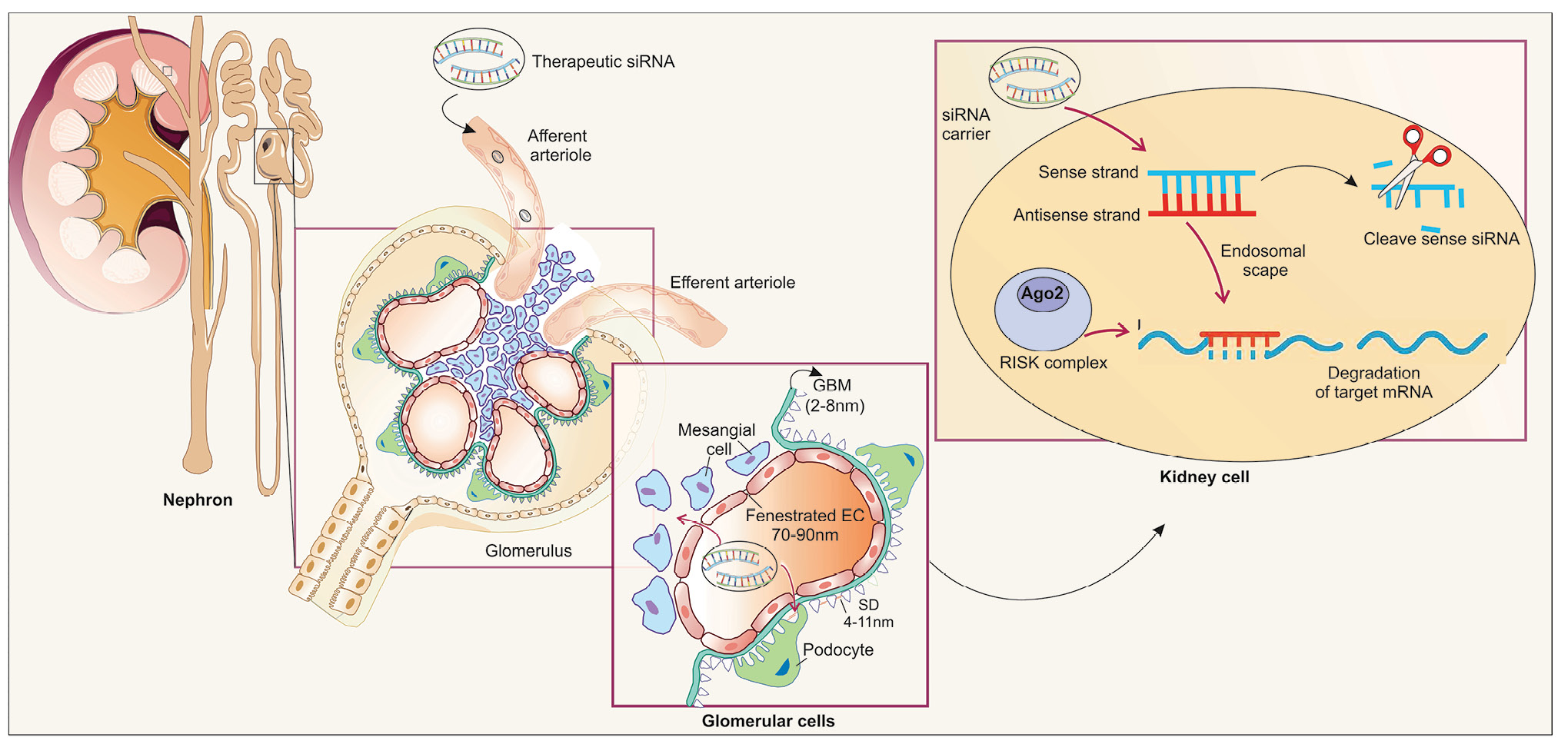
Fig. 1.
siRNA treatment for glomerulonephritis.
.
siRNA treatment for glomerulonephritis.
siRNA has become a valuable and promising therapeutic agent for kidney disorders, owing to its high specificity in targeting disease-associated genes. Clinical trials have demonstrated their potential across diverse renal conditions, including primary hyperoxaluria, hypertension, acute kidney injury (AKI), and IgAN. In primary hyperoxaluria, FDA-approved siRNA drugs like Lumasiran30 and Nedosiran,31 target HAO1 and LDHA, respectively, to reduce urinary oxalate levels and nephrocalcinosis. For hypertension, Zilebesiran32 inhibits angiotensinogen, effectively lowering blood pressure by reducing renin-angiotensin system activity.
siRNA therapy has shown protective effects in patients at high risk of AKI,33 undergoing cardiac surgery by targeting apoptosis-related pathways. Teprasiran, a siRNA targeting p53, demonstrated efficacy in ischemia-reperfusion injury models by reducing tubular apoptosis and preserving renal function. These advancements highlight the potential of siRNA in tackling both acute and chronic kidney conditions.34
The main challenges in delivering siRNAs to glomeruli
The delivery of siRNAs to glomeruli faces significant extracellular and intracellular barriers that hinder their therapeutic potential in GN. Their small molecular weight (~13 kDa) and size (7 nm) result in rapid kidney clearance, further reducing their bioavailability. Additionally, the negative charge of siRNA molecules creates a formidable barrier to membrane penetration, making cellular uptake inefficient without appropriate delivery systems. Systemic barriers are another problem in delivering siRNAs. Upon systemic administration, unmodified siRNAs are rapidly degraded by serum RNases and endonucleases, resulting in their short half-life (about 5-10 minutes) in circulation. Even after siRNAs reach the glomerular compartment, they encounter intracellular barriers, including endosomal entrapment and potential off-target effects, which can compromise both safety and efficacy.35,36
Glomerular siRNA delivery is also hindered by the glomerular filtration barrier’s (GFB’s) size selectivity, rapid clearance, and cellular complexity. Cellular complexity is the main problem. The kidneys contain at least 26 cell types, posing challenges for optimizing and delivering drugs to specific cell types. The glomerulus has a GFB comprising endothelial fenestrae (70–90 nm openings), the glomerular basement membrane (2–8 nm pores), and podocyte slit diaphragms (4–11 nm gaps). These act as obstacles to siRNA drug delivery, allowing only small molecules with diameters less than 6 nm to pass through the GFB. Advances in nanocarrier design and chemical modifications are critical to overcoming these barriers.
Another vital part of the nephron is the tubular system, which reabsorbs endogenous substances. Generally, proximal tubular cells are targeted in RNA studies. While delivering siRNAs to tubular cells is challenging, various strategies have been explored, including encapsulating oligonucleotides in nanoparticles, especially in the context of glomerular injury. Direct injection methods into the kidneys have been used in preclinical studies to target the kidney locally and avoid liver accumulation; their use in humans has been limited by invasiveness and injection difficulty.37
Mesangial cells within the glomerulus are the primary targets for siRNA delivery.4,38-40 To target these cells, siRNA drugs must exceed 6 nm in size to prevent filtration by the urinary tract, yet remain below 70–90 nm to be retained by the glomerulus and facilitate passage through the endothelial fenestra.38 By using synthetic compounds to form complexes with siRNA, the size of the delivery nanoparticle can be increased, thereby preventing glomerular filtration in the kidneys (Fig. 1).
Delivery systems
The most important hurdles to the therapeutic efficacy of siRNA administration in the treatment of GN are the instability of siRNA within the living organism, arising from rapid degradation by blood and tissue enzymes, off-target effects, and immune responses. Additionally, other obstacles include poor cellular uptake due to limited cell membrane penetration and the challenge of delivering siRNA to the site of action.41,42 Therefore, it is essential to develop delivery systems that can effectively transport siRNAs to target tissues. While siRNAs linked to lipids or encapsulated in liposomes have been shown to accumulate in the liver and silence specific genes, their presence in the kidneys may indicate degradation in tubular cells or elimination into the tubular lumen. This process reduces the targeting efficiency of siRNAs to renal tissues. Renal glomerular endothelium has a unique state. The glomerular capillary is distinguished by its fenestrated endothelium, which lacks a diaphragmatic covering. This feature contrasts with the fenestrated capillaries found in the villi of the intestine and in endocrine glands. On the other hand, the hydraulic pressure within the glomerular capillary is estimated to be between 45 and 70 mm Hg, which is considerably higher than that in the peripheral capillaries or portal venules.39
This variation may indicate that effective drug delivery to the glomeruli can be achieved while bypassing other tissues.
TGF-β is a recognized profibrotic driver of kidney disease progression in mesangial cells. TGF-β1 activates MAPK1 with synergistic interaction between TGF-β1-induced MAPK1 and Smad signaling. Additionally, in cultured mesangial cells, MAPK1 is known to initiate or modulate TGF-β1 expression, which is triggered by multiple factors, including angiotensin II, renin, mechanical stretch, and high glucose levels. Effective inhibition of intraglomerular gene expression has been achieved using siRNA conjugates with nanocarriers containing polyethylene glycol-poly l-lysine (PEG-PLL). This setup provides a valuable tool for investigating the molecular process driving glomerular diseases.39 Wang and his team employed liposomal nanocarriers, approximately 110 nm in size, containing p38α MAPK and p65 siRNA. Polyethylenimine (PEI), as a synthetic branched or linear polymer with a positive charge, can protect siRNAs from degradation. These complexes facilitate cellular absorption, provide efficient lysosomal safeguarding, and enhance the release of siRNAs into the cytosol. In Shimizu’s study of experimental models of nephritis, intraperitoneal administration of siRNA/PEG-PLL complexes targeting mitogen-activated protein kinase 1 (MAPK1) decreased glomerular MAPK1 mRNA and protein expression, leading to reduced proteinuria and glomerular inflammation.4
Nanoparticle-based therapies offer advantages, such as improved therapeutic efficacy and reduced toxicity at higher doses. Chitosan, a biodegradable, biocompatible, non-toxic, and non-immunostimulatory cationic polymer, has been widely studied for its potential to deliver therapeutic compounds to the kidneys. Several studies have demonstrated the effectiveness of low-molecular-weight chitosan in this context. Alan et al evaluated chitosan/siRNA nanoplexes for silencing the PDGF-B and PDGFR-β genes in the kidney, specifically targeting mesangial cell growth and matrix buildup in a Mesangial Proliferative Glomerulonephritis (MsPGN) model triggered by anti-Thy-1.1 antibody. The findings showed chitosan/siPDGF-B + siPDGFR-β nanoplexes as an innovative therapeutic approach to reduce mesangial proliferation and fibrosis in anti-Thy1 GN rat models.43 GalNAc-siRNAs are another example of ligand-targeted conjugates that have been shown to suppress hepatic expression of complement components (C3, C5) in complement-mediated GN models, thereby indirectly reducing renal immune damage. Overall, engineered carriers and compounds have made significant progress and can significantly enhance renal siRNA delivery. Further optimization of biodistribution and endosomal escape is suggested.
siRNA in different glomerulonephritis
siRNA therapy is emerging as a promising targeted approach for GN, addressing pathogenic pathways such as inflammation, fibrosis, and immune dysregulation. The upcoming sections highlight the role of siRNAs in targeting key pathological pathways in different GNs.
C3 glomerulopathy
Complement 3 glomerulopathy (C3G) is an uncommon and complex kidney disorder characterized by significant C3 deposition in the glomeruli, with minimal to no immunoglobulin. This condition leads to damage to the glomerular structure. The pivotal events in the pathogenesis of the disease include mutations in complement factors and regulators, such as C3, FB, FH, FI, and CFHRs, as well as autoantibodies targeting complement components, such as C3 and FB. Additionally, nephritic elements that reinforce C3 and/or C5 convertase may also play a role.44 Approximately 50-70% of patients with primary C3G exhibit inherited and/or acquired irregularities, and each of these irregularities can result in inappropriate activation or inadequate regulation of the signaling pathway.45 The clinical progression of C3G is variable and typically unfavorable, with about 50% of patients experiencing rapidly progressive glomerulonephritis that leads to end-stage renal failure in the first decade after being diagnosed. At present, there are no approved treatments available for C3G, and clinical management primarily focuses on supportive care aimed at managing proteinuria and hypertension. This may include immunosuppressive therapy and, in some cases, plasma exchange.46 Given the fundamental importance of the complement system in the development of C3 glomerulopathy, consideration has been directed to anti-complement agents, including eculizumab, which is a monoclonal antibody against C5, but unfortunately, the therapeutic response to eculizumab is very heterogeneous, indicating the activity of other dependent upstream pathways to C3-convertase, which cannot be blocked by eculizumab.47 The study by Zanchi et al evaluated the effect of GalNAc SLN501, a siRNA, on liver C3 production in factor H (FH) deficiency (Cfh + /- mice). They demonstrate that SLN501 and SLN500 can cause a dose-dependent reduction in hepatic C3 mRNA (up to ~91–95% silencing), a greater reduction of serum C3, abolish C3 activation fragments, decrease FB activation, and restore plasma C5 levels to near wild-type levels. In the glomerulus, high-dose SLN501 (8 mg/kg) and SLN500 (5 mg/kg) significantly reduced C3d deposition (~52–53% reduction)—a slower development of mesangial and subendothelial electron-dense deposits in the kidneys was also observed.
The current findings suggest that RNAi-mediated inhibition of C3 in the liver could be a promising therapeutic approach for patients with C3 glomerulopathy associated with partial or complete loss of factor H function.48 Although there are currently no treatments designed explicitly for C3 glomerulopathy, inhibiting liver-produced C3 with RNAi offers promising new therapeutic options. Preclinical studies in mouse models of C3G have demonstrated that this approach can effectively reduce fluid-phase complement dysregulation, limit glomerular C3 deposition, and slow the progression of ultrastructural kidney damage. Nonetheless, additional clinical research is needed to determine the safety profile of RNAi therapy for C3G in humans, particularly potential side effects such as increased susceptibility to infections and liver Toxicity in human subjects.
Membranous nephropathy (MN)
Membranous nephropathy is an autoimmune kidney disease characterized by the thickening of the glomerular capillary walls attributable to the deposition of immune complexes. In primary MN, antibodies often target the phospholipase A2 receptor (PLA2R) on the surface of podocytes, leading to the formation of immune complex deposits that trigger local complement activation and ultimately cause podocyte destruction and damage to the glomerular basement membrane. The classical pathway initiates harmful complement activation in MN. In an autoimmune mouse model of MN, targeting C3 with N-acetyl-galactosamine (GalNAc)- conjugated siRNAs for 4 weeks after the onset of significant proteinuria reduced disease. This treatment led to the complete elimination of glomerular C3 and the downstream C5b-9 complex.49
siRNA in lupus nephritis
Systemic lupus erythematosus (SLE) is a complex autoimmune disease characterized by the production of autoantibodies, such as those targeting double-stranded DNA (dsDNA), which cause inflammation and tissue damage. One of the most serious complications related to SLE includes lupus nephritis (LN), which can lead to severe proteinuria, chronic renal failure, and end-stage kidney disease. Recent studies have explored the therapeutic potential of siRNA as a new approach to modulating immune responses in SLE.
Elevated levels of circulating interferon (IFN)-α indicate immune system activation in SLE and correlate with the severity of the disease. Typically, type I IFN levels are heightened in SLE; however, this increase is frequently associated with abnormal gene expression within the IFN signaling pathway. This gene expression profile, known as the ‘IFN signature’, serves as a marker of more severe disease presentation, particularly when it impacts the kidneys.50 Type I interferons stimulate dendritic cells (DCs), leading to enhanced expression of costimulatory molecules, major histocompatibility complex class I and II (MHC class I and II) proteins, as well as chemokines and their receptors. Moreover, IFNs promote the activation of T and B cells, which increases antibody production.51
Sirtuin 1 (SIRT1), a NAD+ -dependent deacetylase, plays an essential role in numerous biological functions, including immune regulation. Regarding SLE, SIRT1 is linked to the diversity and extension of follicular helper T (Tfh) cells. SIRT1, by promoting Tfh cell expansion, appears to be a promising therapeutic target for SLE. Studies have shown that suppressing SIRT1 expression with siRNA or blocking its function with agents such as EX527 or nicotinamide can hinder Tfh cell expansion in experimental settings.52
The complement structure plays a dual role in LN development. The contribution of kidney-secreted complement C3 to the circulating pool in humans is significant,53 indicating that modulating complement activity can affect the course of LN. The CD40-CD40L pathway is essential for the pathogenesis of SLE, the generation of autoantibodies, and their deposition in the kidneys, leading to renal injury in patients with LN. Ripoll et al found that glomerular C3 deposits were significantly reduced in mice treated with cholesterol-conjugated anti-CD40 siRNA, resulting in decreased C3 gene expression in the kidneys. CD40 silencing also led to a decrease in infiltrating T-cells, suggesting a change in the subsequent local inflammatory response.54 These results support the potential therapeutic effects of selective CD40 blockade in LN.
In LN, hyperactivation of B lymphocytes and abnormal cytokine expression are essential mechanisms. Inflammatory cytokines such as IL-6, IL-10, and IL-21 are known to stimulate B cell maturation, thereby contributing to the progression of lupus.55 Research has shown that the excessive expression of B cell-activating factor (BLYSS) by type I IFNs plays a role in the progression of SLE, and its blocking may postpone the disease in mouse models. A recent study utilizing dual blockade therapy with engineered siRNAs targeting BLYSS and interferon regulatory factor 5 (IRF5) in NZB/WF1 mice with conventional autoimmune nephritis demonstrated a decline in circulating anti-dsDNA antibodies and decreased immune complex deposition within renal histopathological lesions.56
Dihydroartemisinin (DHA), an artemisinin byproduct, exhibits significant anti-malarial activity with anti-inflammatory and immunoregulatory properties. The Artemisinin derivative SM934DHA has also been shown to alleviate symptoms of lupus in BXSB mice by downregulating TLR4 signaling.57 Nevertheless, the focus of old-style antimalarial artemisinin medicines on a single target limits their use in the treatment of autoimmune diseases. TAT-CLs-DHA/siRNA is a therapeutic delivery system that utilizes a combination of TAT peptide-modified cationic liposomes, DHA, a metabolite of artemisinin, and specific siRNA. TAT-CLs-DHA/siRNA targets explicitly Toll-like receptor 4 (TLR4) signaling in lupus nephritis through a dual mechanism involving the delivery of siRNA that silences high-mobility group box 1 (HMGB1) and the anti-inflammatory effects of DHA. HMGB1 is a proinflammatory cytokine that binds to TLR4, triggering inflammatory responses. This system aims to inhibit the signaling pathway of TLR4, a receptor that plays a significant role in the inflammatory response. The research showed that administering dihydroartemisinin and HMGB1 siRNAs via parenteral routes every 4 days using (TAT)-modified cationic liposomes (TAT-CLs-DHA/siRNA) effectively inhibited B-cell proliferation and activation in lupus-prone MRL/lpr mice by targeting the TLR4 signaling pathway. Also, there was a decrease in proteinuria, serum anti-dsDNA antibody levels, and the secretion of interleukins IL-6, IL-10, IL-17, and IL-21.58
In MRL/lpr mice, a well-established model of LN, repeated intraperitoneal injections of MAPK1 siRNA resulted in significant suppression of MAPK1 mRNA and protein expression in glomeruli. MAPK1 siRNA delivered via poly (ethylene glycol)-poly (l-lysine) nanocarriers. The siRNA/nanocarrier complex (~10–20 nm) effectively crossed endothelial fenestrations to target mesangial cells. This therapy reduced glomerular inflammation, proteinuria, and fibrosis, improving kidney function.4
siRNA in immunoglobulin A nephropathy
Immunoglobulin A nephropathy is the most prevalent type of glomerulonephritis and carries a high risk of kidney failure. The pathogenesis of IgAN involves several key events (the “four-hit” hypothesis) comprising the creation of galactose-deficient IgA1 (Gd-IgA1), which acts as an autoantigen, the creation of autoantibodies against Gd-IgA1, formation of immune complexes between the autoantibodies and Gd-IgA1, and the deposition of these immune complexes in the glomerular mesangium, leading to glomerular injury. The development of siRNAs targeting specific components of the immune response, inflammation, or complement system could lead to more effective and safer treatments for IgAN.
Toll-like receptor 9 (TLR9), IL-6, and APRIL (A proliferation-inducing ligand) are involved in the synthesis of Gd-IgA1.59 A recent study showed that siRNA knockdown of APRIL completely blocked the IL-6-induced overproduction of Gd-IgA1. Using siRNA or other inhibitory strategies targeting APRIL and IL-6 may reduce nephritogenic Gd-IgA1 levels and thereby mitigate kidney damage. Understanding individual variations in TLR9 expression and response could lead to personalized treatment strategies for patients with IgAN.59 Immune complex formation causes the activation of the complement system. The complement system can be activated through three different pathways: the lectin, classical, and alternative pathways. Key components such as C3 and C5 play pivotal roles in these pathways. Evidence supports the lectin pathway’s influence on IgAN progression and worse outcomes. MASP-2 is an essential enzyme that initiates the lectin pathway of the complement system, making it a suitable target for drug development.60 In a study by Ling and his colleagues, they used RNL288, a MASP2-targeting siRNA, for blocking the lectin pathway. They demonstrated reduced MASP2 expression in primary human hepatocytes and cynomolgus monkey hepatocytes. Furthermore, in humanized mice, serum MASP2 levels were reduced by up to 94%. In non-human primates, MASP2 levels remained suppressed for 3 months without any adverse effects noted in toxicity assessments. These results indicate that RNK288 may be a feasible treatment alternative for individuals with IgAN by inhibiting lectin pathway activation, thereby potentially improving disease outcomes.61
Complement activation leads to the production of inflammatory mediators, such as C3a and C5a, which enhance tissue damage and inflammation. Meanwhile, C5b contributes to tissue damage in IgAN by creating the membrane attack complex (MAC), disrupting cellular membranes, and exacerbating glomerular injury.62 Given the roles of C5 and other components of the complement system, they represent potential therapeutic targets. A randomized clinical trial used Cemdisiran, an RNA interference therapy targeting hepatic C5 production, in IgAN patients to reduce proteinuria. The treatment achieved a 98.7% reduction in serum C5 levels and a 37.4% reduction in the urinary protein-to-creatinine ratio (UPCR). These results suggest that Cemdisiran may help reduce proteinuria and potentially slow the progression to kidney failure and mortality. The therapy was well tolerated, with the most common treatment-related side effect being mild and transient injection site reactions. Additionally, a mild increase of alanine transaminase and aspartate transaminase was observed in 9% of patients, which normalized without any intervention.63
Both p38α MAPK and p65 are crucial regulators in inflammatory signaling pathways, particularly those involving p38 MAPK and NF-κB. Their over-activation contributes significantly to the inflammatory processes observed in IgAN. By silencing these genes, the liposomal nanoparticles aim to reduce inflammation, thereby protecting renal function and delaying the progression of kidney disease. Recent research has focused on the co-delivery of p38α MAPK and p65 siRNA using innovative liposomal nanoparticles specifically designed to target the glomerulus, presenting a promising therapeutic strategy for IgAN. The co-delivery system not only effectively silenced the targeted genes but also alleviated key IgAN-associated symptoms, such as proteinuria and renal inflammation. 39 This suggests that the approach could be a versatile platform for addressing novel strategies of glomerulus-directing and a hopeful calming way for other inflammatory diseases.
Obesity-related glomerulopathy
Obesity presents a remarkable public health challenge, with its prevalence rising on a global scale. One considerable renal complication linked to obesity is obesity-related glomerulopathy (ORG), characterized by proteinuria and glomerulomegaly in obese individuals, without any clinical and histopathological signs of the other renal diseases.64 The pathogenesis of ORG is associated with adipose tissue dysfunction, such as elevated inflammation, rather than alterations in adipose tissue volume, suggesting that body mass index (BMI) may not be a reliable indicator of metabolic syndrome.65 In contrast to healthy individuals, the renal tissues of patients with ORG exhibited elevated levels of tumor necrosis factor-α, interleukin (IL)-1, and IL-6. This elevation in pro-inflammatory cytokines causes glomerular sclerosis and subsequent deterioration of renal organization and function. Podocytes are specialized visceral epithelial cells that demonstrate limited renewing capacity. The quantity and structural integrity of podocytes are essential for sustaining normal glomerular filtration. Adiponectin is an adipocytokine predominantly produced by adipose tissue, with anti-atherosclerotic, anti-inflammatory, and anti-diabetic properties. It is crucial in the instruction of glucose and lipid metabolism and demonstrates an inverse relationship with obesity.66 The silencing of the adiponectin gene using siRNA led to increased ROS production, which, in turn, triggered the NLRP3 inflammasome and phosphorylated NF-κB in podocytes.
Critical perspective and future directions
The results of siRNA therapies to date have been promising in experimental models of glomerulonephritis, such as GalNAc-linked siRNAs targeting hepatic C3, which are liver-specific and efficient in gene silencing. A few clinical studies have also shown promising results, including Cemdisiran, which targets hepatic C5 synthesis and has demonstrated clinical efficacy, including significant reductions in proteinuria and complement activity in IgA nephropathy with a well-defined immune profile. These strategies are promising and offer the highest translational potential, leveraging established drug-delivery platforms and favorable pharmacokinetics.
In contrast, approaches that directly target intraglomerular cells (e.g., mesangial, endothelial, or podocyte-specific delivery using nanoparticles or liposomes) remain largely preclinical. While studies using PEG-PLL or chitosan nanoplexes have demonstrated effective gene silencing in animal models, challenges in achieving selective glomerular uptake, preventing endosomal degradation, and minimizing immune activation continue to hinder clinical application.
In an overview of the mechanisms of siRNA therapies, targeting simultaneous activation of the complement (C3, C5, MASP-2) and fibrosis/inflammation (p38 MAPK, NF-κB, TGF-β) pathways appears promising. Dual-targeting by siRNA systems, such as BLYSS + IRF5 inhibition in lupus nephritis, may be more effective in improving efficacy by addressing the multifactorial nature of immune damage.
However, the limitation of using siRNA in the treatment of glomerulonephritis is that it can bind unintended genes and silence them, or unmodified siRNAs can trigger innate immune responses via toll-like receptors (TLR3, TLR7, TLR8), leading to type I interferon induction. Recent clinical trials (e.g., Cemdisiran in IgAN) generally had favorable safety profiles. Most adverse events were mild and transient, such as injection-site reactions or mild liver enzyme elevations. Serious events were rare. Long-term monitoring is ongoing, particularly for immune-related adverse events and hepatic toxicity. Compared with conventional therapy, unlike broad immunosuppressants, siRNA therapies provide targeted gene silencing, which may reduce systemic immunosuppression and associated risks, such as infections or bone marrow suppression.
Although siRNA therapies for GN show promise in preclinical studies, they are still far from clinical implementation and inclusion in guidelines. Key challenges include high costs, limited accessibility in resource-limited settings, and potential health disparities. Cost-effectiveness analyses comparing siRNA with conventional therapies, along with strategies such as tiered pricing, insurance coverage, local manufacturing, and public-private partnerships, will be essential to enabling equitable global access.
Other alternatives to siRNAs include antisense oligonucleotides (ASOs), which bind to target mRNA via single-stranded interactions and are degraded by RNase H. They have easier access to nuclear targets and simpler chemical structures. However, they are generally weaker in gene silencing and less stable than siRNAs.67 Unlike siRNA, which has reversible effects, the CRISPR/Cas method permanently modifies DNA sequences, raising safety and ethical concerns about off-target and irreversible mutations.68 Furthermore, monoclonal antibodies (mAbs), which are currently the mainstay of immunological therapy in glomerulonephritis (e.g., rituximab, eculizumab), act at the protein level. In contrast, siRNA therapies can inhibit the production of pathogenic proteins altogether, potentially providing longer-lasting effects at lower doses.69 Overall, siRNA therapies represent a good example of a biologic drug and ggene silencing technology, with their target-specific performance and good safety supporting their translational potential in kidney diseases.
The development of siRNA-based therapies for glomerulonephritis should focus on translating preclinical studies into clinical trials and long-term follow-up. The use of molecular and genetic biomarkers in parallel with siRNA therapy (e.g., complement activation profiles, interferon signatures, or podocyte injury markers) could be a promising approach for personalized therapeutic strategies.
Conclusion
To sum up, siRNA therapy demonstrated significant potential in treating GN. siRNA therapies offer a promising, targeted approach to treating glomerulonephritis by targeting the disease’s underlying molecular mechanisms. While traditional treatments provide broad immunosuppression, they often come with significant side effects and do not target specific disease pathways. The development of effective siRNA therapies for GN is still in its early stages. Still, ongoing research and clinical trials may pave the way for more precise and potent therapies with reduced adverse effects than traditional approaches. siRNA therapies show promise in modulating key signaling pathways and silencing intraglomerular genes, improving kidney function, reducing proteinuria, and glomerular damage, inflammation, and fibrosis. Still, more research is needed to fully understand their long-term effects and ensure their safety and efficacy.
Competing Interests
The authors state that they have no conflicts of interest to disclose.
Data Availability Statement
All the data produced during this study are incorporated within this published article.
Declaration of AI-Assisted Tools in the Writing Procedure
We want to acknowledge the use of AI-based tools for grammar and language editing in the preparation of this manuscript.
Ethical Approval
Not applicable.
Acknowledgements
We would like to appreciate of the cooperation of Clinical Research Development Unit, Imam Reza General Hospital, and Tabriz, Iran in conducting of this research.
References
- Kazi AM, Hashmi MF. Glomerulonephritis. In: StatPearls [Internet]. Treasure Island, FL: StatPearls Publishing; 2025. Available from: https://www.ncbi.nlm.nih.gov/books/NBK560657/.
- Hosseiniyan Khatibi SM, Ardalan M, Abediazar S, Zununi Vahed S. The impact of steroids on the injured podocytes in nephrotic syndrome. J Steroid Biochem Mol Biol 2020; 196:105490. doi: 10.1016/j.jsbmb.2019.105490 [Crossref] [ Google Scholar]
- Hejazian SM, Zununi Vahed S, Moghaddas Sani H, Nariman-Saleh-Fam Z, Bastami M, Hosseiniyan Khatibi SM. Steroid-resistant nephrotic syndrome: pharmacogenetics and epigenetic points and views. Expert Rev Clin Pharmacol 2020; 13:147-56. doi: 10.1080/17512433.2020.1702877 [Crossref] [ Google Scholar]
- Shimizu H, Hori Y, Kaname S, Yamada K, Nishiyama N, Matsumoto S. siRNA-based therapy ameliorates glomerulonephritis. J Am Soc Nephrol 2010; 21:622-33. doi: 10.1681/asn.2009030295 [Crossref] [ Google Scholar]
- Rovin BH, Adler SG, Barratt J, Bridoux F, Burdge KA, Chan TM. Executive summary of the KDIGO 2021 Guideline for the Management of Glomerular Diseases. Kidney Int 2021; 100:753-79. doi: 10.1016/j.kint.2021.05.015 [Crossref] [ Google Scholar]
- Weng Y, Xiao H, Zhang J, Liang XJ, Huang Y. RNAi therapeutic and its innovative biotechnological evolution. Biotechnol Adv 2019; 37:801-25. doi: 10.1016/j.biotechadv.2019.04.012 [Crossref] [ Google Scholar]
- Setten RL, Rossi JJ, Han SP. The current state and future directions of RNAi-based therapeutics. Nat Rev Drug Discov 2019; 18:421-46. doi: 10.1038/s41573-019-0017-4 [Crossref] [ Google Scholar]
- Schumacher A, Rookmaaker MB, Joles JA, Kramann R, Nguyen TQ, van Griensven M. Defining the variety of cell types in developing and adult human kidneys by single-cell RNA sequencing. NPJ Regen Med 2021; 6:45. doi: 10.1038/s41536-021-00156-w [Crossref] [ Google Scholar]
- Ahmadian E, Rahbar Saadat Y, Dalir Abdolahinia E, Bastami M, Shoja MM, Zununi Vahed S. The role of cytokines in nephrotic syndrome. Mediators Inflamm 2022; 2022:6499668. doi: 10.1155/2022/6499668 [Crossref] [ Google Scholar]
- Zununi Vahed S, Hosseiniyan Khatibi SM, Ardalan M. Canonical effects of cytokines on glomerulonephritis: a new outlook in nephrology. Med Res Rev 2025; 45:144-63. doi: 10.1002/med.22074 [Crossref] [ Google Scholar]
- Xiong Y, Li W, Jin S, Wan S, Wu S. Inflammation in glomerular diseases. Front Immunol 2025; 16:1526285. doi: 10.3389/fimmu.2025.1526285 [Crossref] [ Google Scholar]
- Iyengar A, Kamath N, Radhakrishnan J, Estebanez BT. Infection-related glomerulonephritis in children and adults. Semin Nephrol 2023; 43:151469. doi: 10.1016/j.semnephrol.2023.151469 [Crossref] [ Google Scholar]
- Ardalan M, Ahmadian E, Hosseiniyan Khatibi SM, Rahbar Saadat Y, Bastami M, Bagheri Y. Microbiota and glomerulonephritis: an immunological point of view. Am J Med Sci 2022; 364:695-705. doi: 10.1016/j.amjms.2022.05.025 [Crossref] [ Google Scholar]
- Wetmore JB, Guo H, Liu J, Collins AJ, Gilbertson DT. The incidence, prevalence, and outcomes of glomerulonephritis derived from a large retrospective analysis. Kidney Int 2016; 90:853-60. doi: 10.1016/j.kint.2016.04.026 [Crossref] [ Google Scholar]
-
Daehn IS, Duffield JS. The glomerular filtration barrier: a structural target for novel kidney therapies. Nat Rev Drug Discov 2021. 20: 770-88. doi: 10.1038/s41573-021-00242-0.
- Romagnani P, Kitching AR, Leung N, Anders HJ. The five types of glomerulonephritis classified by pathogenesis, activity and chronicity (GN-AC). Nephrol Dial Transplant 2023; 38:ii3-10. doi: 10.1093/ndt/gfad067 [Crossref] [ Google Scholar]
- Sethi S, Haas M, Markowitz GS, D’Agati VD, Rennke HG, Jennette JC. Mayo clinic/renal pathology society consensus report on pathologic classification, diagnosis, and reporting of GN. J Am Soc Nephrol 2016; 27:1278-87. doi: 10.1681/asn.2015060612 [Crossref] [ Google Scholar]
- Hejazian SM, Rahbar Saadat Y, Bahmanpour Z, Hosseiniyan Khatibi SM, Ardalan M, Zununi Vahed S. Dicer and Drosha expression in patients with nephrotic syndrome. Biofactors 2020; 46:645-52. doi: 10.1002/biof.1638 [Crossref] [ Google Scholar]
- Hejazian SM, Rahbar Saadat Y, Hosseiniyan Khatibi SM, Farnood F, Farzamikia N, Hejazian SS. Circular RNAs as novel biomarkers in glomerular diseases. Arch Physiol Biochem 2024; 130:568-80. doi: 10.1080/13813455.2023.2212328 [Crossref] [ Google Scholar]
- Moghaddas Sani H, Hejazian M, Hosseinian Khatibi SM, Ardalan M, Zununi Vahed S. Long non-coding RNAs: An essential emerging field in kidney pathogenesis. Biomed Pharmacother 2018; 99:755-65. doi: 10.1016/j.biopha.2018.01.122 [Crossref] [ Google Scholar]
- Nakhjavani M, Etemadi J, Pourlak T, Mirhosaini Z, Zununi Vahed S, Abediazar S. Plasma levels of miR-21, miR-150, miR-423 in patients with lupus nephritis. Iran J Kidney Dis 2019; 13:198-206. [ Google Scholar]
- Zununi Vahed S, Nakhjavani M, Etemadi J, Jamshidi H, Jadidian N, Pourlak T. Altered levels of immune-regulatory microRNAs in plasma samples of patients with lupus nephritis. Bioimpacts 2018; 8:177-83. doi: 10.15171/bi.2018.20 [Crossref] [ Google Scholar]
- Wang LP, Gao YZ, Song B, Yu G, Chen H, Zhang ZW. MicroRNAs in the progress of diabetic nephropathy: a systematic review and meta-analysis. Evid Based Complement Alternat Med 2019; 2019:3513179. doi: 10.1155/2019/3513179 [Crossref] [ Google Scholar]
- Hejazian SM, Ardalan M, Shoja MM, Samadi N, Zununi Vahed S. Expression levels of miR-30c and miR-186 in adult patients with membranous glomerulonephritis and focal segmental glomerulosclerosis. Int J Nephrol Renovasc Dis 2020; 13:193-201. doi: 10.2147/ijnrd.S258624 [Crossref] [ Google Scholar]
- Ardalan M, Hejazian SM, Fazlazar Sharabiyani H, Farnood F, Ghafari Aghdam A, Bastami M. Dysregulated levels of glycogen synthase kinase-3β (GSK-3β) and miR-135 in peripheral blood samples of cases with nephrotic syndrome. PeerJ 2020; 8:e10377. doi: 10.7717/peerj.10377 [Crossref] [ Google Scholar]
- Rahbar Saadat Y, Hejazian SM, Nariman-Saleh-Fam Z, Bastami M, Poursheikhani A, Shoja MM. Glucocorticoid receptors and their upstream epigenetic regulators in adults with steroid-resistant nephrotic syndrome. Biofactors 2020; 46:995-1005. doi: 10.1002/biof.1680 [Crossref] [ Google Scholar]
- Szeto CC, Li PK. MicroRNAs in IgA nephropathy. Nat Rev Nephrol 2014; 10:249-56. doi: 10.1038/nrneph.2014.50 [Crossref] [ Google Scholar]
-
Chakole VR, Dutta T, Sen P. siRNA therapeutics for effective management of rheumatoid arthritis. Next Nanotechnol 2025. 7: 100135. doi: 10.1016/j.nxnano.2025.100135.
-
Sioud M. RNA interference: story and mechanisms. In: Ditzel HJ, Tuttolomondo M, Kauppinen S, eds. Design and Delivery of SiRNA Therapeutics. New York, NY: Springer. 2021. p. 1-15. doi: 10.1007/978-1-0716-1298-9_1.
- Kang C. Lumasiran: a review in primary hyperoxaluria type 1. Drugs 2024; 84:219-26. doi: 10.1007/s40265-023-01987-1 [Crossref] [ Google Scholar]
- Hoppe B, Koch A, Cochat P, Garrelfs SF, Baum MA, Groothoff JW. Safety, pharmacodynamics, and exposure-response modeling results from a first-in-human phase 1 study of nedosiran (PHYOX1) in primary hyperoxaluria. Kidney Int 2022; 101:626-34. doi: 10.1016/j.kint.2021.08.015 [Crossref] [ Google Scholar]
- Lemine M, Almuzainy S, Aljubeh R, Alilo A. Zilebesiran and hypertension: a systematic review and meta-analysis. J Saudi Heart Assoc 2024; 36:420-30. doi: 10.37616/2212-5043.1408 [Crossref] [ Google Scholar]
- Thielmann M, Corteville D, Szabo G, Swaminathan M, Lamy A, Lehner LJ. Teprasiran, a small interfering RNA, for the prevention of acute kidney injury in high-risk patients undergoing cardiac surgery: a randomized clinical study. Circulation 2021; 144:1133-44. doi: 10.1161/circulationaha.120.053029 [Crossref] [ Google Scholar]
- Losito A, Solano G. Small interfering RNA in kidney diseases: promises and limitations. Kidney Dial 2024; 5:1. doi: 10.3390/kidneydial5010001 [Crossref] [ Google Scholar]
- Sajid MI, Moazzam M, Kato S, Yeseom Cho K, Tiwari RK. Overcoming barriers for siRNA therapeutics: from bench to bedside. Pharmaceuticals (Basel) 2020; 13:294. doi: 10.3390/ph13100294 [Crossref] [ Google Scholar]
- Rajeev A, Siby A, Koottungal MJ, George J, John F. Knocking down barriers: advances in siRNA delivery. ChemistrySelect 2021; 6:13350-62. doi: 10.1002/slct.202103288 [Crossref] [ Google Scholar]
- Ahn I, Kang CS, Han J. Where should siRNAs go: applicable organs for siRNA drugs. Exp Mol Med 2023; 55:1283-92. doi: 10.1038/s12276-023-00998-y [Crossref] [ Google Scholar]
- Wang J, Masehi-Lano JJ, Chung EJ. Peptide and antibody ligands for renal targeting: nanomedicine strategies for kidney disease. Biomater Sci 2017; 5:1450-9. doi: 10.1039/c7bm00271h [Crossref] [ Google Scholar]
- Wang Y, Wu Q, Wang J, Li L, Sun X, Zhang Z. Co-delivery of p38α MAPK and p65 siRNA by novel liposomal glomerulus-targeting nano carriers for effective immunoglobulin a nephropathy treatment. J Control Release 2020; 320:457-68. doi: 10.1016/j.jconrel.2020.01.024 [Crossref] [ Google Scholar]
- Takabatake Y, Isaka Y, Imai E. In vivo transfer of small interfering RNA or small hairpin RNA targeting glomeruli. Methods Mol Biol 2009; 466:251-63. doi: 10.1007/978-1-59745-352-3_18 [Crossref] [ Google Scholar]
- Higuchi Y, Kawakami S, Hashida M. Strategies for in vivo delivery of siRNAs: recent progress. BioDrugs 2010; 24:195-205. doi: 10.2165/11534450-000000000-00000 [Crossref] [ Google Scholar]
- Sargazi S, Arshad R, Ghamari R, Rahdar A, Bakhshi A, Fathi Karkan S. siRNA-based nanotherapeutics as emerging modalities for immune-mediated diseases: a preliminary review. Cell Biol Int 2022; 46:1320-44. doi: 10.1002/cbin.11841 [Crossref] [ Google Scholar]
- Alan S, Şalva E, Yılmaz İ, Turan S, Akbuğa J. The effectiveness of chitosan-mediated silencing of PDGF-B and PDGFR-β in the mesangial proliferative glomerulonephritis therapy. Exp Mol Pathol 2019; 110:104280. doi: 10.1016/j.yexmp.2019.104280 [Crossref] [ Google Scholar]
- Smith RJH, Appel GB, Blom AM, Cook HT, D’Agati VD, Fakhouri F. C3 glomerulopathy - understanding a rare complement-driven renal disease. Nat Rev Nephrol 2019; 15:129-43. doi: 10.1038/s41581-018-0107-2 [Crossref] [ Google Scholar]
- Daina E, Cortinovis M, Remuzzi G. Kidney diseases. Immunol Rev 2023; 313:239-61. doi: 10.1111/imr.13167 [Crossref] [ Google Scholar]
- Nester CM, Smith RJ. Treatment options for C3 glomerulopathy. Curr Opin Nephrol Hypertens 2013; 22:231-7. doi: 10.1097/MNH.0b013e32835da24c [Crossref] [ Google Scholar]
- Le Quintrec M, Lapeyraque AL, Lionet A, Sellier-Leclerc AL, Delmas Y, Baudouin V. Patterns of clinical response to eculizumab in patients with C3 glomerulopathy. Am J Kidney Dis 2018; 72:84-92. doi: 10.1053/j.ajkd.2017.11.019 [Crossref] [ Google Scholar]
- Zanchi C, Locatelli M, Cerullo D, Aumiller V, Corna D, Rottoli D. Efficacy of GalNAc C3 siRNAs in factor H-deficient mice with C3 glomerulopathy. Mol Immunol 2024; 168:10-6. doi: 10.1016/j.molimm.2024.02.010 [Crossref] [ Google Scholar]
- Seifert L, Zahner G, Meyer-Schwesinger C, Hickstein N, Dehde S, Wulf S. The classical pathway triggers pathogenic complement activation in membranous nephropathy. Nat Commun 2023; 14:473. doi: 10.1038/s41467-023-36068-0 [Crossref] [ Google Scholar]
- Baechler EC, Batliwalla FM, Karypis G, Gaffney PM, Ortmann WA, Espe KJ. Interferon-inducible gene expression signature in peripheral blood cells of patients with severe lupus. Proc Natl Acad Sci U S A 2003; 100:2610-5. doi: 10.1073/pnas.0337679100 [Crossref] [ Google Scholar]
- Le Bon A, Thompson C, Kamphuis E, Durand V, Rossmann C, Kalinke U. Cutting edge: enhancement of antibody responses through direct stimulation of B and T cells by type I IFN. J Immunol 2006; 176:2074-8. doi: 10.4049/jimmunol.176.4.2074 [Crossref] [ Google Scholar]
- He L, Liao W, Wang X, Wang L, Liang Q, Jiang L. Sirtuin 1 overexpression contributes to the expansion of follicular helper T cells in systemic lupus erythematosus and may serve as an accessible therapeutic target. Rheumatology (Oxford) 2024; 63:1699-709. doi: 10.1093/rheumatology/kead453 [Crossref] [ Google Scholar]
- Tang S, Zhou W, Sheerin NS, Vaughan RW, Sacks SH. Contribution of renal secreted complement C3 to the circulating pool in humans. J Immunol 1999; 162:4336-41. doi: 10.4049/jimmunol.162.7.4336 [Crossref] [ Google Scholar]
- Ripoll È, Merino A, Herrero-Fresneda I, Aran JM, Goma M, Bolaños N. CD40 gene silencing reduces the progression of experimental lupus nephritis modulating local milieu and systemic mechanisms. PLoS One 2013; 8:e65068. doi: 10.1371/journal.pone.0065068 [Crossref] [ Google Scholar]
- Ohl K, Tenbrock K. Inflammatory cytokines in systemic lupus erythematosus. J Biomed Biotechnol 2011; 2011:432595. doi: 10.1155/2011/432595 [Crossref] [ Google Scholar]
- Guiteras J, Ripoll É, Bolaños N, De Ramon L, Fontova P, Lloberas N. The gene silencing of IRF5 and BLYSS effectively modulates the outcome of experimental lupus nephritis. Mol Ther Nucleic Acids 2021; 24:807-21. doi: 10.1016/j.omtn.2021.03.019 [Crossref] [ Google Scholar]
- Diao L, Tao J, Wang Y, Hu Y, He W. Co-delivery of dihydroartemisinin and HMGB1 siRNA by TAT-modified cationic liposomes through the TLR4 signaling pathway for treatment of lupus nephritis. Int J Nanomedicine 2019; 14:8627-45. doi: 10.2147/ijn.S220754 [Crossref] [ Google Scholar]
- Diao L, Li M, Tao J, Xu X, Wang Y, Hu Y. Therapeutic effects of cationic liposomes on lupus-prone MRL/lpr mice are mediated via inhibition of TLR4-triggered B-cell activation. Nanomedicine 2022; 40:102491. doi: 10.1016/j.nano.2021.102491 [Crossref] [ Google Scholar]
- Makita Y, Suzuki H, Kano T, Takahata A, Julian BA, Novak J. TLR9 activation induces aberrant IgA glycosylation via APRIL- and IL-6-mediated pathways in IgA nephropathy. Kidney Int 2020; 97:340-9. doi: 10.1016/j.kint.2019.08.022 [Crossref] [ Google Scholar]
- Lafayette RA, Rovin BH, Reich HN, Tumlin JA, Floege J, Barratt J. Safety, tolerability and efficacy of narsoplimab, a novel MASP-2 inhibitor for the treatment of IgA nephropathy. Kidney Int Rep 2020; 5:2032-41. doi: 10.1016/j.ekir.2020.08.003 [Crossref] [ Google Scholar]
- Pan L, Meng G, Liu W, Huang C, Xiaoyan Y, Cai G. RNK288: a MASP2-targeting small interfering RNA (siRNA) for the treatment of IgA nephropathy by blocking the activation of lectin pathway: SA-PO729. J Am Soc Nephrol 2024; 35:10-681. doi: 10.1681/ASN.2024rbnftmnz [Crossref] [ Google Scholar]
- Caravaca-Fontán F, Gutiérrez E, Sevillano ÁM, Praga M. Targeting complement in IgA nephropathy. Clin Kidney J 2023; 16:ii28-39. doi: 10.1093/ckj/sfad198 [Crossref] [ Google Scholar]
- Barratt J, Liew A, Yeo SC, Fernström A, Barbour SJ, Sperati CJ. Phase 2 trial of cemdisiran in adult patients with IgA nephropathy: a randomized controlled trial. Clin J Am Soc Nephrol 2024; 19:452-62. doi: 10.2215/cjn.0000000000000384 [Crossref] [ Google Scholar]
- Tsuboi N, Koike K, Hirano K, Utsunomiya Y, Kawamura T, Hosoya T. Clinical features and long-term renal outcomes of Japanese patients with obesity-related glomerulopathy. Clin Exp Nephrol 2013; 17:379-85. doi: 10.1007/s10157-012-0719-y [Crossref] [ Google Scholar]
- Akoumianakis I, Antoniades C. The interplay between adipose tissue and the cardiovascular system: is fat always bad?. Cardiovasc Res 2017; 113:999-1008. doi: 10.1093/cvr/cvx111 [Crossref] [ Google Scholar]
- Fogo AB. Causes and pathogenesis of focal segmental glomerulosclerosis. Nat Rev Nephrol 2015; 11:76-87. doi: 10.1038/nrneph.2014.216 [Crossref] [ Google Scholar]
- Bondue T, van den Heuvel L, Levtchenko E, Brock R. The potential of RNA-based therapy for kidney diseases. Pediatr Nephrol 2023; 38:327-44. doi: 10.1007/s00467-021-05352-w [Crossref] [ Google Scholar]
- Uddin F, Rudin CM, Sen T. CRISPR gene therapy: applications, limitations, and implications for the future. Front Oncol 2020; 10:1387. doi: 10.3389/fonc.2020.01387 [Crossref] [ Google Scholar]
- Manrique J, Cravedi P. Role of monoclonal antibodies in the treatment of immune-mediated glomerular diseases. Nefrologia 2014; 34:388-97. doi: 10.3265/Nefrologia.pre2014.Feb.12506 [Crossref] [ Google Scholar]