Bioimpacts. 16:33404.
doi: 10.34172/bi.33404
Original Article
Naphthoquinones mediate differentiation of human umbilical cord derived mesenchymal stem cells into insulin producing cells through regulation of Wnt and BMP pathways
Javeria Masnoon Data curation, Formal analysis, Funding acquisition, Investigation, Methodology, Validation, Visualization, Writing – original draft, 1 
Aisha Ishaque Formal analysis, Investigation, Methodology, Validation, Writing – review & editing, 1, 2
Irfan Khan Resources, 1, 3
Zaheer Ul-Haq Formal analysis, Resources, 1
Asmat Salim Conceptualization, Formal analysis, Funding acquisition, Project administration, Supervision, Writing – review & editing, 1, *
Author information:
1Dr. Panjwani Center for Molecular Medicine and Drug Research, International Center for Chemical and Biological Sciences, University of Karachi, Karachi-75270, Pakistan
2College of Molecular Medicine, Ziauddin University, Karachi-75600, Pakistan
3Centre for Regenerative Medicine and Stem Cell Research, The Aga Khan University, Karachi-74800, Pakistan
Abstract
Introduction:
Diabetes mellitus (DM) being a chronic metabolic disorder, causes a major concern for the healthcare system. Among different types, type 1 DM (T1DM) results in the destruction of insulin-producing pancreatic β cells, mediated by the immune system. Studies have demonstrated that human umbilical cord derived mesenchymal stem cells (hUMSCs) exhibit great potential to regenerate β-cells. Moreover, in order to enhance the regenerative potential of MSCs, several strategies are being utilized, including preconditioning with bioactive compounds. Among these, naphthoquinones can be used for MSC preconditioning in order to augment their therapeutic potential for β-cell regeneration, as these compounds possess anti‐inflammatory and anti-diabetic properties.
Methods:
hUMSCs were isolated, characterized, and treated with non-cytotoxic concentrations of lawsone, lapachol, or their combination. The preconditioned cells were subsequently analyzed for pancreatic β-cell differentiation at gene and protein levels. The study also explores the role of Wnt and BMP signaling pathways during the differentiation process through gene expression analysis. Binding patterns of these compounds with their respective receptors were analyzed using in silico studies.
Results:
Gene expression profiling showed overexpression of pancreatic β-cell–specific markers in the Law + hUMSC group, whereas downregulation of Neurogenin-3 (NGN3) was observed in all treatment groups. Immunocytochemical analysis also showed enhanced expression of insulin in Law + hUMSCs, relative to other groups. Transcriptional analysis of the wingless/integrated (Wnt) and bone morphogenetic protein (BMP) pathways showed increased Wnt and decreased BMP expression across all treatment groups. In silico analyses showed that the binding patterns of lawsone or lapachol with frizzled (FZD) and activin-like kinase 1 (ALK1) receptors share comparable sequence similarity, facilitating their binding to these receptors and regulating downstream Wnt and BMP signaling.
Conclusion:
The study concludes that the regulatory role of lawsone and lapachol can be exploited for preconditioning of MSCs for improved pancreatic β-cell differentiation.
Graphical Abstract
Keywords: BMP, Differentiation, Insulin producing cells, MSCs, Naphthoquinone, Wnt
Copyright and License Information
© 2026 The Author(s).
This work is published by BioImpacts as an open access article distributed under the terms of the Creative Commons Attribution Non-Commercial License (
http://creativecommons.org/licenses/by-nc/4.0/). Non-commercial uses of the work are permitted, provided the original work is properly cited.
Funding Statement
This project is financially supported by PhD Indigenous Fellowship provided by Higher Education Commission, Pakistan to Dr. Javeria Masnoon.
Introduction
Diabetes is a chronic metabolic disorder defined by defective insulin production and/or resistance, resulting in persistent hyperglycemia. The prevalence of diabetes has been constantly on the rise during the past decades, which highlights its growing public health burden across the globe.1 It is now predicted to be ranked among the world's leading causes of mortality by 2030. Moreover, it is estimated that by 2040, diabetes will affect 642 million individuals worldwide. Diabetes is primarily classified into type 1, type 2, and gestational forms. Among them, type 1 diabetes mellitus (T1DM) accounts for about 9.5% of all cases. T1DM develops because of an autoimmune response resulting in pancreatic β-cell destruction. Individuals with T1DM depend on exogenous insulin for the management of hyperglycemia, however, diabetes associated complications cannot be prevented. In recent years, β-cell replacement through islet transplantation has emerged as a promising treatment option, however, limited availability of islet donors and constant immunosuppression is a major obstacle.2
The replacement of degenerated β-cells through regenerative therapies may provide hope for treating not only T1DM but also its associated complications.3 Many studies have reported positive outcomes of these cell-based therapies. Embryonic stem cell (ESC)-derived insulin producing cells (IPCs) have shown remarkable hypoglycemic effect in streptozotocin (STZ)-induced diabetic mice.4 Similarly, studies reported successful differentiation of mice ESCs into insulin producing cells using induction media and other factors.5-7 Protocols for the differentiation of human ESCs (hESCs) toward the pancreatic endocrine lineage were established based on these findings.8 Despite promising results, the application of hESC-derived IPCs in preclinical and clinical settings remains ethically debatable.9 MSCs, on the other hand, are the adult, fibroblast-like non-hematopoietic multi-potent cells, having remarkable plasticity and less ethical concerns. Furthermore, the use of MSCs for therapeutic applications is preferred mainly due to their immunomodulatory activities. In vitro differentiation of MSCs into IPCs has also been extensively investigated using various protocols.10-13 A study demonstrated the differentiation of human adipose tissue MSCs (hAD-MSCs) into IPCs using conditioned medium augmented with albumin, fibroblast-growth factor, and antibiotics in the presence of collagenase I. These extrinsic factors successfully differentiated hAD-MSCs into IPCs under in vitro conditions.14 Similarly, a two-step protocol was shown to differentiate hAD-MSCs into IPCs using low-glucose DMEM, nicotinamide and β-mercaptoethanol followed by high-glucose DMEM, β-mercaptoethanol, exendin-4, and nicotinamide.15
Although naphthoquinones have been well-documented for antidiabetic and β-cell protective effects in vivo, their role as direct molecular modulators of lineage-specific signaling pathways during MSC differentiation remains unexplored. To date, no study has investigated a single small-molecule intervention to study MSC differentiation into pancreatic β-cell lineage16 as a potential therapeutic strategy for the management of β-cell damage in T1DM.
In this study, two compounds from the naphthoquinone class, lawsone and lapachol were selected based on their quinone-based redox-active structures to precondition hUMSCs. Naphthoquinones are known for their diverse biological activities, i.e., anti-inflammatory, antioxidant, anticancer, and neuroprotective effects. They have been reported previously for the reversal of T1DM in mice 17 and/or rats.18,19 MSCs preconditioned with lawsone, have also been demonstrated for pancreatic β-cell regeneration potential in STZ-induced diabetic rats.19 Considering the bioactive properties of naphthoquinones, this study is designed to evaluate the capability of hUMSCs, pretreated with lapachol, lawsone, or their combination, to differentiate into IPCs under controlled in vitro environment. The study also focused on evaluating the role of preconditioned cells in modulating Wnt and BMP signaling pathways during MSC commitment to IPCs. Furthermore, the study determined the binding pattern of lawsone or lapachol with Wnt and BMP receptors through in silico approach. The strategy to develop IPCs may serve as the treatment modality for the management of T1DM and its associated systemic complications.
Materials and Methods
Materials
The reagents/materials used in the study are as follows: Antibodies: anti-CD29, anti-CD117/c-kit, anti-vimentin (Sigma-Aldrich, USA); anti-CD90 (Cedarlane CELLutions Biosystems, Canada); anti-CD45 (BD Pharmingen, USA); anti-F-actin, Alexa fluor 488 and 546 goat anti-mouse IgG (Santa Cruz Biotechnology Inc., USA); anti-stro1 antibody, TRIzol reagent, anti-insulin, anti-glucagon, Alexa fluor 546 rat anti-rabbit IgG (Invitrogen, USA); Bovine serum albumin (BSA) (Merck, Millipore, USA); BrightGreen 2X qPCR MasterMix (Applied Biological Materials Inc., Canada); DAPI (MP biomedical Inc, USA); Dulbecco's modified eagle medium (DMEM), penicillin/streptomycin, Trypsin EDTA (Gibco, USA); DMSO, lawsone, lapachol, MTT, formaldehyde (Sigma-Aldrich, USA), fetal bovine serum (FBS), sodium pyruvate (Biowest, USA); RevertAid First Strand cDNA synthesis kit, Triton X-100 (Invitrogen, USA); Ethylenediaminetetraacetic acid (EDTA), Ethanol (Thermo Fisher Scientific, USA); Tween 20, Trypan Blue (MP biomedical, Inc, USA).
Ethical statement and sample collection
This study protocol was reviewed and approved by the Institutional Ethics Committee (IEC) at International Center for Chemical and Biological Sciences (ICCBS) with the assigned protocol number, “ICCBS/IEC-037-HT-2018/Protocol/1.0”. Cord tissue samples were obtained from healthy donors undergoing full-term cesarean deliveries at the Zainab Panjwani Memorial Hospital, Karachi, after obtaining their written informed consent. Collected samples were immediately placed in sterile glass containers (0.5% EDTA in PBS) and transported under cold/aseptic conditions to the cell culture laboratory at the ICCBS for processing.
Processing of umbilical cord tissue and explant culture
Cord tissues were processed under aseptic conditions under biosafety cabinet using mechanical/explant method, as previously described.18 In brief, the cord tissue was rinsed with PBS to remove residual debris and blood clots. Explants of approximately 2-3 mm in size were transferred into T-75 culture flasks having DMEM supplemented with 10% FBS, 1 mM sodium pyruvate, 100 U/mL penicillin and 100 µg/mL streptomycin. The culture flasks were kept at 37 °C in the 5% CO₂ incubator. Culture medium was refreshed every 24-72 h to maintain optimal growth conditions. After 10-15 days, cells migrated out of the explants, adhered to the flask surface, and started to proliferate. At around 80–90% confluence, the cells were sub-cultured to subsequent passages using trypsin EDTA. For all experiments, MSCs of passages 3 -5 were used.
hUMSC characterization
hUMSCs were characterized using immunocytochemistry and tri-lineage differentiation as reported elsewhere.19
Immunocytochemistry
Presence of MSC surface markers was analyzed via immunocytochemistry. hUMSCs were cultured on sterile cover slips placed in a 24-well plate and kept in a CO2 incubator for 24 h. Cells were fixed using 4% paraformaldehyde solution for 10 min at room temperature and subsequently washed with PBS. Triton X-100 (0.1%) was used to permeabilize the cells for 10 min, followed by washing with PBS. To prevent non-specific binding, blocking solution (2% BSA) was added to each well for 30 min at 37°C. Primary antibodies against CD90, Stro1, Vimentin, or CD45 were added to the corresponding wells at a dilution of 1:100 and incubated overnight at 4 °C. Next day, the primary antibody solutions were removed, and the cells were rinsed with PBS. Alexa Fluor 546 goat anti-mouse secondary antibody (1:200) was added to each well for 1 h at 37 °C. Nuclei were stained with 0.5 µg/mL DAPI for 10 min. Cells were washed five times with PBS. The cover slips were mounted with fluorescent mounting medium, and analyzed under a fluorescent microscope (Eclipse NiE, Nikon).
Tri-lineage differentiation potential
hUMSCs were cultured in six-well plates under optimized in vitro growth conditions. Upon reaching full confluence, the basal culture medium was replaced with lineage-specific i.e. osteogenic, chondrogenic, or adipogenic induction medium, and maintained for 21 days. MSC differentiation was confirmed with Alizarin Red S, Alcian Blue, and Oil Red O staining, respectively. Representative images were acquired under a bright-field microscope (Eclipse NiE, Nikon).
3-(4,5-Dimethylthiazol-2-yl)-2,5-diphenyltetrazolium bromide (MTT) assay
Cytotoxic effects of lawsone, lapachol, or their combination were evaluated at different concentrations by MTT assay as previously reported.18 Briefly, hUMSCs were cultured in a 96‐well plate and kept for incubation at 37 °C for 24 h. Different concentrations of each compound i.e., lawsone (5, 10, 25, 50 and 75 µM), lapachol (5, 10, 25, 50 and 75 µM), and combination (5, 10, and 25 µM of each compound) were added to the assigned wells and incubated for 24 h at 37 °C. Each concentration was tested in triplicate and untreated cells were used as control. Next day, MTT dye was added to each well for 4 h. MTT dye was removed, Dimethyl sulfoxide (DMSO) was added, and analyzed at an absorbance of 570 nm via a spectrophotometer (Multiskan Go; Thermo Fisher Scientific).
In vitro differentiation of hUMSCs into insulin producing cells (IPCs)
hUMSCs were categorized into four groups to observe their differentiation toward IPCs, as follows:
-
Group 1: Untreated hUMSCs (Control)
-
Group 2: Lawsone treated hUMSCs (Law + hUMSCs)
-
Group 3: Lapachol treated hUMSCs (Lap + hUMSCs)
-
Group 4: Lawsone and Lapachol treated hUMSCs (Law + Lap + hUMSCs)
hUMSCs were treated with the optimized concentration i.e. 10 µM of lawsone, lapachol, and their combination (Law + Lap) separately, in serum-free DMEM for 24 h.18 The following day, compound containing medium was replaced with 10% FBS supplemented DMEM and cells were incubated at 37 °C for further 7 days. The same protocol was repeated for another week. After a two-week treatment, the cells were trypsinized for gene expression analysis.
Gene expression analysis
RNA isolation and cDNA synthesis
RNA was extracted from control and treatment groups using Trizol method according to manufacturer’s instructions. The concentration and purity of RNA was determined at 260 nm via NanoDrop (Thermo Fisher Scientific,). Complementary DNA (cDNA) was synthesized using the RevertAid cDNA synthesis kit, following the instructions provided by the manufacturer.
Quantitative polymerase chain reaction (qPCR)
qPCR was performed to analyze the expression of pancreatic β-cell markers. Furthermore, genes specific to the Wnt and BMP pathways were also evaluated. The reactions were performed in triplicate, starting with an initial denaturation step at 95 °C for 10 min, followed by 40 cycles of denaturation at 95°C for 15 seconds, and annealing at 58 °C for 1 min. The Ct values were used to calculate the fold change and glyceraldehyde-3-phosphate dehydrogenase (GAPDH) was used as the standard internal control to normalize the expression of genes. The primer sequences for the targeted genes are provided in Table S1 (Supplementary file 1).
Protein expression analysis
Immunocytochemistry was performed to analyze the expression of insulin and glucagon in all the experimental groups, as mentioned above. The cells were visualized under a fluorescent microscope (Eclipse NiE, Nikon).
Statistical analysis
The experimental data were analyzed using IBM-SPSS statistics software. The values are reported as mean ± SEM, with an observation number (n) = 3 and a significance threshold of P < 0.05 (P < 0.05*, P < 0.01**, and P < 0.001***). The analysis utilized one-way ANOVA for multiple group comparisons with a post hoc Bonferroni test.
In silico analysis
Computational analyses were conducted to evaluate the molecular binding interactions of lawsone and lapachol with signaling receptors of the Wnt and BMP pathways, i.e., FZD and ALK1, respectively. Three-dimensional (3D) molecular structures of lawsone and lapachol were retrieved from the PubChem database. The structure of FZL protein was obtained from the RCSB database (PDB ID: 7EVW), whereas ALK-1 structure was created via SWISS modeling, using the ALK-1 template from the RCSB database (PDB ID: 7YRU). The models underwent three iterations of refining using Gibb's free energy and evaluated using MolProbity. The final versions of FZL and ALK-1 were subjected to blind docking with lawsone and lapachol using CB-dock using default parameters.
Results
hUMSC culture and passaging
After about 10-15 days of culture, hUMSCs moved out of the explants and started to adhere to the flask surface. The adherent cells multiplied and formed monolayer, exhibiting fibroblast-like appearance (P0). Once, the cells reached 80-90% confluence, they were sub-cultured to subsequent passages for continued expansion (Fig. 1A).
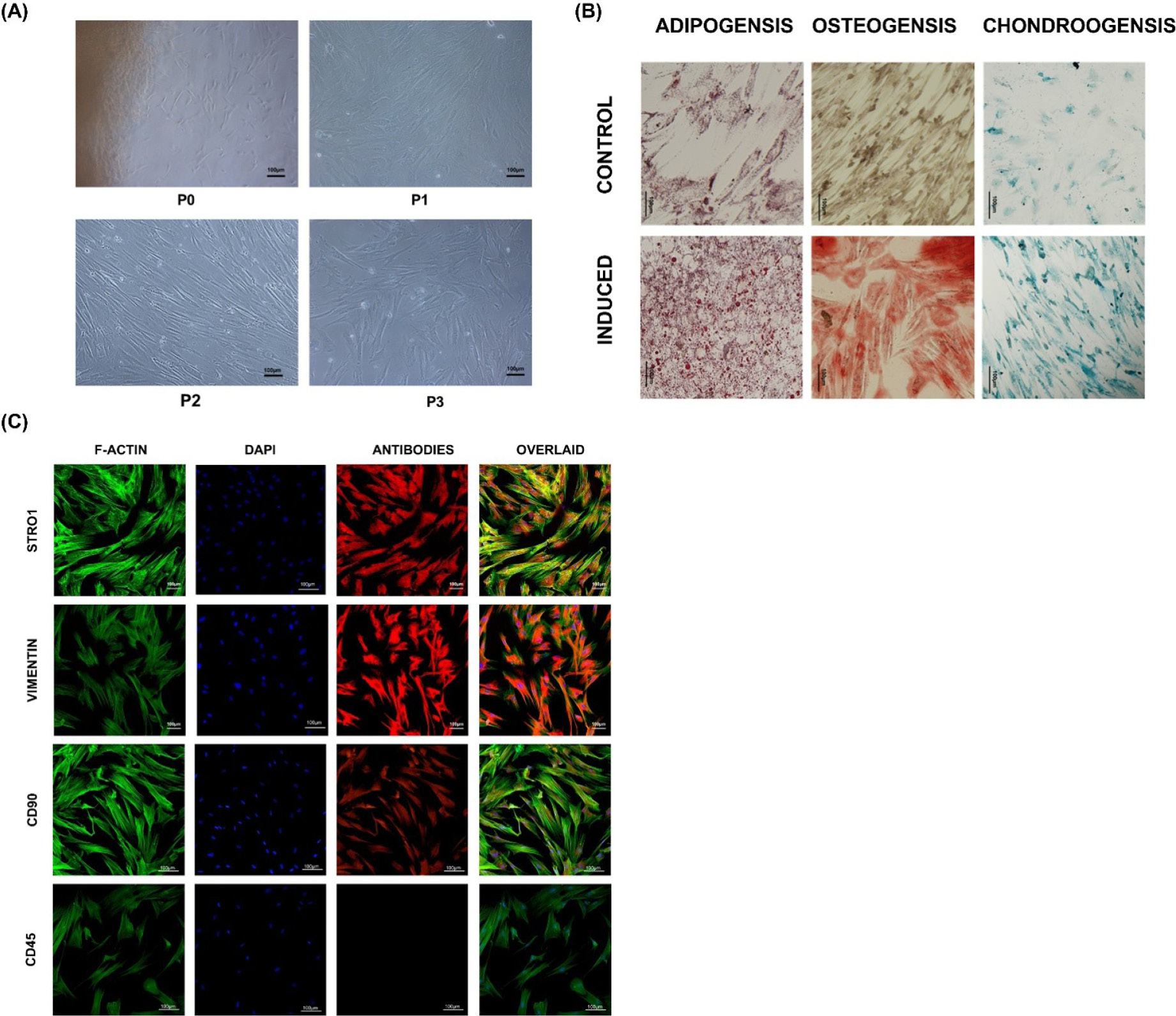
Fig. 1.
Characterization of hUMSCs: (A) Morphological characteristics of hUMSCs at different passages, P0 - P3. (B) Tri-lineage differentiation of hUMSCs; Adipocytes (stained with Oil Red O), Osteocytes (stained with Alizarin Red), and Chondrocytes (stained with Alcian Blue). (C) hUMSCs demonstrating positive expression of MSC-specific markers (Stro1, Vimentin, and CD90), whereas, hematopoietic marker i.e., CD45was not expressed, indicated by immunocytochemical analysis. Nuclei were stained with DAPI and cytoskeleton was stained with F-actin. All images were taken under fluorescent microscope.
.
Characterization of hUMSCs: (A) Morphological characteristics of hUMSCs at different passages, P0 - P3. (B) Tri-lineage differentiation of hUMSCs; Adipocytes (stained with Oil Red O), Osteocytes (stained with Alizarin Red), and Chondrocytes (stained with Alcian Blue). (C) hUMSCs demonstrating positive expression of MSC-specific markers (Stro1, Vimentin, and CD90), whereas, hematopoietic marker i.e., CD45was not expressed, indicated by immunocytochemical analysis. Nuclei were stained with DAPI and cytoskeleton was stained with F-actin. All images were taken under fluorescent microscope.
hUMSC characterization
Immunocytochemistry
Immunocytochemical analysis of hUMSCs showed positive expression of Vimentin, Stro1, and CD90, specific to MSCs, while the hematopoietic marker CD45 was not detected (Fig. 1B).
Tri-lineage differentiation
Treated hUMSCs showed osteogenic, chondrogenic and adipogenic differentiation after 3 weeks in the lineage specific induction media, with distinct characteristics i.e., mineral deposition, proteoglycans and oil droplets, respectively (Fig. 1C).
Differentiation of hUMSCs
Treatment of hUMSCs with the non-cytotoxic concentration of lawsone, lapachol (10 µM each), or their combination in serum free DMEM, showed differentiation toward IPCs after 14 days. The treated cells showed the formation of islet-like clusters (Fig. 2).
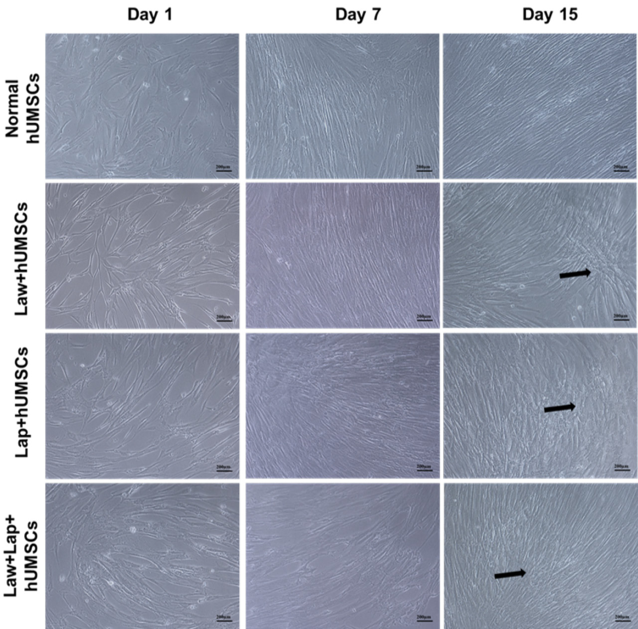
Fig. 2.
hUMSC differentiation into IPCs in vitro: hUMSCs differentiated into insulin producing cells (IPCs) following their treatment with lawsone (Law), lapachol (Lap), or their combination (Law + Lap) after 15 days. Law + hUMSC group showed more clusters representing islet formation (black arrows) compared to other groups. All images were taken under phase contrast microscope.
.
hUMSC differentiation into IPCs in vitro: hUMSCs differentiated into insulin producing cells (IPCs) following their treatment with lawsone (Law), lapachol (Lap), or their combination (Law + Lap) after 15 days. Law + hUMSC group showed more clusters representing islet formation (black arrows) compared to other groups. All images were taken under phase contrast microscope.
Gene expression analysis
qPCR analysis was performed to check the expression of genes specific to β-cells, and Wnt and BMP pathways. Law + hUMSCs showed significant upregulation of pancreatic β-cell markers, i.e., insulin (INS-1), MAF bZIP transcription factor A 1 (MAFA1), pancreatic and duodenal homeobox 1 (PDX1), forkhead box A2 (FOXA2), and NKX6 homeobox 1 (NKX6.1), compared to the untreated control. However, the expression of these markers was found to be slightly higher but statistically non-significant in both Lap + hUMSC and combined treatment groups. Furthermore, the expression of NGN3 was found to be reduced in all the treated groups as compared to the control group (Fig. 3).
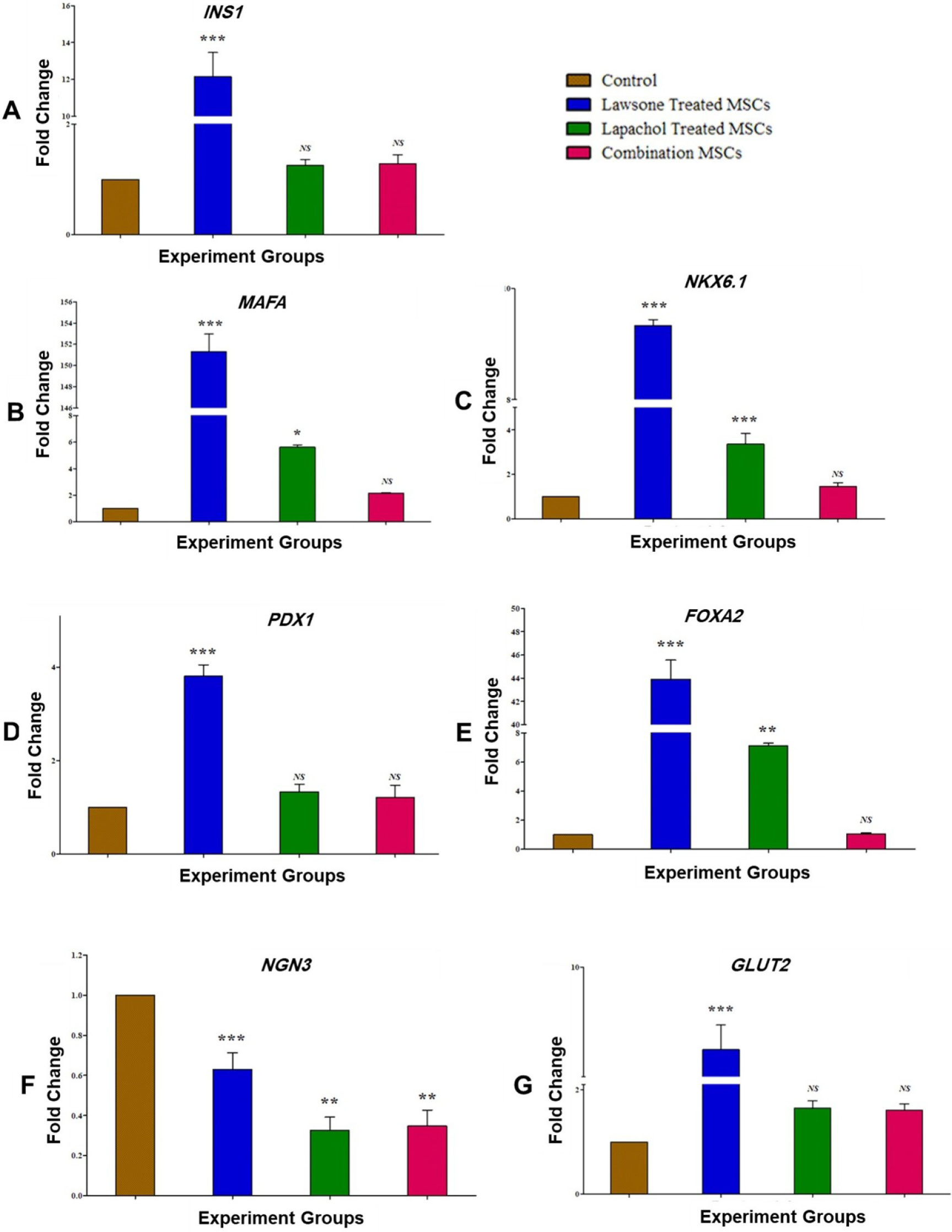
Fig. 3.
Gene expression analysis of pancreatic β-cell markers:hUMSCs treated with lawsone (Law), lapachol (Lap), or their combination (Law + Lap) were analyzed for the gene expression of (A)INS-1, (B) MAFA, (C) PDX1, (D) FOXA2, (E)NKX6.1, (F) GLUT2, and(G) NGN3, at day 15. Statistical comparisons were performed by one-way ANOVA with a post hoc Bonferroni test. The statistical values are reported as mean ± SEM with n = 3 and a significance value of P < 0.05 (non-significant NS, P < 0.05*, P < 0.01**, and P < 0.001***).
.
Gene expression analysis of pancreatic β-cell markers:hUMSCs treated with lawsone (Law), lapachol (Lap), or their combination (Law + Lap) were analyzed for the gene expression of (A)INS-1, (B) MAFA, (C) PDX1, (D) FOXA2, (E)NKX6.1, (F) GLUT2, and(G) NGN3, at day 15. Statistical comparisons were performed by one-way ANOVA with a post hoc Bonferroni test. The statistical values are reported as mean ± SEM with n = 3 and a significance value of P < 0.05 (non-significant NS, P < 0.05*, P < 0.01**, and P < 0.001***).
Transcriptional dynamics of the Wnt pathway in lawsone- or lapachol-treated hUMSC groups revealed increased expression of Wnt mediators, including Wnt family member 2 (WNT2), catenin beta 1 (CTNNB1, encoding β-catenin), MYC proto-oncogene (c-MYC), Jun proto-oncogene (c-JUN), and cyclin D1 (CCND1). Conversely, the expression of Wnt pathway inhibitors, glycogen synthase kinase 3 beta (GSK3β) and axin 1 (AXIN1), was reduced compared to the control group (Fig. 4). Furthermore, expression of BMP pathway mediators, such as inhibitor of DNA binding 1 (ID1) and members of the Suppressor of Mothers Against Decapentaplegic (SMAD) family (SMAD1, SMAD5, SMAD6, and SMAD7), was consistently reduced in all treatment groups compared to the control (Fig. 5).
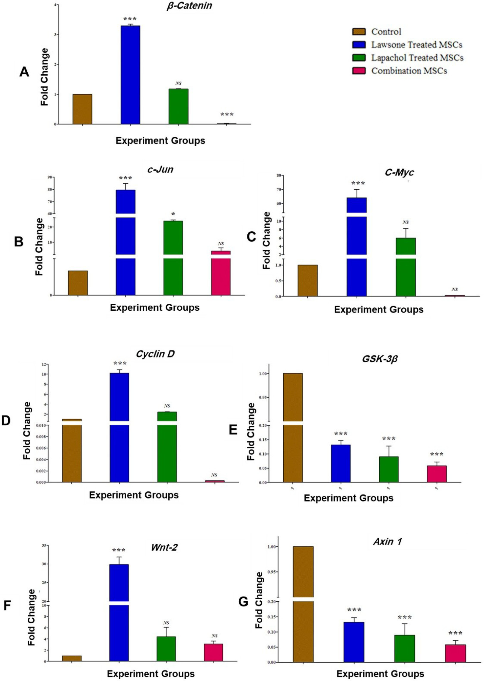
Fig. 4.
Gene expression analysis of Wnt pathway mediators: hUMSCs treated with lawsone (Law), lapachol (Lap), or their combination (Law + Lap) were analyzed for the gene expression of (A) Wnt-2, (B)c-Myc,(C) c-Jun, (D) Cyclin D, (E) β-Catenin, (F) GSK-3β, and (G) Axin1,at day 15. Statistical comparisons were performed by one-way ANOVA with a post hoc Bonferroni test. The statistical values are reported as mean ± SEM with n = 3 and a significance value of P < 0.05 (non-significant NS, P < 0.05*, and P < 0.001***).
.
Gene expression analysis of Wnt pathway mediators: hUMSCs treated with lawsone (Law), lapachol (Lap), or their combination (Law + Lap) were analyzed for the gene expression of (A) Wnt-2, (B)c-Myc,(C) c-Jun, (D) Cyclin D, (E) β-Catenin, (F) GSK-3β, and (G) Axin1,at day 15. Statistical comparisons were performed by one-way ANOVA with a post hoc Bonferroni test. The statistical values are reported as mean ± SEM with n = 3 and a significance value of P < 0.05 (non-significant NS, P < 0.05*, and P < 0.001***).
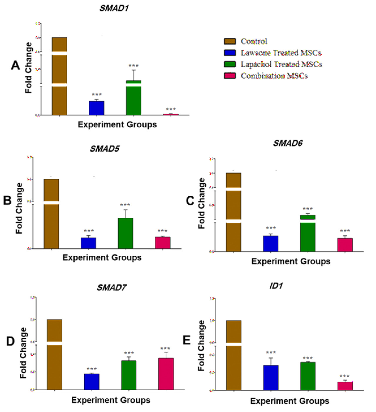
Fig. 5.
Gene expression analysis of BMP pathway mediators: hUMSCs treated with lawsone (Law), lapachol (Lap), or their combination (Law + Lap) were analyzed for the gene expression of (A) SMAD1, (B) SMAD5, (C) SMAD6, (D) SMAD7, and(E)ID1,at day 15. Statistical comparisons were performed by one-way ANOVA with a post hoc Bonferroni test. The statistical values are reported as mean ± SEM with n = 3 and a significance value of P < 0.05 (P < 0.001***).
.
Gene expression analysis of BMP pathway mediators: hUMSCs treated with lawsone (Law), lapachol (Lap), or their combination (Law + Lap) were analyzed for the gene expression of (A) SMAD1, (B) SMAD5, (C) SMAD6, (D) SMAD7, and(E)ID1,at day 15. Statistical comparisons were performed by one-way ANOVA with a post hoc Bonferroni test. The statistical values are reported as mean ± SEM with n = 3 and a significance value of P < 0.05 (P < 0.001***).
Protein expression analysis
Immunocytochemical analysis revealed the expression of insulin in each treated group, compared to control group (Fig. 6A). Law + hUMSCs showed the highest expression of insulin, compared to Lap + hUMSCs and the combined treatment groups (Fig. 6C). Furthermore, the differentiated hUMSCs did not show the expression of glucagon, an α-cell specific marker (Fig. 6B, D).
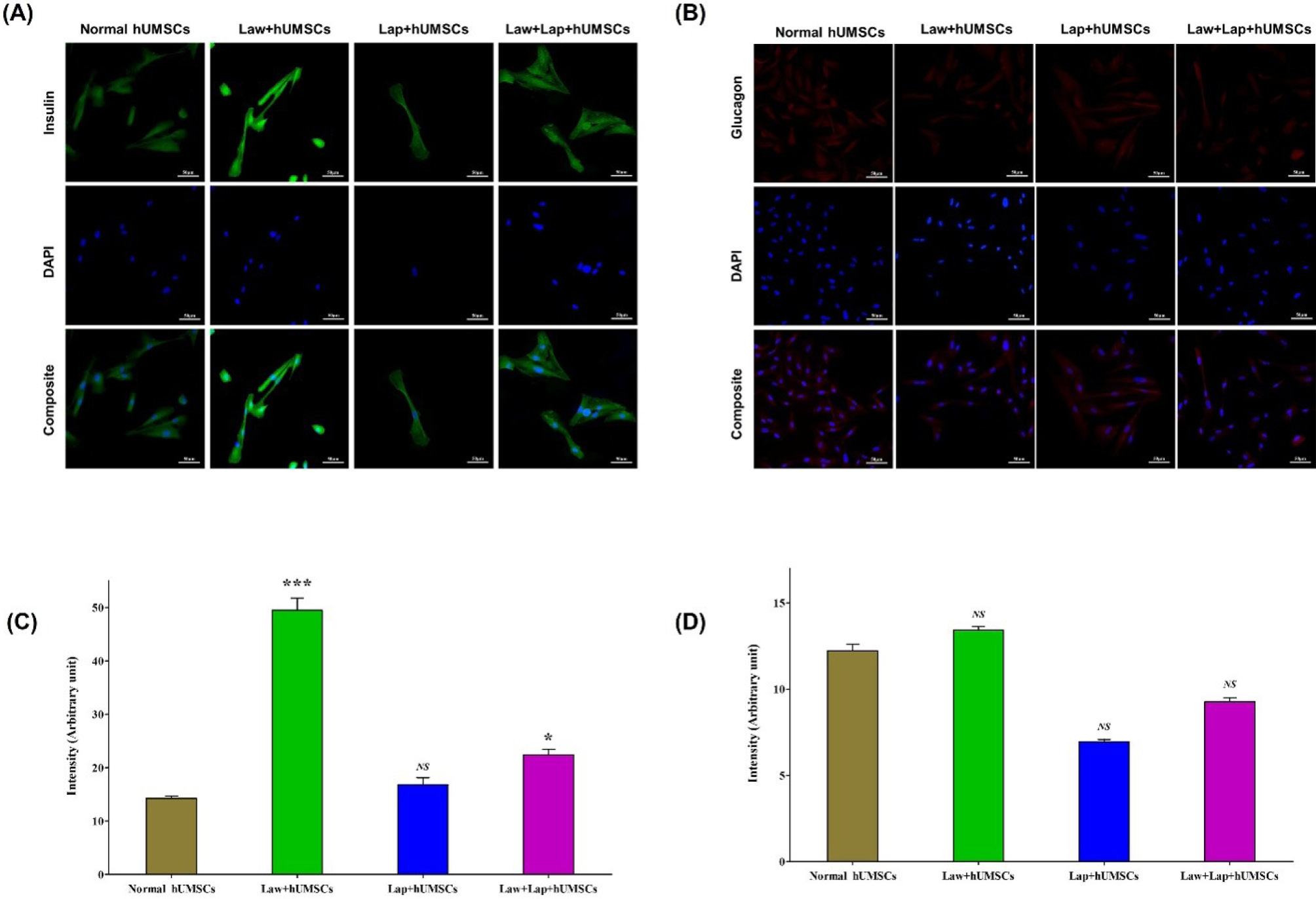
Fig. 6.
Insulin and glucagon expression in treated hUMSCs: hUMSCs treated with lawsone (Law), lapachol (Lap), or their combination (Law + Lap), were analyzed for the protein expression of (A) insulin and (B) glucagon, compared to the control group. Images were taken under fluorescent microscope. (C) Quantitative analysis showed fluorescent intensities of insulin indicating positive expression. (D) All the treated groups showed negative expression of glucagon, an α-cell specific marker. Statistical comparisons were performed by one-way ANOVA with a post-hoc Bonferroni test. The statistical values are reported as mean ± SEM with n = 3 and a significance value of P < 0.05 (non-significant NS, P < 0.05*, and P < 0.001***).
.
Insulin and glucagon expression in treated hUMSCs: hUMSCs treated with lawsone (Law), lapachol (Lap), or their combination (Law + Lap), were analyzed for the protein expression of (A) insulin and (B) glucagon, compared to the control group. Images were taken under fluorescent microscope. (C) Quantitative analysis showed fluorescent intensities of insulin indicating positive expression. (D) All the treated groups showed negative expression of glucagon, an α-cell specific marker. Statistical comparisons were performed by one-way ANOVA with a post-hoc Bonferroni test. The statistical values are reported as mean ± SEM with n = 3 and a significance value of P < 0.05 (non-significant NS, P < 0.05*, and P < 0.001***).
In silico analysis
Molecular docking simulations showed that both lawsone and lapachol formed hydrogen bonds within the seven-transmembrane domain of the FZD receptor, with binding energies of -5.7 kJ/mol and -6.9 kJ/mol, respectively. Docking with the ALK-1 receptor indicated that these compounds formed hydrogen bonds within the serine/threonine kinase domain, with similar maximum binding energies of -5.7 kJ/mol for lawsone and -6.9 kJ/mol for lapachol. The cumulative atomic-level interactions yielded weighted binding scores of -7.2 for lawsone and -6.9 for lapachol (Fig. 7), showing their potential affinity for key components of the Wnt and BMP signaling cascades.
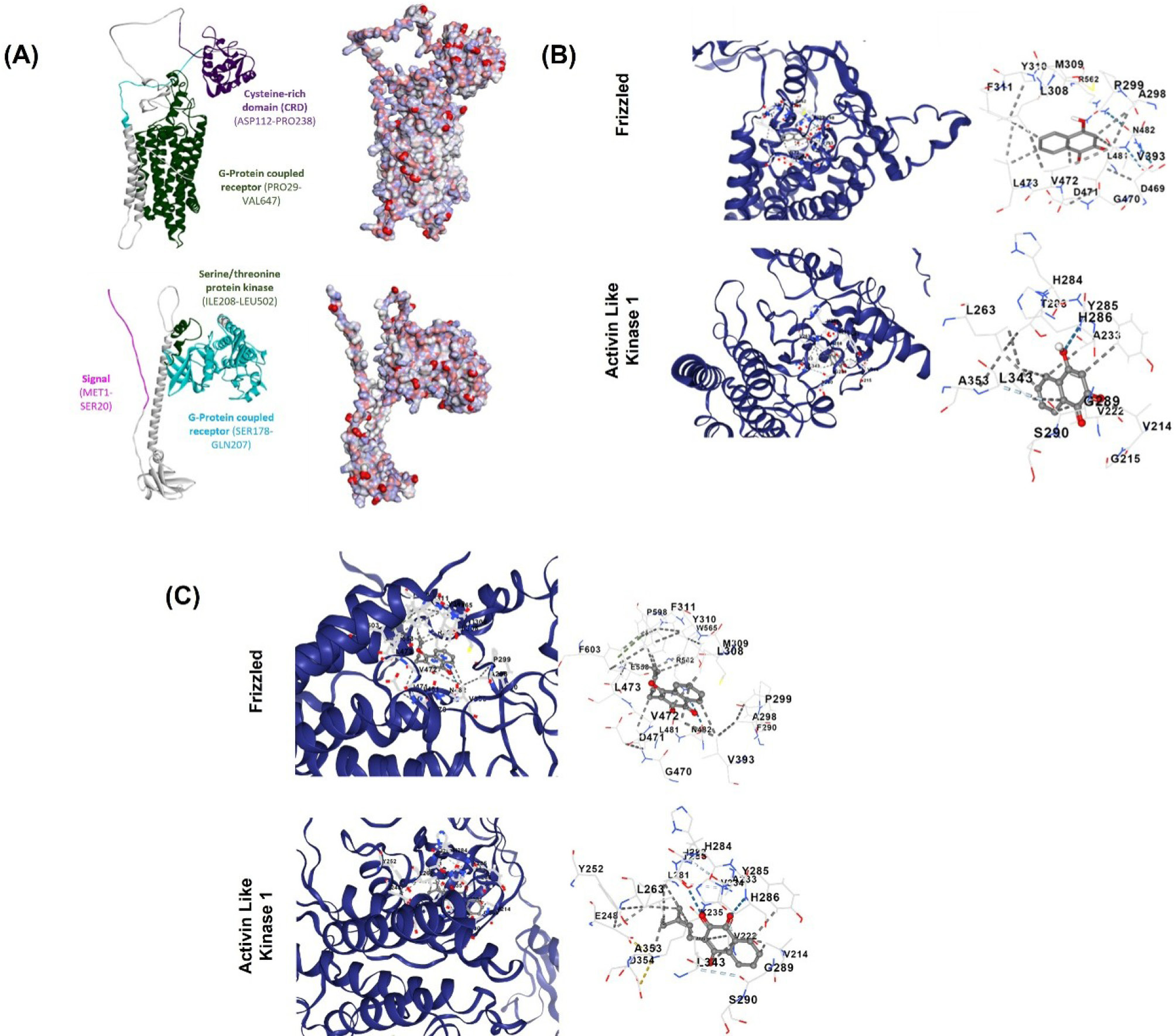
Fig. 7.
In silico analysis of lawsone and lapachol with FZD and ALK1.(A) Three-dimensional ribbon structure and electrostatic surface topology of FZD (top) and ALK1 (bottom) receptors. The active domains present in the receptors are also highlighted. (B) Binding patterns reveal that both ligands occupy well-defined pockets within the receptor cavities, stabilized by a network of hydrogen bonds and hydrophobic interactions. Lawsone exhibits moderate binding affinity (-5.7 kcal/mol), forming key polar contacts with active site residues in FZD and ALK1. (C) Lapachol demonstrates stronger binding affinity (-6.9 kcal/mol), characterized by hydrophobic interactions and additional stabilizing contacts within the receptor pocket, suggesting improved binding stability. The interaction plots further describe the specific amino acid residues involved in ligand coordination, emphasizing critical hydrogen bonding and nonpolar interactions that direct ligand-receptor complex formation. All docking simulations were performed using CB-Dock under standard parameters with a blind docking approach.
.
In silico analysis of lawsone and lapachol with FZD and ALK1.(A) Three-dimensional ribbon structure and electrostatic surface topology of FZD (top) and ALK1 (bottom) receptors. The active domains present in the receptors are also highlighted. (B) Binding patterns reveal that both ligands occupy well-defined pockets within the receptor cavities, stabilized by a network of hydrogen bonds and hydrophobic interactions. Lawsone exhibits moderate binding affinity (-5.7 kcal/mol), forming key polar contacts with active site residues in FZD and ALK1. (C) Lapachol demonstrates stronger binding affinity (-6.9 kcal/mol), characterized by hydrophobic interactions and additional stabilizing contacts within the receptor pocket, suggesting improved binding stability. The interaction plots further describe the specific amino acid residues involved in ligand coordination, emphasizing critical hydrogen bonding and nonpolar interactions that direct ligand-receptor complex formation. All docking simulations were performed using CB-Dock under standard parameters with a blind docking approach.
Discussion
Naphthoquinones and their structural analogs have been widely investigated for their potential therapeutic effects in diabetes treatments.20,21 It is reported that naphthoquinone rich crude extracts of medicinal plants including Lawsonia inermis22,23 and Tabebuia avellanedae24,25 possess anti-diabetic activity.26-29 Several studies have shown that MSCs can differentiate into IPCs in vitro using conditioned media, varied culture conditions,30,31 and/or specific transcription factors.32 Many investigators have evaluated the use of conditioned media in combination with the augmented mixture of non-essential amino acids, high-glucose, nicotinamide, activin A, and β-cellulin cocktail for the differentiation of IPCs as an alternate strategy for T1DM management.33-35 This study examined the role of naphthoquinones, lawsone and lapachol for their potential to differentiate hUMSCs into IPCs. The optimized concentrations of these compounds individually and in combination were used to treat hUMSCs for 2 weeks in serum free DMEM. Transcriptional analysis showed the overexpression of mature pancreatic β-cell markers MAFA, INS-1, PDX1, FOXA2, GLUT2, and NKX6.1, in all treated groups. However, lawsone treated group showed pronounced expression of these genes. Furthermore, NGN3 expression was decreased in all groups.
The combined function of MAFA, PDX1, and NGN3 is widely recognized in the pancreatic β-cell formation.36,37 However, NGN3 specifies the early-stage of pancreatic development and is absent in mature β-cells.38 Conversely, MAFA and PDX1 are recognized as key mediators of β-cell function and remain actively expressed in fully mature β-cells.39 FOXA2 is crucial not only for β-cell formation40 but also for their maturation and the regulation of functional activity.41 NKX6.1 has a fundamental role in regulating the development, maturity, and proliferation of pancreatic β-cells.42 The combination of both PDX1+ /NKX6.1+ and PDX1-/NKX6.1+ genes has been found to effectively transform human pluripotent stem cells into IPCs.43 GLUT2 functions as a glucose transporter and is specifically found in fully developed β-cells.44 According to some literature, GLUT2 may not have a major effect on insulin secretion or glucose uptake.45,46 However, other investigations indicate that their role in diabetes is greatly enhanced.47,48
We also evaluated the differentiation of IPCs via immunocytochemistry and our findings showed increased expression of insulin, a key protein produced by β-cells,49 in all the treated groups, with the highest expression in the lawsone treated group. Additionally, glucagon, which is a protein exclusive to α-cells, showed minimal to no expression.
The main cause of T1DM is the dysfunction of pancreatic β-cells. These cells are highly regulated by a network of intrinsic and extrinsic signals that maintain their normal activity and promote proliferation in response to high insulin demands. Among various signaling pathways, Wnt pathway plays a vital role in the development and function of β-cells,50-52 and its activation has been shown to enhance β-cell proliferation and functional maturation.53-55 Our study also assessed the role of Wnt signaling for the commitment of hUMSCs into IPCs, under in vitro settings. Gene expression analysis revealed a significant downregulation of GSK3β and Axin1 levels across all treatment groups. In comparison, the lawsone-treated group revealed overexpression of Wnt-2, c-MYC, Cyclin D, c-JUN, and β-catenin relative to the control and other treatment groups. These findings align with previous studies showing over-suppression of GSK3β expression. A study also reported higher expression of β-catenin, Cyclin D, and c-MYC, both in vitro and in vivo STZ-induced diabetic mice after gymnemic acid treatment.56 Several other studies have also reported increased pancreatic β-cell proliferation when GSK3β is downregulated.57,58 Similar observations have been shown that inhibition of GSK3β through pharmacological agents or genetic modification promoted β-cell survival and expansion through stabilization of β-catenin.59-61
Numerous studies suggest that inhibition of SMAD proteins62 and ID163 increase pancreatic β-cell proliferation, indicating that their overexpression is involved in the development of diabetes.64,65 Findings from our study showed inhibition of SMADs and ID1 at the transcriptional level, corresponding to the suppression of the BMP pathway, thereby promoting pancreatic β-cell proliferation. Previous studies have also demonstrated that stage-specific inhibition of BMP signaling enhances the efficiency of stem cell differentiation into insulin-producing cells supporting our observations.66-68
We have also analyzed the interaction of lawsone and lapachol with Wnt and BMP receptors via in silico analysis. Blind docking demonstrated the binding of the ligands i.e., lawsone or lapachol to the seven-transmembrane domain of FZL at the N-terminal region. Numerous studies have demonstrated that Wnt2 has a strong affinity for the N-terminus of FZL; therefore, their binding leads to the activation of the downstream signaling cascade through the Wnt canonical pathway. It has been shown that there was a substantial reduction in β-cell mass in rats when cells were exposed to TCF7L2, a canonical Wnt pathway inhibitor. This demonstrates the significance of the canonical Wnt pathway in paracrine functions.69-72 Our study revealed that Law + FZL and Lap + FZL had comparable amino acid sequences, both involved in the binding of the Wnt2 ligand to the FZL receptor.73
Lawsone and lapachol have been reported to bind to the serine/threonine kinase domain of the BMP pathway receptor, ALK-1. Type 1 kinases, upon activation through the same domain, phosphorylate the SMAD proteins.74,75 Previous studies have shown that inhibiting BMP type I receptors can reduce BMP-induced phosphorylation of SMAD1/5/8, resulting in differentiation of stem cells.76 Small-molecule inhibitors such as K02288 have been reported to effectively block the kinase activity of ALK receptors, leading to suppression of downstream SMAD-mediated transcriptional signaling.77 Regulation of the BMP pathways is therefore considered an important mechanism in stem cell fate decisions, since it can modulate signaling pathways that maintain progenitor characteristics and control lineage differentiation.78 Interestingly, our investigation demonstrated that the Law + ALK-1 and Lap + ALK-1 complexes involve amino acid residues similar to those reported for the K02288 + ALK-1 interaction,77 suggesting a comparable inhibitory mechanism that may contribute to the differentiation of hUMSCs into IPCs.
Limitations of the study
Functional validation of insulin-producing cells including glucose-stimulated insulin secretion, C-peptide release, and insulin content analyses, is required to support our findings. The long-term stability, maturation, and functional competence of the differentiated β-like cells should also be established and will be evaluated in future studies.
Conclusion
The current study demonstrated the role of naphthoquinones i.e., lawsone and lapachol, in the differentiation of hUMSCs into IPCs. Among all groups analyzed, lawsone treated group showed significant overexpression of β-cell specific markers, showing its potential for hUMSC commitment toward IPCs both at gene and protein levels. Although, lapachol treated hUMSCs showed reduced expression of β-cell markers as compared to the lawsone treated cells, their expression was still higher in comparison to the untreated control. Optimization of culture conditions is required to enhance the potential of lapachol for efficient differentiation of hUMSCs. Additionally, the study has reported the potential of naphthoquinones as a potent regulator of Wnt and BMP signaling pathways which was evident by the modulation of the specific key molecules at the gene level. In silico analyses also showed similar binding patterns of lawsone or lapachol with respective receptors of Wnt and BMP pathways. Therefore, regulatory role of these compounds can be exploited in the pancreatic β-cell regenerative applications for the management of T1D.
Research Highlights
What is the current knowledge?
-
Lack of standardized single-step differentiation approaches limits the reproducibility of MSC-derived IPC generation.
-
Insulin expression in MSC-derived IPCs is often low, heterogeneous, and insufficient for functional applications.
What is new here?
-
A single-molecule preconditioning strategy using lawsone/lapachol biases hUMSC fate toward a β-cell like phenotype.
-
Simultaneous activation of Wnt signaling and suppression of BMP signaling promotes coordinated β-cell lineage commitment.
Competing Interests
The authors declare no conflict of interest.
Data Availability Statement
The data that support the findings of this study are available from the corresponding author upon request.
Declaration of AI-assisted Tools in the Writing Procedure
None.
Ethical Approval
The study approved by the Institutional Ethics Committee with the assigned protocol number, “ICCBS/IEC-037-HT-2018/Protocol/1.0”. Cord tissue samples were obtained from healthy donors undergoing full-term cesarean deliveries at the Zainab Panjwani Memorial Hospital, Karachi, after obtaining their written informed consent.
Supplementary files
Supplementary file 1 contains Table S1.
(xlsx)
Acknowledgements
This project is financially supported by PhD Indigenous Fellowship provided by Higher Education Commission, Pakistan to Dr. Javeria Masnoon. We are also thankful to Zainab Panjwani Memorial Hospital, Karachi, for providing us the umbilical cord tissue samples.
References
- Mirzaei M, Rahmaninan M, Mirzaei M, Nadjarzadeh A, Dehghani Tafti AA. Epidemiology of diabetes mellitus, pre-diabetes, undiagnosed and uncontrolled diabetes in Central Iran: results from Yazd health study. BMC Public Health 2020; 20:166. doi: 10.1186/s12889-020-8267-y [Crossref] [ Google Scholar]
- Triolo TM, Bellin MD. Lessons from human islet transplantation inform stem cell-based approaches in the treatment of diabetes. Front Endocrinol (Lausanne) 2021; 12:636824. doi: 10.3389/fendo.2021.636824 [Crossref] [ Google Scholar]
-
Khan J, Alexander A, Agrawal M, Ajazuddin, Dubey SK, Siddique S, et al. Stem cell-based therapies: a new ray of hope for diabetic patients. Curr Stem Cell Res Ther 2019. 14: 146-51. doi: 10.2174/1574888x13666181002154110.
- Soria B, Roche E, Berná G, León-Quinto T, Reig JA, Martín F. Insulin-secreting cells derived from embryonic stem cells normalize glycemia in streptozotocin-induced diabetic mice. Diabetes 2000; 49:157-62. doi: 10.2337/diabetes.49.2.157 [Crossref] [ Google Scholar]
- Lumelsky N, Blondel O, Laeng P, Velasco I, Ravin R, McKay R. Differentiation of embryonic stem cells to insulin-secreting structures similar to pancreatic islets. Science 2001; 292:1389-94. doi: 10.1126/science.1058866 [Crossref] [ Google Scholar]
- Rajagopal J, Anderson WJ, Kume S, Martinez OI, Melton DA. Insulin staining of ES cell progeny from insulin uptake. Science 2003; 299:363. doi: 10.1126/science.1077838 [Crossref] [ Google Scholar]
- Kubo A, Shinozaki K, Shannon JM, Kouskoff V, Kennedy M, Woo S. Development of definitive endoderm from embryonic stem cells in culture. Development 2004; 131:1651-62. doi: 10.1242/dev.01044 [Crossref] [ Google Scholar]
-
Rezania A, Bruin JE, Arora P, Rubin A, Batushansky I, Asadi A, et al. Reversal of diabetes with insulin-producing cells derived in vitro from human pluripotent stem cells. Nat Biotechnol 2014. 32: 1121-33. doi: 10.1038/nbt.3033.
- Quiskamp N, Bruin JE, Kieffer TJ. Differentiation of human pluripotent stem cells into β-cells: potential and challenges. Best Pract Res Clin Endocrinol Metab 2015; 29:833-47. doi: 10.1016/j.beem.2015.10.011 [Crossref] [ Google Scholar]
- Karaoz E, Okcu A, Ünal ZS, Subasi C, Saglam O, Duruksu G. Adipose tissue-derived mesenchymal stromal cells efficiently differentiate into insulin-producing cells in pancreatic islet microenvironment both in vitro and in vivo. Cytotherapy 2013; 15:557-70. doi: 10.1016/j.jcyt.2013.01.005 [Crossref] [ Google Scholar]
- Sun Y, Chen L, Hou XG, Hou WK, Dong JJ, Sun L. Differentiation of bone marrow-derived mesenchymal stem cells from diabetic patients into insulin-producing cells in vitro. Chin Med J (Engl) 2007; 120:771-6. doi: 10.1097/00029330-200705010-00007 [Crossref] [ Google Scholar]
- Enderami SE, Soleimani M, Mortazavi Y, Nadri S, Salimi A. Generation of insulin-producing cells from human adipose-derived mesenchymal stem cells on PVA scaffold by optimized differentiation protocol. J Cell Physiol 2018; 233:4327-37. doi: 10.1002/jcp.26266 [Crossref] [ Google Scholar]
- Dave SD, Vanikar AV, Trivedi HL. Extrinsic factors promoting in vitro differentiation of insulin-secreting cells from human adipose tissue-derived mesenchymal stem cells. Appl Biochem Biotechnol 2013; 170:962-71. doi: 10.1007/s12010-013-0250-y [Crossref] [ Google Scholar]
- Sanajou D, Bahrambeigi S, Aslani S. β-LAPachone is renoprotective in streptozotocin-induced diabetic mice via regulating the PI3K/Akt/mTOR signaling pathway. Iran J Basic Med Sci 2021; 24:650-6. doi: 10.22038/ijbms.2021.55565.12422 [Crossref] [ Google Scholar]
- Rahnamay Moshtagh P, Hojati Emami S, Sharifi AM. Differentiation of human adipose-derived mesenchymal stem cell into insulin-producing cells: an in vitro study. J Physiol Biochem 2013; 69:451-8. doi: 10.1007/s13105-012-0228-1 [Crossref] [ Google Scholar]
- Zhang T, Li M, Lu J, Wang J, Zhang M, Panichayupakaranant P. Insights into the sources, structure, and action mechanisms of quinones on diabetes: a review. Molecules 2025; 30:665. doi: 10.3390/molecules30030665 [Crossref] [ Google Scholar]
- Khan M, Shah MA, Kamal M, Ola MS, Ali M, Panichayupakaranant P. Comparative antihyperglycemic and antihyperlipidemic effects of lawsone methyl ether and lawsone in nicotinamide-streptozotocin-induced diabetic rats. Metabolites 2023; 13:863. doi: 10.3390/metabo13070863 [Crossref] [ Google Scholar]
- Masnoon J, Ishaque A, Khan I, Salim A, Kabir N. Effect of lawsone-preconditioned mesenchymal stem cells on the regeneration of pancreatic β cells in type 1 diabetic rats. Cell Biochem Funct 2023; 41:833-44. doi: 10.1002/cbf.3833 [Crossref] [ Google Scholar]
- Jameel F, Irfan F, Salim A, Khan I, Khalil EA. Alpha terpineol preconditioning enhances regenerative potential of mesenchymal stem cells in full thickness acid burn wounds. Regen Ther 2024; 26:188-202. doi: 10.1016/j.reth.2024.05.008 [Crossref] [ Google Scholar]
- Shah MA, Keach JE, Panichayupakaranant P. Antidiabetic naphthoquinones and their plant resources in Thailand. Chem Pharm Bull (Tokyo) 2018; 66:483-92. doi: 10.1248/cpb.c17-00529 [Crossref] [ Google Scholar]
- Shah MA, Reanmongkol W, Radenahmad N, Khalil R, Ul-Haq Z, Panichayupakaranant P. Anti-hyperglycemic and anti-hyperlipidemic effects of rhinacanthins-rich extract from Rhinacanthus nasutus leaves in nicotinamide-streptozotocin induced diabetic rats. Biomed Pharmacother 2019; 113:108702. doi: 10.1016/j.biopha.2019.108702 [Crossref] [ Google Scholar]
- Aremu A, Oridupa OO, Akorede GJ, Basiru A, Ahmed AO. In vivo bioactivity-guided fractions and compounds isolation of antidiabetic activities of Lawsonia Inermis Linn leaves in streptozocine induced diabetic Wistar rats. Indiana J Agric Life Sci 2022; 2:53-61. doi: 10.5281/zenodo.7267429 [Crossref] [ Google Scholar]
- Kapoor N, Kandwal P, Sharma G, Gambhir L. Redox ticklers and beyond: naphthoquinone repository in the spotlight against inflammation and associated maladies. Pharmacol Res 2021; 174:105968. doi: 10.1016/j.phrs.2021.105968 [Crossref] [ Google Scholar]
- Varma SB, Jaybhaye DL. Antihyperglycemic activity of Tectona grandis Linn bark extract on alloxan induced diabetes in rats. Int J Ayurveda Res 2010; 1:163-6. doi: 10.4103/0974-7788.72488 [Crossref] [ Google Scholar]
- Mansouri A, Esmaeili F, Nejatpour A, Houshmand F, Shabani L, Ebrahimie E. Differentiation of P19 embryonal carcinoma stem cells into insulin-producing cells promoted by pancreas-conditioned medium. J Tissue Eng Regen Med 2016; 10:600-12. doi: 10.1002/term.1927 [Crossref] [ Google Scholar]
- Ahmad T, Suzuki YJ. Juglone in oxidative stress and cell signaling. Antioxidants (Basel) 2019; 8:91. doi: 10.3390/antiox8040091 [Crossref] [ Google Scholar]
- Durán AG, Chinchilla N, Simonet AM, Gutiérrez MT, Bolívar J, Valdivia MM. Biological activity of naphthoquinones derivatives in the search of anticancer lead compounds. Toxins (Basel) 2023; 15:348. doi: 10.3390/toxins15050348 [Crossref] [ Google Scholar]
- Cahyana AH, Nazhifah N, Liandi AR. One-pot four-component synthesis of phenazine derivative using 2-hydroxy-1,4-naphthoquinone. IOP Conf Ser Mater Sci Eng 2020; 902:012020. doi: 10.1088/1757-899x/902/1/012020 [Crossref] [ Google Scholar]
- Ojewunmi OO, Oshodi T, Ogundele OI, Micah C, Adenekan S. In vitro antioxidant, antihyperglycaemic and antihyperlipidaemic activities of ethanol extract of Lawsonia inermis leaves. Br J Pharm Res 2014; 4:301-14. doi: 10.9734/bjpr/2014/6359 [Crossref] [ Google Scholar]
- Waseem M, Khan I, Iqbal H, Eijaz S, Usman S, Ahmed N. Hypoxic preconditioning improves the therapeutic potential of aging bone marrow mesenchymal stem cells in streptozotocin-induced type-1 diabetic mice. Cell Reprogram 2016; 18:344-55. doi: 10.1089/cell.2016.0002 [Crossref] [ Google Scholar]
- Pokrywczynska M, Krzyzanowska S, Jundzill A, Adamowicz J, Drewa T. Differentiation of stem cells into insulin-producing cells: current status and challenges. Arch Immunol Ther Exp (Warsz) 2013; 61:149-58. doi: 10.1007/s00005-012-0213-y [Crossref] [ Google Scholar]
- Gabr MM, Zakaria MM, Refaie AF, Abdel-Rahman EA, Reda AM, Ali SS. From human mesenchymal stem cells to insulin-producing cells: comparison between bone marrow- and adipose tissue-derived cells. Biomed Res Int 2017; 2017:3854232. doi: 10.1155/2017/3854232 [Crossref] [ Google Scholar]
- Gabr MM, Zakaria MM, Refaie AF, Ismail AM, Abou-El-Mahasen MA, Ashamallah SA. Insulin-producing cells from adult human bone marrow mesenchymal stem cells control streptozotocin-induced diabetes in nude mice. Cell Transplant 2013; 22:133-45. doi: 10.3727/096368912x647162 [Crossref] [ Google Scholar]
- Elsayed AK, Younis I, Ali G, Hussain K, Abdelalim EM. Aberrant development of pancreatic beta cells derived from human iPSCs with FOXA2 deficiency. Cell Death Dis 2021; 12:103. doi: 10.1038/s41419-021-03390-8 [Crossref] [ Google Scholar]
- Bastidas-Ponce A, Roscioni SS, Burtscher I, Bader E, Sterr M, Bakhti M. Foxa2 and Pdx1 cooperatively regulate postnatal maturation of pancreatic β-cells. Mol Metab 2017; 6:524-34. doi: 10.1016/j.molmet.2017.03.007 [Crossref] [ Google Scholar]
- Piccand J, Meunier A, Merle C, Jia Z, Barnier JV, Gradwohl G. Pak3 promotes cell cycle exit and differentiation of β-cells in the embryonic pancreas and is necessary to maintain glucose homeostasis in adult mice. Diabetes 2014; 63:203-15. doi: 10.2337/db13-0384 [Crossref] [ Google Scholar]
- Sosa-Larios TC, Ortega-Márquez AL, Rodríguez-Aguilera JR, Vázquez-Martínez ER, Domínguez-López A, Morimoto S. A low-protein maternal diet during gestation affects the expression of key pancreatic β-cell genes and the methylation status of the regulatory region of the MafA gene in the offspring of Wistar rats. Front Vet Sci 2023; 10:1138564. doi: 10.3389/fvets.2023.1138564 [Crossref] [ Google Scholar]
- Tran R, Moraes C, Hoesli CA. Controlled clustering enhances PDX1 and NKX61 expression in pancreatic endoderm cells derived from pluripotent stem cells. Sci Rep 2020; 10:1190. doi: 10.1038/s41598-020-57787-0 [Crossref] [ Google Scholar]
- Memon B, Younis I, Abubaker F, Abdelalim EM. PDX1(-) /NKX61( + ) progenitors derived from human pluripotent stem cells as a novel source of insulin-secreting cells. Diabetes Metab Res Rev 2021; 37:e3400. doi: 10.1002/dmrr.3400 [Crossref] [ Google Scholar]
- Akinci E, Banga A, Greder LV, Dutton JR, Slack JM. Reprogramming of pancreatic exocrine cells towards a beta (β) cell character using Pdx1, Ngn3 and MafA. Biochem J 2012; 442:539-50. doi: 10.1042/bj20111678 [Crossref] [ Google Scholar]
- Xu H, Tsang KS, Chan JC, Yuan P, Fan R, Kaneto H. The combined expression of Pdx1 and MafA with either Ngn3 or NeuroD improves the differentiation efficiency of mouse embryonic stem cells into insulin-producing cells. Cell Transplant 2013; 22:147-58. doi: 10.3727/096368912x653057 [Crossref] [ Google Scholar]
- Beamish CA, Zhang L, Szlapinski SK, Strutt BJ, Hill DJ. An increase in immature β-cells lacking Glut2 precedes the expansion of β-cell mass in the pregnant mouse. PLoS One 2017; 12:e0182256. doi: 10.1371/journal.pone.0182256 [Crossref] [ Google Scholar]
- Kellett GL, Brot-Laroche E. Apical GLUT2: a major pathway of intestinal sugar absorption. Diabetes 2005; 54:3056-62. doi: 10.2337/diabetes.54.10.3056 [Crossref] [ Google Scholar]
- Chadt A, Al-Hasani H. Glucose transporters in adipose tissue, liver, and skeletal muscle in metabolic health and disease. Pflugers Arch 2020; 472:1273-98. doi: 10.1007/s00424-020-02417-x [Crossref] [ Google Scholar]
- Xia CQ, Zhang P, Li S, Yuan L, Xia T, Xie C. C-Abl inhibitor imatinib enhances insulin production by β cells: c-Abl negatively regulates insulin production via interfering with the expression of NKx22 and GLUT-2. PLoS One 2014; 9:e97694. doi: 10.1371/journal.pone.0097694 [Crossref] [ Google Scholar]
- Ablamunits V, Sherry NA, Kushner JA, Herold KC. Autoimmunity and beta cell regeneration in mouse and human type 1 diabetes: the peace is not enough. Ann N Y Acad Sci 2007; 1103:19-32. doi: 10.1196/annals.1394.006 [Crossref] [ Google Scholar]
- Sun H, Zhang A, Gong Y, Sun W, Yan B, Lei S. Improving effect of cordycepin on insulin synthesis and secretion in normal and oxidative-damaged INS-1 cells. Eur J Pharmacol 2022; 920:174843. doi: 10.1016/j.ejphar.2022.174843 [Crossref] [ Google Scholar]
- Hayat R, Manzoor M, Hussain A. Wnt signaling pathway: a comprehensive review. Cell Biol Int 2022; 46:863-77. doi: 10.1002/cbin.11797 [Crossref] [ Google Scholar]
- Salinno C, Cota P, Bastidas-Ponce A, Tarquis-Medina M, Lickert H, Bakhti M. β-cell maturation and identity in health and disease. Int J Mol Sci 2019; 20:5417. doi: 10.3390/ijms20215417 [Crossref] [ Google Scholar]
- Chen J, Ning C, Mu J, Li D, Ma Y, Meng X. Role of Wnt signaling pathways in type 2 diabetes mellitus. Mol Cell Biochem 2021; 476:2219-32. doi: 10.1007/s11010-021-04086-5 [Crossref] [ Google Scholar]
- Kannan P, Raghunathan M, Mohan T, Palanivelu S, Periandavan K. Gymnemic acid ameliorates pancreatic β-cell dysfunction by modulating Pdx1 expression: a possible strategy for β-cell regeneration. Tissue Eng Regen Med 2022; 19:603-16. doi: 10.1007/s13770-022-00435-7 [Crossref] [ Google Scholar]
- Liu YA, Jin Q, Ding Q, Hao X, Mo T, Yan S. A dual inhibitor of DYRK1A and GSK3β for β-cell proliferation: aminopyrazine derivative GNF4877. ChemMedChem 2020; 15:1562-70. doi: 10.1002/cmdc.202000183 [Crossref] [ Google Scholar]
- Napolitano T, Silvano S, Ayachi C, Plaisant M, Sousa-Da-Veiga A, Fofo H. Wnt pathway in pancreatic development and pathophysiology. Cells 2023; 12:565. doi: 10.3390/cells12040565 [Crossref] [ Google Scholar]
- Chmielowiec J, Szlachcic WJ, Yang D, Scavuzzo MA, Wamble K, Sarrion-Perdigones A. Human pancreatic microenvironment promotes β-cell differentiation via non-canonical WNT5A/JNK and BMP signaling. Nat Commun 2022; 13:1952. doi: 10.1038/s41467-022-29646-1 [Crossref] [ Google Scholar]
- Bi H, Ma R, Chen Z, Wang Y, Ding X. The stem cell within the vessel wall: multipotent pericytes modulating β-cell function and diabetic complications. Stem Cell Res Ther 2025; 16:568. doi: 10.1186/s13287-025-04669-9 [Crossref] [ Google Scholar]
- Wang H, Ai J, Shopit A, Niu M, Ahmed N, Tesfaldet T. Protection of pancreatic β-cell by phosphocreatine through mitochondrial improvement via the regulation of dual AKT/IRS-1/GSK-3β and STAT3/Cyp-D signaling pathways. Cell Biol Toxicol 2022; 38:531-51. doi: 10.1007/s10565-021-09644-7 [Crossref] [ Google Scholar]
-
Wang P, Karakose E, Liu H, Swartz E, Ackeifi C, Zlatanic V, et al. Combined inhibition of DYRK1A, SMAD, and trithorax pathways synergizes to induce robust replication in adult human beta cells. Cell Metab 2019; 29: 638-52.e5. doi: 10.1016/j.cmet.2018.12.005.
- Bruun C, Christensen GL, Jacobsen ML, Kanstrup MB, Jensen PR, Fjordvang H. Inhibition of beta cell growth and function by bone morphogenetic proteins. Diabetologia 2014; 57:2546-54. doi: 10.1007/s00125-014-3384-8 [Crossref] [ Google Scholar]
- Ghasemi Gojani E, Rai S, Norouzkhani F, Shujat S, Wang B, Li D. Targeting β-cell plasticity: a promising approach for diabetes treatment. Curr Issues Mol Biol 2024; 46:7621-67. doi: 10.3390/cimb46070453 [Crossref] [ Google Scholar]
- Adeerjiang Y, Sidike A, Gan XX, Li QT, Jiang S. The role of Wnt3a/β-catenin/TCF7L2 pathway in diabetes and cardiorenal complications. Cardiorenal Med 2025; 15:72-82. doi: 10.1159/000543145 [Crossref] [ Google Scholar]
- Balatskyi VV, Sowka A, Dobrzyn P, Piven OO. WNT/β-catenin pathway is a key regulator of cardiac function and energetic metabolism. Acta Physiol (Oxf) 2023; 237:e13912. doi: 10.1111/apha.13912 [Crossref] [ Google Scholar]
- Grgurevic L, Christensen GL, Schulz TJ, Vukicevic S. Bone morphogenetic proteins in inflammation, glucose homeostasis and adipose tissue energy metabolism. Cytokine Growth Factor Rev 2016; 27:105-18. doi: 10.1016/j.cytogfr.2015.12.009 [Crossref] [ Google Scholar]
- Akerfeldt MC, Laybutt DR. Inhibition of Id1 augments insulin secretion and protects against high-fat diet-induced glucose intolerance. Diabetes 2011; 60:2506-14. doi: 10.2337/db11-0083 [Crossref] [ Google Scholar]
- Wang HL, Wang L, Zhao CY, Lan HY. Role of TGF-beta signaling in beta cell proliferation and function in diabetes. Biomolecules 2022; 12:373. doi: 10.3390/biom12030373 [Crossref] [ Google Scholar]
- Sunkara RR, Mehta D, Sarate RM, Waghmare SK. BMP-AKT-GSK3β signaling restores hair follicle stem cells decrease associated with loss of Sfrp1. Stem Cells 2022; 40:802-17. doi: 10.1093/stmcls/sxac041 [Crossref] [ Google Scholar]
- Ogieuhi IJ, Agbo CE, Ajekiigbe VO, Anthony CS, Onyehalu JC, Nwankwo CK. Stem cell-derived pancreatic beta cells: a step closer to functional diabetes treatment?. BMC Endocr Disord 2025; 25:181. doi: 10.1186/s12902-025-01997-y [Crossref] [ Google Scholar]
- Arroyave F, Uscátegui Y, Lizcano F. From iPSCs to pancreatic β cells: unveiling molecular pathways and enhancements with vitamin C and retinoic acid in diabetes research. Int J Mol Sci 2024; 25:9654. doi: 10.3390/ijms25179654 [Crossref] [ Google Scholar]
- Memon B, Abdelalim EM. Stem cell therapy for diabetes: beta cells versus pancreatic progenitors. Cells 2020; 9:283. doi: 10.3390/cells9020283 [Crossref] [ Google Scholar]
- Azbazdar Y, Karabicici M, Erdal E, Ozhan G. Regulation of Wnt signaling pathways at the plasma membrane and their misregulation in cancer. Front Cell Dev Biol 2021; 9:631623. doi: 10.3389/fcell.2021.631623 [Crossref] [ Google Scholar]
- Rim EY, Clevers H, Nusse R. The Wnt pathway: from signaling mechanisms to synthetic modulators. Annu Rev Biochem 2022; 91:571-98. doi: 10.1146/annurev-biochem-040320-103615 [Crossref] [ Google Scholar]
- Najafi SM. Canonical wnt signaling (Wnt/β-catenin pathway): a potential target for cancer prevention and therapy. Iran Biomed J 2020; 24:269-80. doi: 10.29252/ibj.24.5.264 [Crossref] [ Google Scholar]
- Ye Y, Barghouth M, Luan C, Kazim A, Zhou Y, Eliasson L. The TCF7L2-dependent high-voltage activated calcium channel subunit α2δ-1 controls calcium signaling in rodent pancreatic beta-cells. Mol Cell Endocrinol 2020; 502:110673. doi: 10.1016/j.mce.2019.110673 [Crossref] [ Google Scholar]
- Ren Q, Chen J, Liu Y. LRP5 and LRP6 in Wnt signaling: similarity and divergence. Front Cell Dev Biol 2021; 9:670960. doi: 10.3389/fcell.2021.670960 [Crossref] [ Google Scholar]
- Sanchez-Duffhues G, Williams E, Goumans MJ, Heldin CH, Ten Dijke P. Bone morphogenetic protein receptors: structure, function and targeting by selective small molecule kinase inhibitors. Bone 2020; 138:115472. doi: 10.1016/j.bone.2020.115472 [Crossref] [ Google Scholar]
- Olsen OE, Hella H, Elsaadi S, Jacobi C, Martinez-Hackert E, Holien T. Activins as dual specificity TGF-β family molecules: SMAD-activation via activin-and BMP-type 1 receptors. Biomolecules 2020; 10:519. doi: 10.3390/biom10040519 [Crossref] [ Google Scholar]
- Sanvitale CE, Kerr G, Chaikuad A, Ramel MC, Mohedas AH, Reichert S. A new class of small molecule inhibitor of BMP signaling. PLoS One 2013; 8:e62721. doi: 10.1371/journal.pone.0062721 [Crossref] [ Google Scholar]
- Mohedas AH, Xing X, Armstrong KA, Bullock AN, Cuny GD, Yu PB. Development of an ALK2-biased BMP type I receptor kinase inhibitor. ACS Chem Biol 2013; 8:1291-302. doi: 10.1021/cb300655w [Crossref] [ Google Scholar]
- Luo J, Tang M, Huang J, He BC, Gao JL, Chen L. TGFbeta/BMP type I receptors ALK1 and ALK2 are essential for BMP9-induced osteogenic signaling in mesenchymal stem cells. J Biol Chem 2010; 285:29588-98. doi: 10.1074/jbc.M110.130518 [Crossref] [ Google Scholar]