Bioimpacts. 16:32895.
doi: 10.34172/bi.32895
Review
The promising applications of 3D printing technology for diagnosis and therapy of cancer: Recent advances and challenges
Mohsen Farrokhpour Writing – original draft, 1 
Safa Samadzadeh Etehadi Writing – original draft, 2
Pejman Hassanpoor Writing – original draft, 3
Tola Abdulsattar Faraj Writing – original draft, 4
Ayad H. Hasan Writing – original draft, 5, 6
Vesal Abbasian Writing – original draft, 7
Sargol Aminnezhad Conceptualization, Investigation, Writing – original draft, 8
Mohammadreza Azimi Writing – original draft, 9, *
Soheila Kashanian Supervision, Writing – original draft, 10, 11
Mehran Alavi Conceptualization, Investigation, Writing – original draft, Writing – review & editing, 12, *
Author information:
1Firouzgar Hospital, Department of Internal Medicine, School of Medicine, Iran University of Medical Sciences, Tehran, Iran
2Department of Medicine, Tehran University of Medical Sciences, Tehran, Iran
3Department of Microbiology, Faculty of Basic Sciences, Rouzbahan Institute of Higher Education, Sari, Iran
4Department of Medical Analysis, Faculty of Applied Science, Tishk International University, Erbil, Iraq
5Department of Biomedical Sciences, Cihan University-Erbil, Erbil, Kurdistan Region, Iraq
6Department of Medical Microbiology, Faculty of Science and Health, Koya University, Koya, KOY45, Kurdistan Region, Iraq
7School of Medicine, Shahid Beheshti University of Medical Sciences, Tehran, Iran
8Department of Molecular Genetics, Faculty of Biological Sciences, Tarbiat Modares University, Tehran, Iran
9Department of biochemistry, Medical Faculty, Saveh Branch, Islamic Azad University, Saveh, Iran
10Faculty of Chemistry, Razi University, Kermanshah, 6714414971, Iran
11Nanobiotechnology Department, Faculty of Innovative Science and Technology, Razi University, Kermanshah, 6714414971, Iran
12Department of Biological Science, Faculty of Science, University of Kurdistan, Sanandaj, Kurdistan, Iran
Abstract
Using structured materials and special molecules, scientists can create synthetic materials that can change their properties in a controlled and efficient way, resulting in useful functions. These characteristics remind us of the most popular designs found in nature. Specifically, they can produce strong structures when needed, store information, cause movement, and hold and release therapeutic agents. These macromolecular systems can be created using three-dimensional (3D) printing technology that can respond to human physiological conditions. In this review, the reasons for this progress are explained, and the potential future developments for macromolecules with specific functions are discussed. This review delves into the advancements in 3D printing technology and polymeric nanomaterials, particularly in the diagnosis and therapy of cancer. In addition, it highlights the versatility of these materials in creating intricate scaffolds with enhanced therapeutic properties, leveraging nanotechnology to improve cell interactions.
Graphical Abstract
Keywords: 3D printing techniques, Therapeutic agents, Biocompatibility, Polymeric nanomaterials, Cancer therapy, Cancer diagnosis
Copyright and License Information
© 2026 The Author(s).
This work is published by BioImpacts as an open access article distributed under the terms of the Creative Commons Attribution Non-Commercial License (
http://creativecommons.org/licenses/by-nc/4.0/). Non-commercial uses of the work are permitted, provided the original work is properly cited.
Funding Statement
This paper was not funded.
Introduction
Three-dimensional (3D) printing technology generates new purposes for controlled polymeric nanomaterial placement based on biocompatible polymers to create complex 3D scaffolds with high resolution and accuracy.1-3 Isolating and culturing human cells, printing cell-laden scaffolds, and applying the scaffolds for treatment and disease modeling have been indicated in the bioprinting process for therapeutic applications (Fig. 1).4
Three material sources that scaffolds can be made from are synthetic polymers, natural polymers, and polymer derivatives.5 Nanotechnology can provide various materials on the nano-scale with unique therapeutic characteristics and biomaterial-cell interactions suitable for cell health.6-11 When 3D printing tissue engineering scaffolds, nanomaterials improve shape accuracy and printability, and provide various biological functions to bioinks.2,12 In this regard, mechanical and rheological features, printability, and bioactivity may be augmented via the colloidal network between synthetic (polylactic acid [PLA], poly(lactic-co-glycolic acid) [PLGA], and polycaprolactone [PCL]) or natural (cellulose, gelatin, chitosan, and alginate) polymeric nanoparticles (NPs).13-15 For example, shape stability can result from the interactions between the solid NPs, such as nanosilicates.16
During the past decades, natural polymers gained momentum as leading bioactive materials.17,18 Natural biopolymers contain multiple biofunctional molecules that provide both biological activity, regenerative properties, and biomimicry capacity. Living organisms contain natural polymers such as chitosan, cellulose, alginate, dextran, gelatin, and collagen.19-21 Synthetic polymers, which are created by chemical processes, can be adjustable structures and features.22 Some synthetic polymers, as biodegradable materials, can be decomposed by microbes and bodily fluids. Biodegradable synthetic polymers commonly used in industry include the materials PLA as well as PLGA, PCL, polyurethane, and polyvinyl alcohol.23,24 The polymers need to be employed with additional agents because they by themselves fail to fulfill all the requirements needed for creating bioartificial organs applying 3D printing technology. The challenge requires a solution that combines these polymers together with nanomaterials which have both controlled production methods and strong mechanical properties.25 Natural polymer functionalization produces advanced products such as gelatin derivatives as well as chitosan derivatives and cellulose derivatives.26,27 Such functionalized derivatives possess higher sophistication and complexity levels.28
Physicochemical and biomedical properties
Polymeric nanomaterials achieve better mechanical properties and process stability through chemical linkages between adjacent chains, which develop from crosslinking techniques. The outcome of this processing method enables printed structures to maintain long-term durability.29,30 Biomedical applications determine how printed polymeric nanomaterials should be structured. The varying structure of printed polymeric nanomaterials includes porous scaffolds, which serve tissue engineering as well as intricate patterns used for microfluidic devices.31,32
By strategically tailoring both the core structure of the polymer and the peripheral side chain groups, macromolecular assemblies with molecular responsiveness have surfaced as prospective contenders for a wide range of uses, ranging from pharmaceutical delivery to applications in microelectronics.33 Adding controlled dynamic elements to these configurations creates exciting potentials for innovative applications, which include catch-and-release functionality.34 Traditionally, polymeric NPs have been generated by covalently crosslinking single-chain copolymers under sparse conditions, leading to the formation of intramolecularly collapsed polymeric NPs.35 While these systems exhibit fascinating physical attributes, their lack of responsiveness constrains the range of potential applications. The development of supramolecular crosslinking through quadruple-hydrogen-based units serves as a proposed method by Foster et al to revitalize this research and discover new scientific prospects. This strategy enables the reversible condensation of individual macromolecules into crosslinked NPs.36 In a sparse solution, particles were produced by irradiating o-nitrobenzyl-protected 2-ureido-pyrimidinone units, which were attached to the polymer backbone and featured self-complementary hydrogen bonding. The photodeprotection of the o-nitrobenzyl group removed obstacles to hydrogen bonding. In a sparse solution, this led to the creation of transient single-chain polymer NPs.37 The introduction of acidity disrupted the hydrogen-bonded connections, causing the NPs to revert to coiled single-chain macromolecules. Demonstrating their metastable nature, these structures were initially transformed into uniform nanofilms, capitalizing on their high solubility and low viscosity. When the temperature reached a certain level, the single-chain NPs began to "unfold", leading to insoluble extended networks through supra-molecular crosslinking. Significant changes in physical and mechanical characteristics emerged after the transformation of initial NPs occurred. Such nanostructures made from multiple polymer chains, such as micelles and vesicles, represent essential transport carriers of hydrophobic substances through aqueous solutions. The formation of these particles can now be selectively induced through recent methods under varied environmental conditions.33 Amir and colleagues used enzymatic methods to detach phosphate groups from one segment of a diblock copolymer, resulting in amphiphilicity formation. The polymer assembled as colloidal nanostructures after undergoing this method.38
Lee and colleagues introduced a novel micelle disruption method that used photochromic spiropyran units and the isomerization reaction between 365 nm and 620 nm light wavelengths. The spiropyran ring system underwent activation by 365 nm light, which generated a hydrophilic zwitterionic structure that altered the amphiphilic equilibrium of the block copolymer until it dissolved the polymer micelle. The hydrophobic spiropyran ring system underwent conversion back to its original state after 620 nm irradiation, causing its regeneration and restoring the amphiphilic behavior, enabling micellar structure reformation. Molecular rearrangements that modify hydrophilic/hydrophobic properties produce a sequential process that builds and then disassembles nanostructures. This mechanism holds the potential for selectively storing and releasing hydrophobic molecular cargo within the nanostructures. A comparable responsive behavior was engineered into vesicles composed of amphiphilic amide dendrons, incorporating either photocleavable o-nitrobenzyl protecting groups or photoswitchable azobenzene moieties at the core.39 Park and colleagues successfully accomplished the irreversible breakdown of vesicular structures and the liberation of guest molecules by employing the photoinduced removal of the o-nitrobenzyl protecting group. This triggered event perturbed the amphiphilic characteristics of the dendrons. Alternatively, the authors observed that the photoirradiation of vesicles containing azobenzene derivatives at the dendron focal point induced a transient elevation in the permeability of the vesicle walls, thereby facilitating the release of guest molecules.40 Klaikherd et al synergistically attached these collective effects by incorporating multiple orthogonal triggers, leading to the expansion of multi-responsive assembling polymer construction. They integrated temperature-sensitive poly(N-isopropylacrylamide), redox-sensitive (disulfide linkage), and acid-sensitive (acetal) functionalities within a diblock copolymer structure, yielding polymeric micelles capable of disassembling in response to any of the specified inputs. An intriguing future avenue for these multi-responsive systems involves exploring logic operations, where a desired response would only be triggered upon the simultaneous application of multiple stimuli.41 In an advanced fusion of small molecule and macromolecule capabilities, Aldaye and colleagues have developed distinct DNA nanostructures that showcase dynamic responsiveness in terms of assembly and disassembly. The precise structuring of DNA assemblies depends heavily on rigid shape-directing small molecules, which must be integrated into DNA oligomers with single chains.42 By carefully choosing specific base-pairing configurations, the hybridization of DNA structures enables the creation of nanotubes, underscoring the substantial architectural impact afforded by orthogonal handles. Beyond meticulous self-assembly, this research group demonstrated the selective expansion and contraction of nanotube compartments through DNA hybridization events initiated by the introduction of complementary single-stranded DNA. This was exemplified through the encapsulation and release of gold (Au) NPs cargo within the containers formed by DNA nanotubes. A significant forthcoming goal is to establish release mechanisms triggered by DNA/RNA for the precise intracellular delivery of therapeutics in physiological contexts.42 Printed form of polymeric nanobiomaterials in toxicity focuses on the potential toxicity associated with the utilization of printed polymeric nanomaterials in the field of biomaterials.43,44
The evaluation of nanomaterials' toxicity with their environmental and human health side effects becomes essential.45 Extensive research and testing protocols determine the safety of printable polymeric nanomaterials by reducing potential negative consequences. The use of 3D printing technology enables the precise fabrication of polymeric nanomaterials, allowing for the creation of customized structures and designs tailored for biomaterial applications.46 These printed polymeric nanobiomaterials have gained significant attention in various biomedical applications, such as drug delivery carriers, tissue scaffolds, and medical implants due to their unique properties and potential benefits.47 However, one crucial aspect of the application of such materials is the evaluation of their toxicity. Researchers need to thoroughly assess the potential adverse effects and risks associated with the interaction of these materials with living organisms, particularly at the nanoscale.48 Toxicity studies involve assessing cell viability, inflammation, genotoxicity, immunological reactions, and long-term effects under different physicochemical properties and doses of nanomaterials.49,50 Ensuring the biocompatibility of printed polymeric nano-biomaterials is a critical factor for the application in physiological conditions.51 In this way, researchers should design materials that are non-toxic, biodegradable, and capable of promoting the regeneration and healing processes within biological systems.52 By understanding the toxicity of printed polymeric nanobiomaterials, researchers can make informed decisions about their safe and effective use in various biomedical applications.53 Modern understanding of nanotoxicology leads scientists toward developing safer biocompatible materials, which extend opportunities in the healthcare and biotechnology domain.54
In 3D printing applications, the fundamental requirement is to assess the biocompatible nature of the applied materials. ISO 10993 represents an extensive collection of International Organization of Standards evaluation standards for medical device materials biocompatibility assessment. The successful regulation of these standards becomes essential for manufacturers to gain approval from the authorities.55 Standards used in the traditional manufacturing field present special obstacles when implemented in 3D-printed materials. The properties of raw materials may change during 3D printing because the process consists of factors such as cross-linking and melting in addition to creating porosity.56
Relying exclusively on raw material tests does not give sufficient protection for the safety of 3D-printed medical equipment. It becomes imperative to extend assessments to include additives introduced during manufacturing, such as reinforced materials or cross-linker molecules. Assessment of these materials during and after manufacturing is fundamental both for cleaning procedures and keeping ISO 10993 requirements for finished medical devices. Accurate examination of biodegradable 3D printed scaffold degradation needs complete testing through both laboratory and animal studies. In order to evaluate the shelf-life of finished devices, investigators may conduct degradation testing procedures. Emphasizing sterility is pivotal for minimizing the risk of infection associated with implantable medical devices.57 Consequently, 3D-printed implants might require the same validation procedures as any other medical implant, with the results submitted to the Food and Drug Administration (FDA) (ISO 10993–5).
Biocompatibility denotes a material's capacity to interact with living organisms without producing unwanted effects. Researchers can improve the biocompatibility of polymeric nanomaterials by modifying their composition and surface properties. This optimization renders them suitable for various applications, including tissue engineering, drug delivery systems, and medical implants.58
As polymer science advanced during the 1960s, researchers delved into creating synthetic polymeric matrices to address medical challenges. Synthetic polymers have displayed extensive development through numerous creations, which led to extensive scientific evaluation for biomedical purposes. Achieving success for clinical applications of synthetic polymers required overcoming three critical barriers, including poor biocompatibility, as well as toxic synthesis chemicals and deleterious degradation product effects. The advantages of synthetic polymers prove superior to their disadvantages, especially when no alternative systems are available. The development of polymeric materials for biomedical applications brought substantial achievement during the last two decades through the creation of biocompatible and non-toxic materials. Biodegradable synthetic polymers, in particular, have emerged as favored candidates for constructing prosthetic materials, tissue engineering scaffolds, and carriers for controlled-release drug delivery. Each of these applications demands materials with well-defined physical, chemical, biological, biomechanical, and degradation properties to provide effective therapeutic solutions. The subsequent sections present an overview of some of the most biocompatible polymer matrices.59
Hoshino and colleagues crafted artificial polymer NPs, approximately 50 nm in size, to replicate the role of natural antibodies that attach to particular biomolecules. These synthetic antibody-like particles, made of plastic, prove valuable in personalized therapies when the patient is unable to generate antibodies for a known antigen. Due to their cost-effectiveness and safety attributes, as well as their stable nature, these plastic antibodies present versatility as biomaterials for biomedical applications, including biomolecule separation, biosensor development, diagnostic tools, toxic agents, and virus countermeasures. Scientists imprinted binding sites on polymer NP surfaces through molecular interactions that use a target molecule to create them.60,61 Molecular imprinting in polymers originated during the 1950s, which established this scientific approach as historically significant. The application of polymer molecular imprinting for synthesizing synthetic antibodies through nanotechnology reached its discovery point in the late 1980s.62 The synthesis of polymer NPs in water used N-isopropyl acrylamide monomers as backbone, followed by N,N′-methylene bisacrylamide cross-linkers together with various acrylamides that added hydrogen bonding and hydrophobic contact and charge-charge binding capabilities. The synthesis method for polymers excludes both organic solvents and heating, which it can protect the biomacromolecules during imprinting from denaturation.63,64
Overall, the utilization of 3D printing technology in the formation of polymeric nanomaterials provides exciting possibilities for designing and fabricating materials with precise structures, desirable properties, low toxicity, and improved biocompatibility. These developments have the power to completely transform a number of sectors and aid in the creation of creative solutions for a wide variety of applications.
Cancer disease
Cancer exists as a primary global disease, which presents difficulties in medical treatment because of its sophisticated nature and tendency to return. Standard therapies face two major limitations of tumor heterogeneity, together with elevated side effects. Doctors have found 3D bioprinting to be an effective answer, which generates laboratory models with realistic cancer tissue characteristics. The manufacturing process includes three stages that start with model digitalization, then proceed to bio-ink creation through cellular integration with growth factors, followed by exact printing and biological activation of tissues for functionality. Science-based drug screening benefits from this technology, which allows the development of complex tumor models. The technology finds practical uses in surgical education and medical plans for organ transplants as well as procedures involving cancer treatment. 3D printing enables dose control of tablets, which helps the manufacturing process of personalized medicines. The technology serves multiple purposes when developing therapy training simulators and anatomical models while enabling nanomedicine creation. 3D printing technology affects cancer management through multiple operational fields that involve surgery, drug testing, and cancer-on-a-chip testing, along with cancer detection practices and cancer cell metastasis research. 3D printing advances implantable medical device development by improving biocompatibility and drug delivery control while demonstrating its broad application scope in cancer studies and treatment approaches.65,66
Osteosarcoma is the main bone cancer that requires various clinical treatments depending on tumor grade because low-grade lesions require surgery, but high-grade lesions need aggressive approaches. These clinical challenges persist because patients frequently develop resistance to chemotherapy treatment in combination with the difficulties that appear after bone surgery. Insufficient amounts of intrinsic hydrogen peroxide (H2O2), recognized for encouraging osteoclast formation, and the restricted intratumoral penetrability of this molecule in chemotherapeutic-insensitive malignancies contribute to these problems. The exciting solution of 3D printing allows scientists to develop new scaffolds incorporating chemotherapeutics and growth factors, which stimulate bone repair along with precise cancer therapies.67 A recent proposal by Sawaftah and colleagues addresses these challenges by introducing a multifunctional biomaterial embedded in a 3D-printed scaffold utilizing akermanite. This biomaterial integrates calcium peroxide (CaO2) and iron oxide (Fe3O4) NPs. The magnetite NPs act as catalysts for the generation of H2O2 from CaO2, functioning as a reservoir of Ca2+ ions to facilitate bone regeneration. Furthermore, magnetic hyperthermia induced by Fe3O4 NPs under alternative magnetic fields contributes to additional anticancer effects.68
3D Printing strategies
Research on cancer diagnosis and treatment heavily relies on 3D printing methods, as explained in this section. There exists substantial discussion about three-dimensional printing technology, which serves multiple purposes, including tumor modeling and organ development, and implant manufacturing. Table 1 displays how 3D printing consists of multiple approaches, which include extrusion-based printing, inkjet-based printing, SLA-based printing, and laser-induced forward transfer-based printing.66 In addition, Table 2 compares various 3D printing methods employed in cancer diagnosis and therapy, with a focus on identifying knowledge gaps and clinical translation barriers.
Table 1.
The strengths and weaknesses associated with various bio-printing methods
|
3D Bioprinting Technique
|
Advantages
|
Disadvantages
|
Ref.
|
| Stereolithography (SLA) |
-
Elimination of the necessity for X-Y movement is achieved through the concurrent crosslinking of the entire 2D layer.
-
Cell viability is maintained at a level exceeding 85%.
-
A broad range of bioinks can be effectively utilized.
-
Bioprinting attains a high resolution, approximately 1 μm.
|
-
The crosslinking process necessitates a transparent and photosensitive bioink, restricting the options for additives and limiting the cell density to 10^8 cells mL−1.
-
The system is relatively intricate in comparison.
|
69
|
| Inkjet bioprinting |
-
A straightforward bioprinting approach.
-
Cost-effective.
-
Suitable for multi-material bioprinting.
-
Achieves high resolution, approximately 30 μm.
-
Maintains high cell viability within the range of 80–90%.
|
|
70
|
| Laser-assisted bioprinting (LAB) |
|
-
Limited to low cell density ( < 10^6 cells mL−1).
-
Involves an intricate system.
-
Entails relatively high expenses.
|
71
|
| Extrusion-based bioprinting |
-
Capability to print highly viscous bioinks within the range of 30–6 × 10^7 mPa·s.
-
Ability to print high cell densities, including cell spheroids.
-
Applicability for multi-material bioprinting.
-
Involves a relatively simple bioprinting process.
|
-
Relatively low printing speed
-
Low-to-medium resolution, highly dependent on setup
-
Moderate cell viability (40–80%), dependent on setup
|
72
|
| Electrospinning-based bioprinting |
|
|
73
|
Table 2.
A comparative table presenting various 3D printing methods applied in cancer diagnosis and therapy
|
3D Printing methods
|
Application in cancer diagnosis
|
Ref.
|
Application in cancer therapy
|
Ref.
|
Knowledge gaps/clinical translation barriers
|
Ref.
|
| Fused deposition modeling (FDM) |
Applied for producing tumor models for visualization.
Creation of patient-specific anatomical models for surgical planning and preoperative simulation. |
74,75
|
Can be exploited to produce customized drug delivery systems.
Design of scaffolds for tissue engineering. |
76
|
Limited resolution and accuracy for fine details of microstructures.
Inconsistent material properties across different 3D printers.
Challenges in scaling production for clinical application. |
77
|
| SLA |
Generation of highly detailed tumor models for imaging and study.
Potential for bioprinting of tumor tissues for better understanding of the tumor microenvironment. |
78
|
Can print precise structures for organ/tumor therapy.
Development of customized scaffolds for cell growth and drug testing. |
79
|
Long printing times for larger models.
Limited availability of biocompatible materials for SLA printing.
High cost and complexity. |
80
|
| Selective laser sintering (SLS) |
Employed for the production of porous, customized scaffolds for cell culture models to simulate tumor growth.
Development of microfluidic devices for cancer cell analysis. |
81
|
Personalized treatment solutions such as drug-loaded scaffolds.
Printing of thermoplastic structures for supporting cancer cell cultures. |
81
|
Limited resolution for creating highly detailed microstructures.
Complex post-processing steps required.
Need for better material options for in vivo applications. |
81
|
| Inkjet bioprinting |
Production of cell-laden tumor models for drug screening.
Microfluidic devices for early cancer detection (e.g., liquid biopsy). |
82
|
Bioprinting of personalized tumor models for testing therapies.
Printing of cells and biomaterials to create tumor-like structures for in vivo studies. |
83
|
Lack of precise control over cell density and arrangement.
Difficulty in achieving functional tissue structures.
Limited regulatory approval for clinical use. |
84
|
| Extrusion-based bioprinting |
Development of 3D cell models to mimic tumor environments for diagnostic purposes.
Printing of structures that allow for high-throughput drug screening. |
65
|
Creation of bioprinted implants for localized tumor therapy.
Personalized drug delivery systems for targeted therapy. |
85
|
Low resolution and complexity in creating microvascular networks.
Limited understanding of tumor microenvironment interactions.
Material compatibility with biological tissues. |
86
|
| LAB |
Printing of tumor models with high precision.
Fabrication of scaffolds with controlled cell-laden deposition for diagnostic analysis. |
87
|
Forming of functional tissue models for cancer therapy research.
Personalized cancer vaccines or immune cell therapy applications. |
87
|
Technical challenges with laser precision and material properties.
Expensive and technically complex equipment.
Slow translation into clinical practice due to cost and scalability issues. |
88
|
| Digital Light Processing (DLP) |
High-resolution tumor models for preoperative planning.
Application of patient-specific data for better representation of cancerous tissues. |
89
|
Providing custom scaffolds for regenerative cancer therapies.
Bioprinting of tissue-engineered cancer models for research and therapy. |
89
|
Limited availability of biocompatible and functional materials for DLP.
Printing speed is slower compared to other methods.
Issues with the strength and mechanical properties of printed structures. |
90
|
| Multi-material 3D printing |
Enables the printing of tumor models with varying properties for more accurate diagnosis.
Can be used to generate multi-layered cancer tissue models. |
65
|
Complex drug delivery systems where different materials can be used to control release rates.
Personalized cancer treatment devices, such as custom implants or prosthetics. |
65
|
Lack of standardization in multi-material printing processes.
Need for better integration of multiple materials that mimic the tumor microenvironment.
Regulatory hurdles in adopting multi-material approaches in clinical settings. |
91
|
Extrusion-based printing
Extrusion-based bioprinting stands as the most common technique in research for cancer applications because of its cost-effectiveness and adaptability. Employing a temperature-controlled cartridge, this method extrudes bio-ink from a fine nozzle, utilizing various extrusion forces. Noteworthy studies in the context of cancer include the development of cervical cancer models and the creation of stable constructs for tissue biomimetics using hydroxyethyl cellulose-based bio-inks.92
Gospodinova et al conducted research about the extrusion printing of bioinks constructed with high-viscosity thixotropic hydroxyethylcellulose in combination with various sodium alginate amounts, and HeLa cells, which investigated the extrusion printing of bioinks with high viscosity and thixotropic hydroxyethylcellulose containing different sodium alginate concentrations alongside HeLa cells. The research showed that as sodium alginate concentration in relative amounts decreased, cell viability increased up to 81.5% over one day of incubation. The hydrogel matrix showed strong biocompatibility properties which promoted cell multiplication while ensuring a substantial increase in the total cell numbers during seven days of incubation. The method of extrusion printing caused a primarily negative effect on cell survival rates during the hydrogel embedding process when cells were added to the system. Notably, embedding HeLa cells in blends with 1% and 2.5% sodium alginate did not significantly affect cell viability, even with residence times up to 90 minutes prior to the extrusion of the bioink. The printed structures from this process hold potential for use as a cervical tumor model.93
Zou et al created lung cancer tumors through their in vitro three-dimensional model using a 3D bioprinter with a threaded pump, which extruded A549 cells within hydrogel scaffolds. The temperature-sensitive hydrogels allowed easier printing at low temperatures, which improved the success rates of the printing procedure. Lower temperatures also contributed to a reduced metabolic rate of the cells in the hydrogel, promoting higher cell survival post-printing. Following preset parameters, the bio-ink was extruded and deposited onto a grid hydrogel on the scaffold. The spacing between the cellular hydrogel scaffold and the medium remained consistent in the grid arrangement. After the completion of the crosslinking process involving the cellular sodium alginate/gelatine/fibrinogen hydrogel scaffold and the CaCl2/TGesa/thrombin crosslinking agent, the resulting medium could freely permeate the hydrogel scaffold. This facilitated the unrestricted absorption of nutrients and release of metabolites by the cells within the hydrogel scaffold. A 3D-printed biological ink designed for lung cancer modeling used A549 cells in combination with sodium alginate/gelatine/fibrinogen. The 3D-bioprinted lung cancer model developed a cell-friendly porous architecture that presents better resemblance to natural lung conditions than standard 2D cell-line models. The cell survival numbers demonstrated no substantial distinction between the 2D and 3D printer groups. The proliferation rate for cells followed different patterns between 2D and 3D groups and showed initial quick growth in the 2D group and a later reduction and slower continuous growth in the 3D group. The biological regulation of A549 cells suffered changes detected by RNA-seq that identified 3112 altered genes, which could lead to better lung cancer research. The study demonstrates the ability of 3D bioprinting to generate sustainable in vitro tumor models, which serve as a new platform for studying lung cancer and performing anticancer drug analyses according to Figs. 2 and 3.94
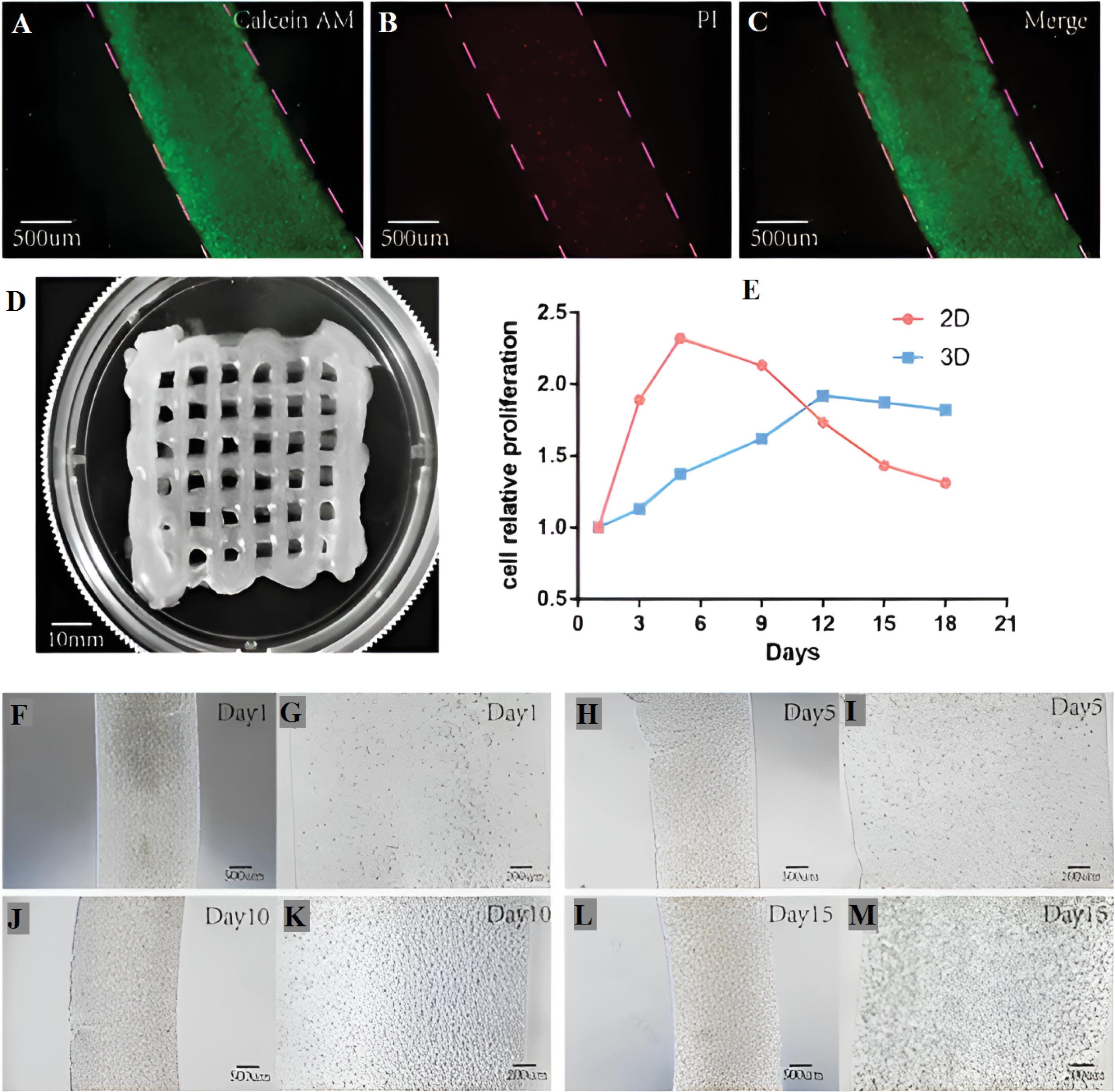
Fig. 2.
The evaluation of viable and non-viable cells within the hydrogel scaffold, housing A549 cells, after 15-day cultivation in the 3D-bioprinted hydrogel is demonstrated. Panels (A–C) exhibit live cells stained with calcein in green and deceased cells stained with PI in red. Panel (D) illustrates the successful printing of a grid-patterned hydrogel scaffold containing cells. Panel (E) provides a comparison of the cell proliferation rate between the 2D and 3D models. Panels (F–M) visually capture the progression of the cellular hydrogel under a light microscope on days 1, 5, 10, and 15. Reprinted under the terms and conditions of the Creative Commons Attribution (CC BY) license (https://creativecommons.org/licenses/by/4.0/).94
.
The evaluation of viable and non-viable cells within the hydrogel scaffold, housing A549 cells, after 15-day cultivation in the 3D-bioprinted hydrogel is demonstrated. Panels (A–C) exhibit live cells stained with calcein in green and deceased cells stained with PI in red. Panel (D) illustrates the successful printing of a grid-patterned hydrogel scaffold containing cells. Panel (E) provides a comparison of the cell proliferation rate between the 2D and 3D models. Panels (F–M) visually capture the progression of the cellular hydrogel under a light microscope on days 1, 5, 10, and 15. Reprinted under the terms and conditions of the Creative Commons Attribution (CC BY) license (https://creativecommons.org/licenses/by/4.0/).94
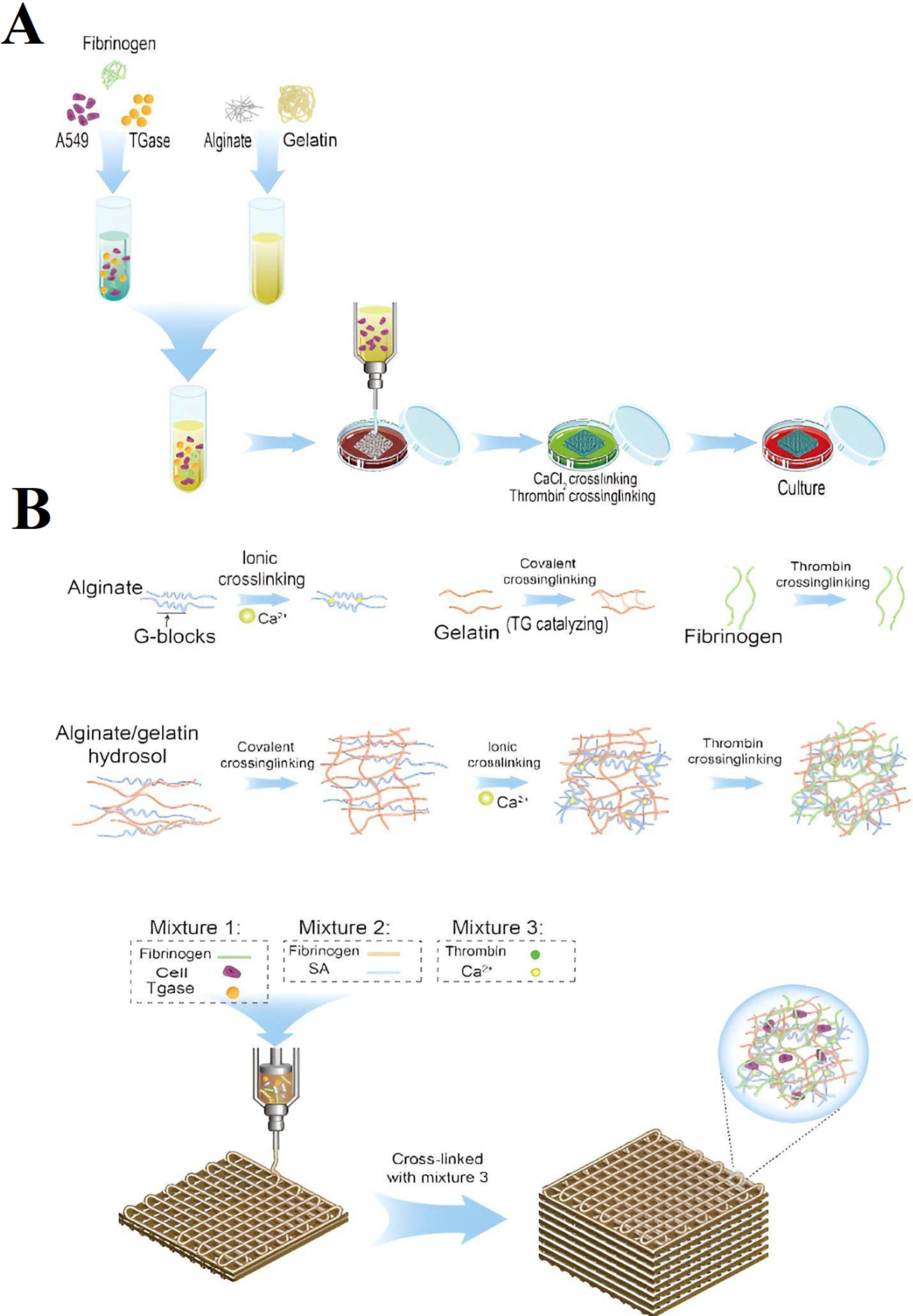
Fig. 3.
(A) Overview of the 3D bioprinting process depicted in a flowchart. (B) Diagram illustrating the crosslinking of hydrogel in the bioprinting procedure. Reprinted under the terms and conditions of the Creative Commons Attribution (CC BY) license (https://creativecommons.org/licenses/by/4.0/).94
.
(A) Overview of the 3D bioprinting process depicted in a flowchart. (B) Diagram illustrating the crosslinking of hydrogel in the bioprinting procedure. Reprinted under the terms and conditions of the Creative Commons Attribution (CC BY) license (https://creativecommons.org/licenses/by/4.0/).94
Laser-assisted 3D bioprinting (LAB)
LAB serves as a bioprinting technology, which utilizes laser energy to develop artificial tissues. The technique performs according to the laser-induced forward-transfer (LIFT) principle to deposit both biomaterials and living cells and enables precise layer construction of scaffold-free 3D cell systems, which lead to sturdy gel structures.17,95
A LAB system incorporates three essential parts, starting with a pulsed laser source, followed by a biological material depository target and concluding with a substrate that receives printed material. A nozzle-free configuration of laser-assisted bioprinting enables accurate delivery of near-infrared pulsed laser beams to biological materials by using a focus system with scanning mirrors. The target receives laser pulses through CAD modeling that builds high-pressure vapor pockets at the designated area. A cell droplet forms as a result of laser-triggered pressure rise so that cells reach the receiving substrate for cross-linking. The technology successfully avoids cell clogging problems while maintaining the health of cells in use. The improved high-throughput operation and reliable reproducibility of LAB enable researchers to produce 3D-printed pre-cancerous and cancerous tissue models.17,96
Hakobyan et al applied LAB to develop a GelMA-based hydrogel model of ADM in AR42J-B-13 rat acinar cells through LAB procedures. The cellular environment of this model functions to assess the transdifferentiated acinar cells during initial PDAC development. GelMA possesses characteristics that allow precise bioprinting through its cell-friendly properties toward cells, resulting in high definition and survival rates of cells. This research investigation demonstrates three distinct growth patterns from acinar and ductal cell structures, as well as coculture spheroids, which exhibit increased ductal cell proliferation, probably due to cellular interaction. Research data that includes Ki67 and EGFR results shows that the established model effectively reproduces early PDAC conditions, thus making it suitable for studying cancer beginnings.87
SLA-based printing
In the realm of 3D bioprinting, SLA has significantly impacted the field since its inception in 1986. Originally used in reconstructive head surgery, it has evolved into a promising method for bioprinting, showcasing improved speed compared to nozzle-based approaches. SLA bioprinters utilize photo-polymerization to solidify cell-laden bio-ink in a layer-by-layer process, allowing the creation of intricate 3D structures without the need for an x-y printhead. This method ensures high cell viability ( > 85%) by minimizing shear stress. Despite the requirement for a transparent liquid, limiting cell density to ~108 cells mL−1, SLA's rapid, shear-free bioprinting with high resolution (~1 μm) holds particular promise in cancer-related research fields. SLA has found applications in the medical field through the creation of drug delivery devices with personalized medicine tablets and synthetic bone models used for surgical practice. Several biomedical applications demonstrate the versatility of SLA because it enables the production of drug-delivering tablets with extended drug delivery and synthetic bone structures with superior strength properties. Medical technology advancement demonstrates the potential of SLA because researchers have created a 3D-printed transdermal microneedle for insulin delivery and developed polyporous scaffolds for treating bone defects.65,66
The research by Parrish presents a versatile platform, which resolves the predictive challenges of standard 2D and small-scale 3D tissue cultures. A platform serves as the proposed solution to resolve three main issues, involving throughput, system complexity, and tissue complexity. The platform features design flexibility that allows usage across diverse construction techniques through its microplate and docking station, which follows ANSI/SLAS standards either from SLA production or precision machine methods. It can support up to 96 samples, equipped with two independently controllable fluid circuits (192 in total), loading access, and a viewing window for imaging during perfusion. To demonstrate its functionalities, ovarian cancer models were bio-fabricated to allow in situ evaluation using microscopy and a perfused resazurin-based metabolic activity assay. The platform provides in situ assessment methods that work with different sample geometries for various tissue constructs. The platform demonstrates its capabilities through a combination of in situ microscopy with perfused metabolic activity assays when building ovarian cancer models. The platform functions to screen drugs by generating dose-response data in real-time, which matches how static well plate cultures operate. The soft tissue components, in addition to hard tissue components can have their quantitative analysis performed using spectral computed tomography scanning technology. The research showed that mechanistic high-throughput screening becomes possible through the use of endothelial and mesenchymal stem cell (HUVEC-MSC) vascular coculture models. Suspension of cells in a gelatin-norbornene hydrogel occurred while they remained inside three-dimensional printed good inserts made from SLA technology. The obtained results demonstrate that the dual perfusion bioreactor platform offers scalable possibilities for tissue construct production of parenchymal and barrier tissues, which serve diverse needs in future multi-organ-on-a-chip systems.97
Inkjet-based printing
The inkjet-based printing method established itself as a crucial innovation of bioprinting technologies after extrusion-based printing. The inkjet printing method consists of essential dual operations: (I) droplet release toward a substrate and (II) droplet-substrate contact. Drop-on-demand Inkjet printing technology provides better printing resolution along with fewer drop outputs than continuous inkjet printing. Continuous inkjet printing involves higher frequencies of drop generation but is accompanied by concerns related to sterility.17,98 An investigation delved into the utilization of matrix materials (Compritol and the model drug Fenofibrate) to formulate loaded inks, either devoid of drugs or combined with a drug, aiming to craft personalized printed dosage forms through hot-melt inkjet printing. The utilization of Compritol, whether a standalone or in conjunction with a drug, resulted in the production of personalized solid dosage forms with multiple materials. The findings illustrated that the release of the drug is contingent upon its localization within the printed formulation. Another inquiry focused on formulating a water-based ink, utilizing polyvinylpyrrolidone and thiamine hydrochloride (as a model drug). Tablets were printed on polyethylene terephthalate (PET) films using inkjet printing, demonstrating rapid drug release without the necessity for solvents and presenting an approach for creating water-soluble drug formulations.99
Cheng et al introduced an innovative method called "inkjet-printed morphogenesis" (iPM) for fabricating tumoroids with a densely packed and mechanically dynamic tumor-stroma interface. Aiming to emulate native tumor characteristics, the approach is grounded in the notion that cellular contractile forces can proficiently reshape a polymer matrix laden with cells, resulting in intricately structured tissue constructs. The study develops two distinct bioinks containing tumor cells as well as cancer-associated fibroblasts (CAFs) that are printed using an inkjet method through a collagen-poly(N-isopropyl acrylamide-co-methyl methacrylate) solvent mix in multi-line or multi-layer sequences. Researchers study how the morphogenesis process changes when they modify the interplay between cellular contractile force and matrix stiffness. The development of "morphogenetic printing" characterizes a breakthrough technology that determines enhanced capacities beyond present-day 3D bioprinting procedures (Fig. 4).100
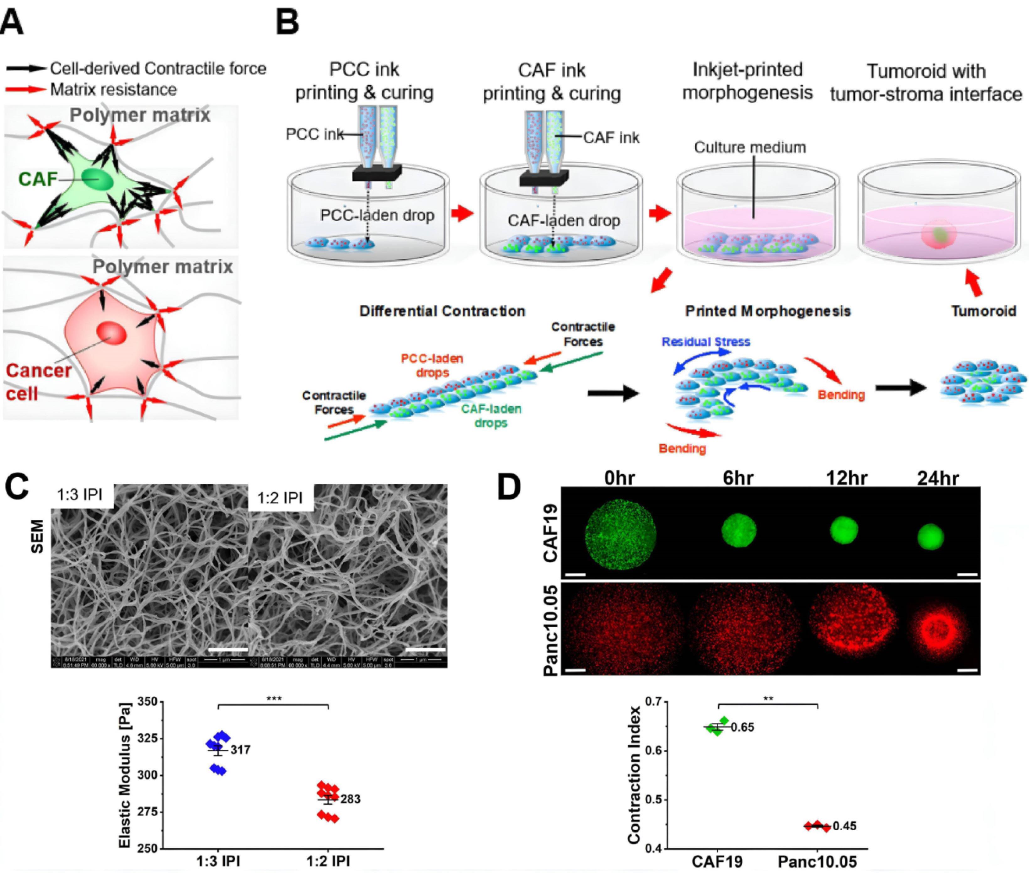
Fig. 4.
The proposed theory for inkjet-printed tumoroid morphogenesis explaining (A) polymer matrix compression through the efforts of cancer-associated fibroblasts (CAFs) and cancer cells and (B) a sequence depicting the inkjet printing technique using cell-rich interpenetrating-polymer inks and The tissue compaction mechanism is explained by the following diagram; (C) SEM images of interpenetrating-polymer inks (IPIs) demonstrate microstructure features and elastic moduli with 1 μm scale bar; (D) Contraction assays analyzed contractile actions by measuring the contraction index of Panc10.05 pancreatic cancer cells and CAF19 pancreatic CAF. Reprinted under the terms and conditions of the Creative Commons Attribution (CC BY) license (https://creativecommons.org/licenses/by/4.0/).100
.
The proposed theory for inkjet-printed tumoroid morphogenesis explaining (A) polymer matrix compression through the efforts of cancer-associated fibroblasts (CAFs) and cancer cells and (B) a sequence depicting the inkjet printing technique using cell-rich interpenetrating-polymer inks and The tissue compaction mechanism is explained by the following diagram; (C) SEM images of interpenetrating-polymer inks (IPIs) demonstrate microstructure features and elastic moduli with 1 μm scale bar; (D) Contraction assays analyzed contractile actions by measuring the contraction index of Panc10.05 pancreatic cancer cells and CAF19 pancreatic CAF. Reprinted under the terms and conditions of the Creative Commons Attribution (CC BY) license (https://creativecommons.org/licenses/by/4.0/).100
Cancer diagnosis
The development of early cancer detection depends on patient outcomes because sensor arrays built with 3D printing technologies offer better diagnostic precision rates.101 3D printing produces objects having dimensional characteristics of height, width, and depth while standing apart from standard 2D printing methods. Digital design information served by this capability results in sequential three-dimensional object production. Three-dimensional printing technology in cancer diagnostics enables medical practitioners to develop innovative personalized diagnostic instruments.102
Researchers used the benefits of 3D printing to associate nanoscale features in biosensors which extended diagnostic opportunities for various clinical conditions. The application of 3D-printed biosensor arrays improves diagnostic instrument sensitivity and specificity which becomes pivotal when searching for small biomarkers especially for cancer detection purposes.103,104
3D printing technologies produce revolutionary changes to cancer applications through their specific operational methods. Multiple design techniques for desktop 3D printers integrate factors related to printing principles and substrate printing capabilities. Each material type requires distinct printing technologies because substrates include three basic categories: materials that can polymerize, thermoplastics, and inks that can be modified through polymerization processes. FDM established itself as a leading cancer diagnosis approach because it enables cost-efficient manufacturing of ABS and PLA-based thermoplastics structures as well as electronic components. The cure mechanism through light exposure of DLP and SLA systems can be illustrated by a study achieving miRNA-21 detection through droplet-based PCR in printed microfluidic devices. Through two-photon polymerization (2PP) engineers gain accurate micrometer-scale capabilities that are useful for developing drug-delivery microneedles. A printing method known as direct ink writing (DIW) lets users adjust their material printing on demand by using inkjet print heads. The SLS technique allows intricate powder beads to melt into complex structures without needing internal support, but it remains scarce for sensor production. The sensitive nature of biological materials finds appropriate solutions through syringe-based extrusion and laser-induced bioprinting methods, which apply to tissue engineering and cancer research applications. These different 3D printing technologies demonstrate their power to transform cancer diagnostics as well as microfluidics and personalized medicine while enabling creative patient-specific solutions for cancer research.103
Tang and colleagues present an economical and automated protein detection method utilizing a 3D-printed unibody microfluidic device. Constructed through SLA, the device incorporates three reagent reservoirs, a three-dimensional mixing network, and a transparent detection chamber employed for the chemiluminescence detection of prostate cancer biomarkers, specifically prostate-specific antigen (PSA) and platelet factor 4. The sandwich-type assay, employing a multi-labeled detection antibody-polyHRP, achieves a remarkable sensitivity, achieving detection limits as low as 0.5 pg/mL and a dynamic range spanning four orders of magnitude. The automated system runs its complete procedure within 30 minutes using a programmable syringe pump to decrease human involvement during operations. The device demonstrates accurate performance in assessing human serum when results are validated when compared to ELISA. The unibody 3D printing approach gives the device superior mechanical properties while reducing leaks, thus making the device an attractive tool for quick analysis of multiple proteins at facilities with limited resources.105
Park et al developed an innovative system that demonstrates advanced ability for blood-based circulating tumor cells (CTCs) detection. The proposed methodology solves the drawbacks of standard ATP luminescence methods through a new 3D-printed immunomagnetic concentrator (3DPIC), which boosts blood-based CTC isolation and accumulation. The structural design of 3DPIC enables fast and efficient cancer cell concentration in blood samples up to 100-fold in 10 mL of blood volume. By enhancing the ATP luminescence assay, this improvement enables observation of 10 cancer cells in a blood sample, while the method surpasses traditional commercial box kits by ten times. The method concludes its operations within 30 minutes, requiring limited operator interaction to demonstrate such efficiency. The 3DPIC proves superior to commercial CTC separation kits when dealing with large blood volumes of up to 100 mL due to its enhanced sensitivity and reliability. Blood sample spiking tests show that the detection limit reaches 10 cells/mL as part of method validation. The integration of 3D printing with immunomagnetic separation and ATP luminescence assay creates a promising platform for fast CTC detection with high sensitivity and specificity, which may become useful for cancer diagnosis and monitoring by analyzing liquid biopsy samples.106
A microfluidic device used to capture CTCs from blood samples was developed by Chen et al through 3D printing. The device improved its CTC capture efficiency through the addition of anti-EpCAM antibodies to tackle low CTC abundance. The optimized 3D printing system enabled researchers to determine channel dimensions and fluid rates, which resulted in capture success rates surpassing 90%. An evaluation of the device utilized MCF-7 breast cancer cells and SW480 colon cancer cells to validate its performance. CTC isolation methods face low capture success rates, which makes researchers pursue 3D printing solutions as an alternative. The device successfully modified its structure and applied antibody interactions to achieve targeted EpCAM-positive tumor cell acquisition with 95% efficiency when testing MCF-7/GFP cells in synthetic blood samples. The strategy that connects 3D printing with antibody functionalization and parameter optimization presents itself as a beneficial method for enhancing CTC detection performance in both diagnostic and therapeutic applications.107
3D bio-printing for cancer models
Innovative bioengineered models, organoid systems, and advanced innovations like 3D bioprinting have brought significant advancements to create more sophisticated in vitro platforms for tumor modeling. The development of clinically advanced 3D in vitro models is imperative to replicate intricate interactions among tumor and stromal cells. These models serve as essential tools for gaining a deeper understanding of molecular mechanisms and for testing the effectiveness of anticancer therapies.108-110
Several experiments that assess anticancer drugs in cancer research continue to use either 2D co-cultures or xenografts or syngeneic mouse models. The simple design of 2D models fails to reproduce the complex dynamic structure found in tumor microenvironments (TME). Cells that grow under this model configuration spread across flat plastic surfaces while residing in monolayers, which may disrupt essential signaling pathways between cells and alter their stimulus responses.111
The authentic shape and the polarization behavior of cells do not persist within 2D cultures as reported in Table 1. The drawback of animal models exists in their high expense, their complex nature, and ethical barriers to using them effectively. These models face challenges related to analyzing effects that fail to represent human-specific events, which restricts their general usability.17,112
Tumors contain a stroma network that includes an extracellular matrix (ECM) together with CAFs and endothelial and immune support cells that engage in active molecular relationships with tumor cells impacting cancer's sophisticated metabolic networks. The experimental techniques, including 2D cocultures and animal models, face criticism because they fail to reproduce the complete intricacies present in TME.17,113
In contrast, multicellular 3D in vitro systems provide a solution to the limitations mentioned earlier, effectively bridging the disparity between experimental feasibility and physiological connection. These 3D models have the ability to mimic crucial mechanical and biochemical cues essential for cancer evolution. This includes factors such as morphology, tissue stiffness, cell-cell/cell–ECM interactions, and particular gradients.111
It is crucial to acknowledge that these models often concentrate on specific connections between one element of the TME and tumor cells. Despite that, current progress in 3D cancer models carries substantial potential to (i) improve drug discovery, (ii) act as robust platforms for drug testing, and (iii) support the creation of personalized cancer treatments.17,114
The research team of Gebeyehu et al created 3D-printed tumor models through the application of polysaccharide hydrogel for chemotherapeutic drug screening. Bioinks at optimized printability from the VitroGel system included Ink H4 and its modified version, Ink H4-RGD, with RGD peptides. The bioinks demonstrated 90% cell viability and earned shear-thinning behavior together with quick shear recovery abilities that allowed extrusion bioprinting without needing UV curing or temperature changes. The 3D-printed models developed with patient-derived non-small-cell lung cancer cells formed spheroids that maintained their stability for 15 days while reaching spheroid formation in one week. The chemotherapeutic resistance of docetaxel, doxorubicin, and erlotinib drugs was higher when tested against 3D spheroids instead of 2D monolayers. The research demonstrates how polysaccharide-based three-dimensional tumor systems facilitate better drug evaluation procedures for achieving patient-specific cancer therapies.115
However, xenograft models from human cancer patients can be applied, but their turnaround time is insufficient for clinical applications. Thus, there is an urgent need for designed ex vivo models that faithfully reproduce in vivo tumor biology. This is particularly crucial for managing PDAC, known for high cancer stem cell (CSC) content and a dense stromal environment. Current ex vivo models, such as organoid and sphere cultures, only partially replicate these features. In a recent investigation, Osuna de la Peña and et al presented an inclusive and highly adaptable ex vivo model of PDAC. This model is established through the 3D co-assembly of peptide amphiphiles (PA) with personalized ECM components. These cultures maintain the distinctive transcriptional profiles of individual patients, demonstrate functionality characteristic of CSC, and exhibit robust tumorigenicity in vivo. Due to its adaptable nature, the system allows researchers to precisely control the niche-linked traits that encompass epithelial-to-mesenchymal transition and matrix deposition. The matrisome and extracellular matrix proteins of self-assembled 3D cultures demonstrate better protein similarities during analysis than organoids. Importantly, the self-assembled cultures more accurately replicate patient-specific in vivo drug responses than other models. The research shows that adjustable self-assembling platforms should be used for cancer studies as they create paths toward future precision medicine methods (Fig. 5I).116
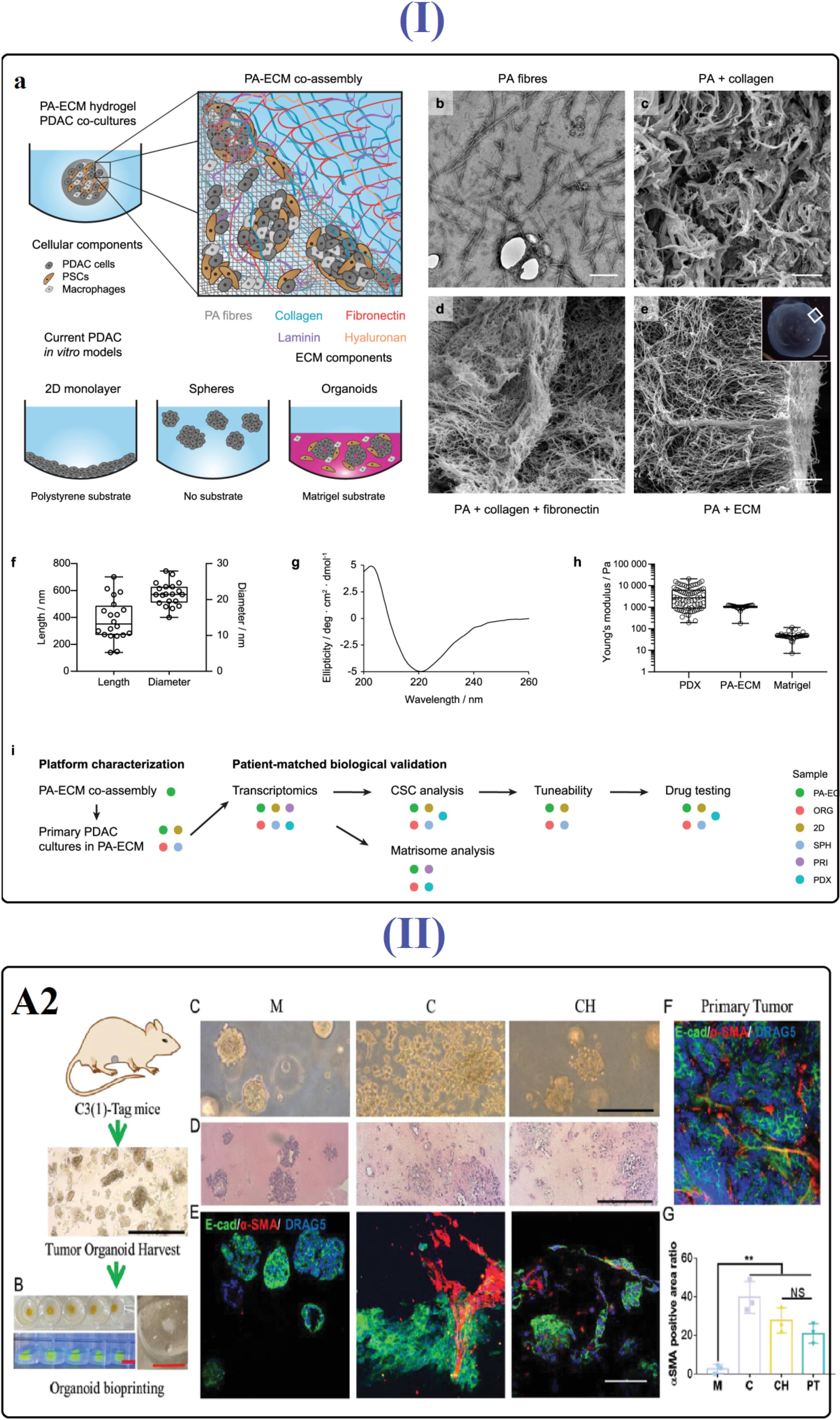
Fig. 5.
(I) (A) PA hydrogel co-assembly with ECM in PDAC modeling. (B) PA fibers depicted in a transmission electron micrograph. (C–E) Scanning electron micrographs of PA hydrogels co-assembled with collagen, fibronectin, and all ECM components. (F) PA fiber size measured from TEM micrographs. (G) PA fiber circular dichroism spectrum. (H) Stiffness comparison with PDX tissue and Matrigel. Reprinted under the terms and conditions of the Creative Commons Attribution (CC BY) license (https://creativecommons.org/licenses/by/4.0/).116 (II) The tumor organoids printed with CH bio-ink accurately replicated the morphology of in vivo tumors. (A) Tumor organoids from the breast, harvested from C3(1)-tag mice. (B) Visualization of FITC-labeled CH bioink within the support bath of the SF hydrogel; side view of CH bioink-loaded organoids under UV light. (C) Evaluation of the morphology of breast tumor organoids on day 1. (D) Hematoxylin and eosin (H&E) staining of breast tumor organoids on day 7. (E&F) Immunofluorescence staining depicting E-cadherin and α-SMA proteins. (G) Quantitative analysis of the α-SMA positive area ratio across three bioinks and the primary tumor. Reprinted under the terms and conditions of the Creative Commons Attribution (CC BY) license (https://creativecommons.org/licenses/by/4.0/).117
.
(I) (A) PA hydrogel co-assembly with ECM in PDAC modeling. (B) PA fibers depicted in a transmission electron micrograph. (C–E) Scanning electron micrographs of PA hydrogels co-assembled with collagen, fibronectin, and all ECM components. (F) PA fiber size measured from TEM micrographs. (G) PA fiber circular dichroism spectrum. (H) Stiffness comparison with PDX tissue and Matrigel. Reprinted under the terms and conditions of the Creative Commons Attribution (CC BY) license (https://creativecommons.org/licenses/by/4.0/).116 (II) The tumor organoids printed with CH bio-ink accurately replicated the morphology of in vivo tumors. (A) Tumor organoids from the breast, harvested from C3(1)-tag mice. (B) Visualization of FITC-labeled CH bioink within the support bath of the SF hydrogel; side view of CH bioink-loaded organoids under UV light. (C) Evaluation of the morphology of breast tumor organoids on day 1. (D) Hematoxylin and eosin (H&E) staining of breast tumor organoids on day 7. (E&F) Immunofluorescence staining depicting E-cadherin and α-SMA proteins. (G) Quantitative analysis of the α-SMA positive area ratio across three bioinks and the primary tumor. Reprinted under the terms and conditions of the Creative Commons Attribution (CC BY) license (https://creativecommons.org/licenses/by/4.0/).117
Conducted by Shi et al, the research delved into the bioprinting of breast tumor cells and organoids using low-concentration collagen-based bioinks. The study explores diverse breast cancer cell subtypes and CAFs, demonstrating successful bioprinting within a complex collagen hydrogel model. Shifting the focus to bioprinting primary tumor tissues, the researchers employed organoids derived from mouse tumor tissue specifically obtained from C3(1)-tag transgenic mice, a model mirroring human basal-like triple-negative breast cancer progression. The embedded bioprinting method facilitates the high-throughput generation of tumor organoid models. Comparative analyses with Matrigel and collagen models reveal the collagen hydrogel bioink's superior maintenance of spatial organization and phenotype, closely resembling in vivo tumor tissue. The study highlights the pivotal role of embedded bioprinting in breast tumor research, providing insights into 3D culture systems for tumor cells and organoids (Fig. 5II).117
Tumor-on-chip
Cancer constitutes an intricate three-dimensional tissue with the ability for dynamic communication with adjacent tissues through various signaling pathways. Consequently, a 3D culture system is better equipped to replicate the architectural complexity of cancer tissue when compared to a 2D culture system (Fig. 6I).118 Nonetheless, 3D cultures face limitations in accurately simulating the dynamic aspects of the TME. In this context, cancer-on-a-chip represents a microfluidic device specifically designed to replicate tumor physiology. It enables the continuous delivery of nutrients or therapeutic compounds, providing a more accurate representation of the dynamic TME.109,119
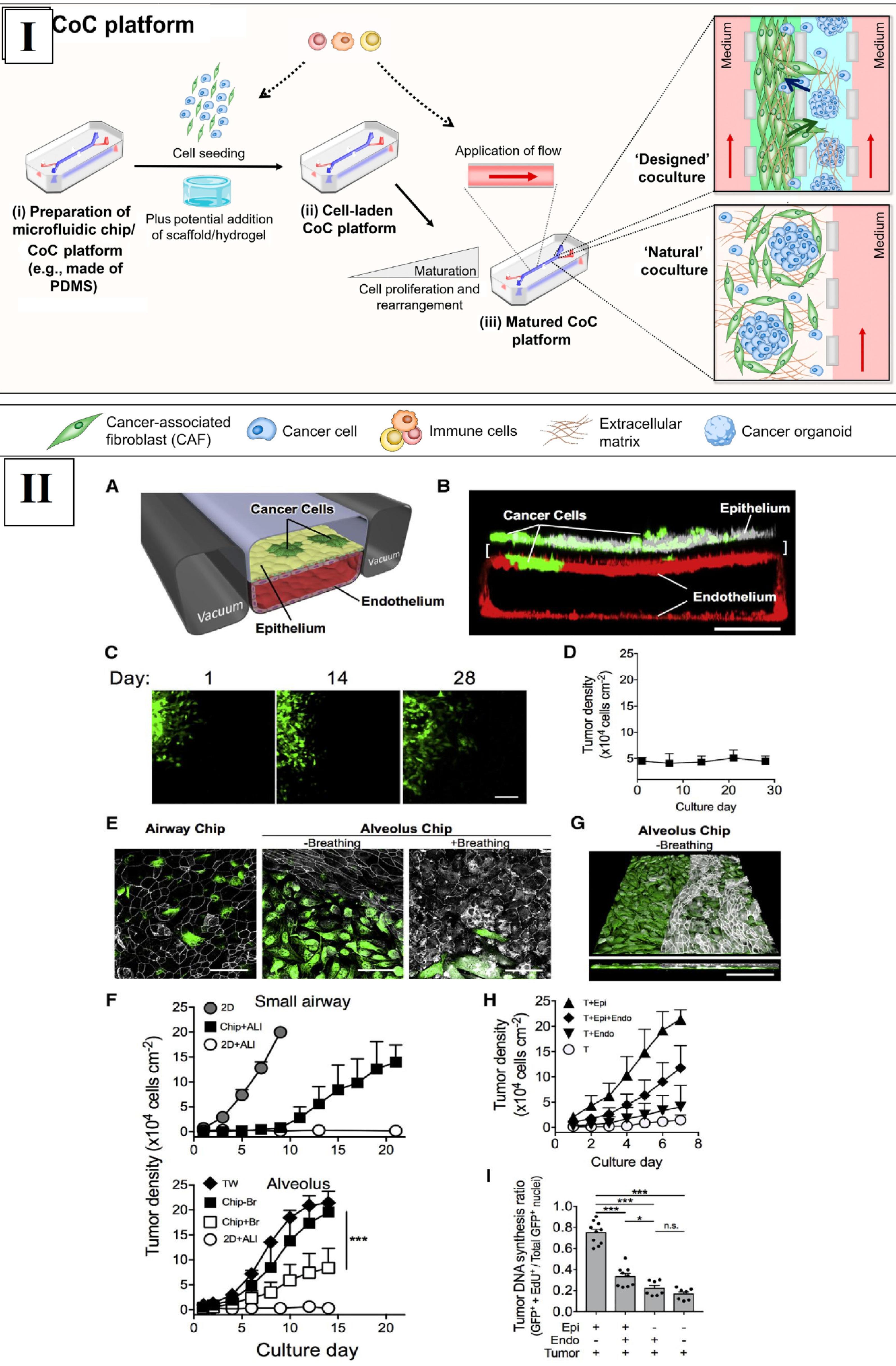
Fig. 6.
(I) The research presents both cell-based 3D models and microfluidic cancer-on-a-chip (CoC) Models. The procedure includes (a and b) cell seeding into CoC devices along with optional hydrogel use and human immune cell attachment capabilities. The introduction of flow happens either during or after maturation to promote cell multiplication as well as cell rearrangement along with immune cell or therapeutic addition. Reprinted under the terms and conditions of the Creative Commons Attribution (CC BY) license (https://creativecommons.org/licenses/by/4.0/).118 (II) Human lung CoC models. The 2-channel chip culture depicts lung cells and NSCLC cells in a microenvironment of porous ECM-coated membrane, which acts as a breathing system in (A). An alveolus chip functions with red fluorescent protein (GFP)-marked lung cancer cells alongside lung epithelial cells and endothelial cells according to the confocal imaging output. (C) GFP-labeled NSCLC cells in an airway chip show minimal proliferation over 28 days. A laboratory analysis measured the density changes of NSCLC cells within the differentiated airway chip platform for an entire month. The growth of GFP-labeled lung cancer cells becomes different when cultured in the airway and alveolar chips with and without breathing present. Lung cancer cells show different levels of growth when grown in a 2D dish, ALI medium, transwell inserts, and microfluidic chips. NTSB-1 transient endogenous lung cancer cells disrupt the alveolar epithelium within a non-breathing alveolus chip according to confocal microscopy. The researchers measure NSCLC tumor cell growth either independently or in combination with alveolar epithelium and microvascular endothelium, or both types of tissues. The experiment determines how lung cancer cell DNA synthesis changes through 5-ethynyl-2′-deoxyuridine (EdU) incorporation assessment while using designated experimental setups. Reprinted under the terms and conditions of the Creative Commons Attribution (CC BY) license (https://creativecommons.org/licenses/by/4.0/).122
.
(I) The research presents both cell-based 3D models and microfluidic cancer-on-a-chip (CoC) Models. The procedure includes (a and b) cell seeding into CoC devices along with optional hydrogel use and human immune cell attachment capabilities. The introduction of flow happens either during or after maturation to promote cell multiplication as well as cell rearrangement along with immune cell or therapeutic addition. Reprinted under the terms and conditions of the Creative Commons Attribution (CC BY) license (https://creativecommons.org/licenses/by/4.0/).118 (II) Human lung CoC models. The 2-channel chip culture depicts lung cells and NSCLC cells in a microenvironment of porous ECM-coated membrane, which acts as a breathing system in (A). An alveolus chip functions with red fluorescent protein (GFP)-marked lung cancer cells alongside lung epithelial cells and endothelial cells according to the confocal imaging output. (C) GFP-labeled NSCLC cells in an airway chip show minimal proliferation over 28 days. A laboratory analysis measured the density changes of NSCLC cells within the differentiated airway chip platform for an entire month. The growth of GFP-labeled lung cancer cells becomes different when cultured in the airway and alveolar chips with and without breathing present. Lung cancer cells show different levels of growth when grown in a 2D dish, ALI medium, transwell inserts, and microfluidic chips. NTSB-1 transient endogenous lung cancer cells disrupt the alveolar epithelium within a non-breathing alveolus chip according to confocal microscopy. The researchers measure NSCLC tumor cell growth either independently or in combination with alveolar epithelium and microvascular endothelium, or both types of tissues. The experiment determines how lung cancer cell DNA synthesis changes through 5-ethynyl-2′-deoxyuridine (EdU) incorporation assessment while using designated experimental setups. Reprinted under the terms and conditions of the Creative Commons Attribution (CC BY) license (https://creativecommons.org/licenses/by/4.0/).122
Aung et al conducted a study introducing a tumor-on-a-chip device for evaluating T-cell recruitment in breast cancer-immune cell interactions. The engineered device features a multi-layered GelMA hydrogel structure with co-cultured cell types, including HUVECs, breast cancer cells, monocytes, and T-cells. Breast cancer cell spheroids are generated using specific techniques. The device serves as a platform to investigate T-cell recruitment dynamics in the context of breast cancer. The study details the fabrication process, including glass surface methacrylation, microfluidic device creation with PAm hydrogels, and additive photopatterning of cell-laden GelMA hydrogels. The device's ability to co-culture diverse cell types within bi-layer hydrogels makes it a versatile tool for studying cellular interactions, offering potential insights for cancer research and therapeutic development in the TME.120
Carvalho and colleagues have pioneered the development of a colorectal tumor-on-a-chip system, a state-of-the-art 3D tool for precision onco-nanomedicine. Employing a rigorous fabrication process, they craft a microfluidic device that faithfully reproduces the complex colorectal TME, surpassing the limitations of conventional 2D cultures. Within this innovative chip, colorectal cancer cells are cultured, culminating in the establishment of a realistic tumor model that closely mimics in vivo conditions. The subsequent analysis focuses on monitoring various parameters, including cell viability, proliferation rates, and the distribution of nanomedicine. Microfluidic channels enable controlled nutrient and oxygen delivery to the tumor model, while advanced imaging techniques visualize real-time interactions. This comprehensive approach provides valuable insights into onco-nanomedicine performance, enhancing our understanding of therapeutic responses in colorectal cancer. The system's innovation lies in its integration of nanomedicine, introducing NPs loaded with therapeutic agents to assess precision and efficacy within the 3D TME.121
Hassell and colleagues developed lung airway and alveolus chip models through a precise fabrication method. This involved casting polydimethylsiloxane (PDMS) for microfluidic devices and cultivating various cell types, including human primary airway and alveolar epithelial cells, lung microvascular endothelial cells, and GFP-labeled non-small cell lung cancer (NSCLC) tumor cells. Co-culture experiments included endothelial cells in Transwell inserts, and organ chips simulated breathing motions with cyclic mechanical deformation. This research emphasizes the profound impact of local lung microenvironmental factors and mechanical breathing motions on the in vitro growth of NSCLC. By employing orthotopic cancer chip models, the study accurately replicates the growth patterns observed in human adenocarcinoma patients, including NSCLC in vivo, emulating resistance development, including the T790M mutation seen in late-stage NSCLC patients unresponsive to third-generation tyrosine kinase inhibitors (TKIs). Moreover, the study mirrors overexpression and phosphorylation of c-Met, observed in EGFR-mutated patients resistant to third-generation TKIs, and replicates clinical trial findings, such as the reduction of interleukin-8 (IL-8) levels in response to TKI treatment. The capability to maintain H1975 cancer cells in a dormant state within an organ-relevant microenvironment offers valuable insights into addressing tumor dormancy and cancer recurrence challenges (Fig. 6II).122
Cancer therapy
The following section details how 3D bioprinting enables cancer treatment through specific drug release methods, as well as multiple drug delivery formats and laboratory tumor three-dimensional models for medication assessment. 3D bioprinting technology is vital for pharmaceutical preparation development because it provides exact drug release precision through managed dosage systems with multiple distribution capabilities. The FDA has endorsed the epilepsy treatment drug Spritam®, which demonstrates the possibilities of this methodology. The research explores oral drug delivery through 3D printing, which permits quick production of solid dosage forms with improved parameters. 3D printing provides solutions to transdermal drug delivery by advancing skin cancer therapy while optimizing drug availability levels. The research discusses implant delivery to show how drug-loaded 3D-printed implants provide sustained localized drug delivery with restrained systemic side effects. Biomimetic scaffolds are built through 3D bioprinting methods for drug screening to create a sophisticated evaluation platform that analyzes tumor cell and benign cell activities within near-realistic three-dimensional media. This method delivers critical findings about medication effectiveness as well as drug-to-drug behavior that supports better cancer treatment methods.74
The research of Uddin et al analyzed the development of transdermal drug delivery through cross-shaped microneedles fabricated by 3D printing as a treatment method for epidermoid skin carcinoma. SLA technology enabled the fabrication of microneedles for inkjet printing, which coated cisplatin along with polyvinyl caprolactam-polyvinyl acetate-polyethylene glycol (SOL). The SLA printing process produced microneedles with 20 microneedles arranged in five rows that could finish in only ten minutes. Optimal inkjet-printed penetration through porcine skin was confirmed by optical coherence tomography imaging. The anti-tumor activity of the cells was measured using mouse models validated with A431 squamous carcinoma cells. The treatment of tumors caused incomplete regression alongside possible microneedle dislodgement, but systemic delivery became possible when placing the non-tumor site first. The treatment of a different area on day eight caused a substantial decrease in tumor dimensions. Research using histopathological techniques validated these results by showing proper cancer cell growth control in the treatments outside tumor areas. Studied findings prove that 3D-printed microneedles used outside tumor sites provide an efficient and risk-free anticancer therapeutic solution, which signifies their prospects in pharmaceutical delivery.123
A breast cancer drug delivery system via electrohydrodynamic jet (E-jet) 3D printing comes from Yang et al. Research investigators produced scaffolds from 5-Fluorouracil (5-FU) and NVP-BEZ235 PLGA that had controlled aperture sizes ranging from 50–200 µm for regulating drug release. The developed drug delivery system demonstrated both uniform drug distribution and adjustable pore size, along with diverse mechanical characteristics. In vitro, tests verified a 30-day period of drug release together with successful MDA-MB-231 cell-blocking effects. The study demonstrated tumor reduction with fewer side effects than standard chemotherapy while testing these methods in living subjects. Breast cancer treatment benefits from the local delivery system as a potential therapeutic method.124
Yi et al invented a 3D-printed biocompatible adhesive patch for the treatment of pancreatic cancer recurrence sites. The PLGA and polycaprolactone device, in combination with 5-FU, delivered drugs precisely at controlled rates. The comprehensive characterization tests verified both the elasticity of the construct and the detailed release pattern of 5-FU during a four-week period, and its successful cancer suppression results in murine subjects. The cytotoxicity tests performed in test tubes indicated that cancer cells died while non-cancerous cells remained unharmed. The P100 patch showed better cancer cell reduction compared to P150 during in vivo experiments, which led to a substantial tumor size decrease. Histological examination revealed apoptosis at the treatment site, while systemic testing confirmed that drug substances spread minimally throughout the body, and the liver experienced very little harm. Targeted cancer therapy shows great potential through the application of 3D-printed patches, according to research findings.125
The study conducted by Souza and team sought to evaluate the relative efficacy of both two-dimensional and three-dimensional cell cultures in assessing the screening, response, and resistance of anti-tumor medications, with a particular emphasis on prostate cancer cell lines (PC3, LNCaP, DU145). The main goal was to determine the most suitable model for conducting preclinical in vitro testing of drugs. The study involved assessing the proliferation, genetic expression, and chemoresistance of cell lines exposed to antineoplastic drugs (paclitaxel and docetaxel) in both 2D and magnetic 3D bioprinting cultures. The findings revealed that 3D cell culture displayed diminished cell proliferation rates, increased resistance to paclitaxel and docetaxel, along with a modified gene expression profile when compared to its 2D counterpart. The derived conclusion emphasized that 3D cell culture is more faithfully replicated in vivo systems, showcasing its potential as a promising and dependable tool for advancing new drug development. The study underscored the significance of this methodology in addressing the constraints of 2D cultures, including the absence of physiologically relevant phenotypes and the complexity inherent in in vivo conditions.126
Kim et al examined how hydrogel application helps NK cells in immunotherapeutic applications through three-dimensional bioprinting for the treatment of leukemia patients as well as solid tumor patients. The researchers employed bioprinting to create a hydrogel micro–macro environment, enhancing NK cell viability with macropores. Micropore formation was controlled by adjusting alginate-to-gelatin ratios, influencing NK cell clusters and proliferation. The hydrogel variant alginate/GEL-2 exhibited superior lytic activity, cytokine secretion, and mRNA expression compared to two-dimensional suspension cells. This hydrogel was applied to genetically modified NK cells targeting EGFR-overexpressing tumor cells. Alginate/GEL-2 facilitated NK cell release after around five days, effectively lysing target cells and displaying high expression levels associated with proliferation and chimeric antigen receptors (CARs) -NK cell-mediated cytotoxicity. Studies confirmed that NKG2D and NKp30 activating receptors increased in number. The combination of hydrogel and three-dimensional bioprinting has developed micro/macropores to resolve immunotherapeutic difficulties in treating various tumors. The findings regarding Alginate/GEL-2 show practical value in stopping cancer recurrence and preventing metastasis after surgical tumor removal (Fig. 7).127
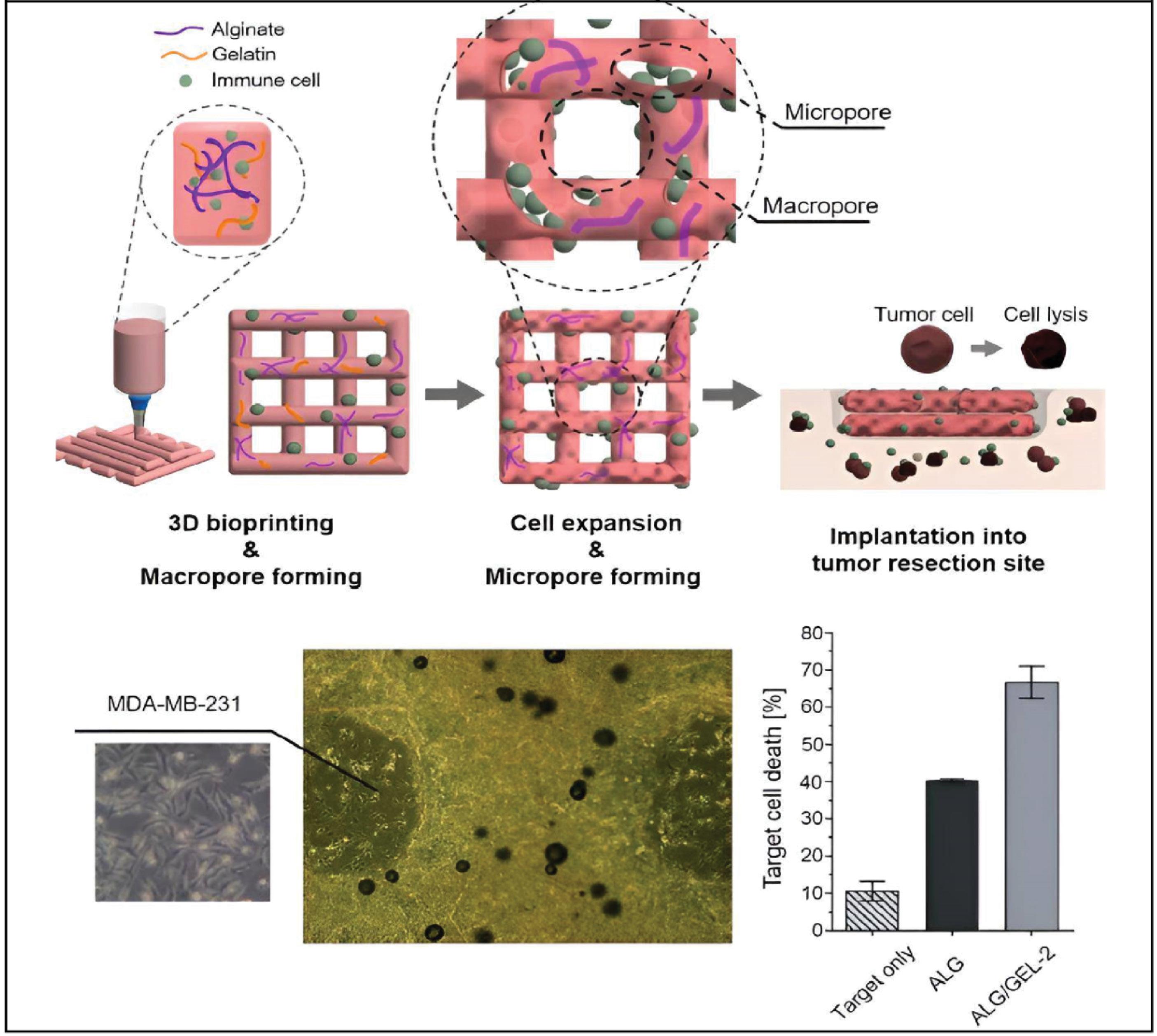
Fig. 7.
(a) This illustration depicts the process of tumoral administration for a micro/macropore-forming hydrogel that contains NK cells generated through 3D bioprinting. The image at (b) shows how pore-forming hydrogels containing CAR-NK cells interact with MDA-MB-231 target cells while using a scale bar that measures 500 μm. (c) Evaluation of target cell death in groups exposed to target cells alone and those treated with hydrogel, as evidenced by flow cytometric data. Reprinted under the terms and conditions of the Creative Commons Attribution (CC BY) license (https://creativecommons.org/licenses/by/4.0/).127
.
(a) This illustration depicts the process of tumoral administration for a micro/macropore-forming hydrogel that contains NK cells generated through 3D bioprinting. The image at (b) shows how pore-forming hydrogels containing CAR-NK cells interact with MDA-MB-231 target cells while using a scale bar that measures 500 μm. (c) Evaluation of target cell death in groups exposed to target cells alone and those treated with hydrogel, as evidenced by flow cytometric data. Reprinted under the terms and conditions of the Creative Commons Attribution (CC BY) license (https://creativecommons.org/licenses/by/4.0/).127
Clinical studies on cancer surgery and 3D printing
The field of cancer surgery has witnessed the development of cutting-edge surgical instruments; however, successfully implementing the necessary surgical methods and perioperative management remains a challenge. Lately, the integration of 3D printing approaches has proven invaluable for surgeons. It facilitates pre-surgical planning, reduces surgery duration, enhances therapeutic outcomes, and underscores the significance of 3D printing in addressing diverse cancer types.17,128 This investigation analyzed virtual 3D modeling for eight patients who received a diagnosis of chondrosarcoma of the petrous apex. Every model included a representation of bone structures along with tumor elements and vessels, with accessory inclusion of pituitary glands and optic nerves. Three different models underwent multicolor 3D printing through the usage of a plaster-based color binder jetting system that demonstrated complex anatomical display ability. A specialized model containing a tumor with an emphasized internal artery demonstrated how volumetric coloring outperformed surface coloring. The 3D-printed physical models provided realistic surgical demonstrations by performing tasks similar to both burr hole surgeries and tumor removal procedures. Using plaster as a material that mimicked bone characteristics enabled precise examination of potential surgery pathways. The application of volumetric coloring methods improved the view of hidden parts containing the internal carotid artery located inside tumors. The technique achieved an accuracy of 0.021 mm for rigid structures based on computer tomography verification, although soft tissue simulation proved challenging.129 Wang and colleagues evaluated the transformative results when a non-co-planar template from 3D printing joins CT-guided 125I seed implantation procedures for locally recurrent rectal cancer. The medical field of surgery depends heavily on the 3D-printed non-co-planar template as it enables precise seed implantation. The customized template helps medical professionals design detailed treatment plans that ensure precise spatial positioning of 125I seeds using careful methods. The combination of 3D printing technology enables surgeons to utilize precise prints, which help them track local rectal cancer anatomy to achieve better surgical outcomes with decreased procedural invasiveness. The achievements of cancer treatments support the success of this innovative solution. The combination of CT-guided 125I seed implantation with a 3D-printed template leads to a major tumor decrease, establishing the targeted strength of this procedure. The purpose of dosimetry analysis is to explore how the template enhances the delivery of therapeutic radiation doses to cancer cells without harming healthy structures as an essential factor for patient treatment success. The combination of 3D-printed non-co-planar templates and CT-guided 125I seed implantation stands as noteworthy progress in surgical recurrence management of rectal cancer because it provides superior accuracy and effectiveness.130
Challenges and solutions
Several limitations, including very complex regulations, difficult scalability, lack of standardization, and unpredictable costs, have been indicated for 3D printing applications in diagnostics and treatment of cancer tumors.4 Current investigations are focusing mostly on case reports and pre-clinical studies, with few clinical studies involving small samples. In next-generation 3D printing technology, comprehensive clinical studies are required to completely evaluate the efficacy and application of 3D printing technology in larger patient groups. In this way, the prediction of the cost of medical materials, the validation of pharmacoeconomic models, or the development of reliable economic simulations should be considered. For scalability and regulatory purposes, interdisciplinary cooperation, cost-effective materials, intellectual property, and medical products are needed. In addition, uniform protocols, quality control standards, and validation in clinical trials should be adopted to standardize 3D printing technology for cancer diagnosis and therapy.131
The logical flow from material science to clinical application in 3D printing technology for cancer therapy and diagnosis involves several key stages, each of which bridges advancements in material design, technology, and medical practices. In this regard, I) collaboration between material scientists, engineers, and clinicians, II) customization for patients, and III) interdisciplinary research are critical factors.132 For effective implementation, close collaboration between researchers, engineers, and clinicians is required to ensure the designs meet medical standards and patient needs. A major advantage of 3D printing is the ability to provide patient-specific treatments. Personalized therapies, such as custom implants or drug delivery systems, can improve patient outcomes. Current interdisciplinary research is vital for developing new materials, improving manufacturing techniques, and overcoming challenges such as scalability, reproducibility, and cost.65
Conclusion
3D printing technology shows excellent potential to transform medical cancer practices in diagnostic procedures and therapeutic methods, along with surgical operations. Its development promotes accurate tissue engineering procedures for medical regeneration applications and authentic tumor modeling systems for pharmaceutical drug trials, along with patient-specific treatments.
Early cancer detection, advanced biomarker discovery, and circulating tumor cell collection can be possible through 3D printing technology owing to its scalable features. The development of computer-controlled drug delivery systems using tailored tumor models through 3D printing allows better patient care outcomes.
The surgical application of 3D printing supports surgical planning before operations with patient-oriented models for better treatment results. The revolutionary nature of 3D printing applications in cancer care becomes clear as it changes how clinicians diagnose diseases and treat patients and perform surgeries, which will create superior personalized cancer therapies. The field of 3D printing for cancer therapy and diagnosis is still evolving, and several advancements, including printing with stem cells or organoids, advanced imaging, artificial intelligence (AI) integration, and wearable and implantable devices, are being made. Future research may focus on 3D printing with stem cells or organoids to recreate functional tissues for both diagnostic and therapeutic purposes. AI-driven models integrated with 3D printed diagnostic tools could revolutionize early detection and treatment planning for cancer. In the future, 3D-printed wearable devices may continuously monitor cancer biomarkers in real time, enabling constant surveillance and personalized treatment adjustments.
Review Highlights
What is the current knowledge?
-
3D printing technology provides excellent potential to transform medical cancer practices in diagnostic procedures and therapeutic methods with surgical operations.
-
Development in 3D printing technology promotes accurate tissue engineering procedures for authentic tumor modeling systems for pharmaceutical drug trials, along with patient-specific treatments.
-
Current investigations are focusing mostly on case reports and pre-clinical studies, with few clinical studies involving small samples.
What is new here?
-
Early cancer detection, advanced biomarker discovery, and circulating tumor cell collection can be possible through 3D printing technology.
-
The surgical application of 3D printing supports surgical planning before operations.
-
The revolutionary nature of 3D printing applications based on next-generation bioinks using nanomaterials in cancer care becomes clear as it changes how clinicians diagnose diseases and treat patients.
-
In next-generation 3D printing technology, comprehensive clinical studies are needed to fully evaluate the efficacy of 3D printing technology in larger patient groups.
Competing Interests
The authors declare that they have no competing interests.
Data Availability Statement
No data was applied for the research described in the article.
Declaration of AI-Assisted Tools in the Writing Procedure
The authors declare that generative AI was applied in preparing some sections of this manuscript. After using this tool/service, the authors reviewed and edited the content as required and take full responsibility for the content of the published article.
Ethical Approval
Not applicable.
References
- Wang F, Liu J, Liu J, Zhang S, Yang M, Kang K. Comparison of the Effects of 3D Printing Bioactive Porous Titanium Alloy Scaffolds and Nano-biology for Direct Treatment of Bone Defects. Cell Mol Biol (Noisy-le-grand) 2022; 68:86-95. doi: 10.14715/cmb/2022.68.3.11 [Crossref] [ Google Scholar]
- Zhang S, Chen X, Shan M, Hao Z, Zhang X, Meng L. Convergence of 3D Bioprinting and Nanotechnology in Tissue Engineering Scaffolds. Biomimetics (Basel) 2023; 8:94. doi: 10.3390/biomimetics8010094 [Crossref] [ Google Scholar]
- Tao Q, Wu C, Li X, Chen W, Sun K, Zhang P. Three-dimensional bio-printed constructs consisting of human umbilical-derived mesenchymal stem cells promote cell viability, proliferation, and differentiation in vitro. Cell Mol Biol (Noisy-le-grand) 2020; 66:165-71. [ Google Scholar]
- Liu S, Jin P. Advances and Challenges in 3D Bioprinted Cancer Models: Opportunities for Personalized Medicine and Tissue Engineering. Polymers (Basel) 2025; 17:948. doi: 10.3390/polym17070948 [Crossref] [ Google Scholar]
- Ouyang L, Yao R, Zhao Y, Sun W. Effect of bioink properties on printability and cell viability for 3D bioplotting of embryonic stem cells. Biofabrication 2016; 8:035020. doi: 10.1088/1758-5090/8/3/035020 [Crossref] [ Google Scholar]
- Zhang T, Wang G. Inhibition of RXRA-mediated PLA2G2A improves delirium in COPD mice by regulating endoplasmic reticulum stress pathway and inhibiting cell apoptosis. Cell Mol Biol (Noisy-le-grand) 2024; 70:226-32. doi: 10.14715/cmb/2024.70.1.31 [Crossref] [ Google Scholar]
- Alavi M, Webster TJ, Li L. Theranostic safe quantum dots for anticancer and bioimaging applications. Micro Nano Bio Aspects 2022; 1:1-11. doi: 10.22034/mnba.2022.154865 [Crossref] [ Google Scholar]
- Mahdavi Dehkharghani F, Ghahremanlou M, Zandi Z, Jalili M, Mozafari MR, Mardani P. Future energy and therapeutic perspectives of green nano-technology: recent advances and challenges. Nano Micro Biosystems 2023; 2:11-21. doi: 10.22034/nmbj.2023.385185.1013 [Crossref] [ Google Scholar]
- Alavi M, Mozafari MR, Hamblin MR, Hamidi M, Hajimolaali M, Katouzian I. Industrial-scale methods for the manufacture of liposomes and nanoliposomes: pharmaceutical, cosmetic, and nutraceutical aspects. Micro Nano Bio Aspects 2022; 1:26-35. doi: 10.22034/mnba.2022.159371 [Crossref] [ Google Scholar]
- Goztepe M, Eroglu O. Research of the unrecognised functions of miR-375 in prostate cancer cells. Cell Mol Biol (Noisy-le-grand) 2024; 70:212-8. doi: 10.14715/cmb/2024.70.3.32 [Crossref] [ Google Scholar]
- Zhu Y, Duan C, Gui Y, Chen D, Su X. Exosomal circMACF1 drives PI3K/AKT/mTOR-mediated autophagy suppression in laryngeal squamous cell carcinoma. Cell Mol Biol (Noisy-le-grand) 2024; 70:179-85. doi: 10.14715/cmb/2024.70.1.24 [Crossref] [ Google Scholar]
- Kim SH, Yeon YK, Lee JM, Chao JR, Lee YJ, Seo YB. Precisely printable and biocompatible silk fibroin bioink for digital light processing 3D printing. Nat Commun 2018; 9:1620. doi: 10.1038/s41467-018-03759-y [Crossref] [ Google Scholar]
- Han S, Fisher JP, Mikos AG, Hogan KJ. Polymeric nanomaterials in 3D bioprinting for tissue engineering and drug delivery applications. Bioprinting 2024; 40:e00345. doi: 10.1016/j.bprint.2024.e00345 [Crossref] [ Google Scholar]
- Zhang L, Huang X, Cole T, Lu H, Hang J, Li W. 3D-printed liquid metal polymer composites as NIR-responsive 4D printing soft robot. Nat Commun 2023; 14:7815. doi: 10.1038/s41467-023-43667-4 [Crossref] [ Google Scholar]
- Zhang N, Wang Z, Zhao Z, Zhang D, Feng J, Yu L. 3D printing of micro-nano devices and their applications. Microsystems & Nanoengineering 2025; 11:35. doi: 10.1038/s41378-024-00812-3 [Crossref] [ Google Scholar]
- Schwab A, Levato R, D'Este M, Piluso S, Eglin D, Malda J. Printability and Shape Fidelity of Bioinks in 3D Bioprinting. Chem Rev 2020; 120:11028-55. doi: 10.1021/acs.chemrev.0c00084 [Crossref] [ Google Scholar]
- Metzger Filho O, Giobbie-Hurder A, Mallon E, Gusterson B, Viale G, Winer EP. Relative Effectiveness of Letrozole Compared With Tamoxifen for Patients With Lobular Carcinoma in the BIG 1-98 Trial. J Clin Oncol 2015; 33:2772-9. doi: 10.1200/jco.2015.60.8133 [Crossref] [ Google Scholar]
- Wang C, Yokota T, Someya T. Natural Biopolymer-Based Biocompatible Conductors for Stretchable Bioelectronics. Chem Rev 2021; 121:2109-46. doi: 10.1021/acs.chemrev.0c00897 [Crossref] [ Google Scholar]
-
Alavi M, Rai M. Chapter 11 - Antibacterial and wound healing activities of micro/nanocarriers based on carboxymethyl and quaternized chitosan derivatives. In: Rai M, CA dos Santos, editors. Biopolymer-Based Nano Films: Elsevier; 2021. p. 191–201. doi: 10.1016/B978-0-12-823381-8.00009-0.
- Samrot AV, Sathiyasree M, Rahim SBA, Renitta RE, Kasipandian K, Krithika Shree S. Scaffold Using Chitosan, Agarose, Cellulose, Dextran and Protein for Tissue Engineering-A Review. Polymers (Basel) 2023; 15:1525. doi: 10.3390/polym15061525 [Crossref] [ Google Scholar]
- Alavi M. Applications of Chitosan and Nanochitosan in Formulation of Novel Antibacterial and Wound Healing Agents. In: Rai M, editor. Nanotechnology in Skin, Soft Tissue, and Bone Infections. Cham: Springer International Publishing; 2020. p. 169-81.
- Parnianchi F, Kashanian S, Nazari M, Peacock M, Omidfar K, Varmira K. Ultrasensitive electrochemical sensor based on molecular imprinted polymer and ferromagnetic nanocomposite for bilirubin analysis in the saliva and serum of newborns. Microchemical Journal 2022; 179:107474. doi: 10.1016/j.microc.2022.107474 [Crossref] [ Google Scholar]
- Kirillova A, Yeazel TR, Asheghali D, Petersen SR, Dort S, Gall K. Fabrication of Biomedical Scaffolds Using Biodegradable Polymers. Chem Rev 2021; 121:11238-304. doi: 10.1021/acs.chemrev.0c01200 [Crossref] [ Google Scholar]
- Alavi M, Webster TJ. Recent progress and challenges for polymeric microsphere compared to nanosphere drug release systems: Is there a real difference?. Bioorg Med Chem 2021; 33:116028. doi: 10.1016/j.bmc.2021.116028 [Crossref] [ Google Scholar]
- Ligon SC, Liska R, Stampfl J, Gurr M, Mülhaupt R. Polymers for 3D Printing and Customized Additive Manufacturing. Chem Rev 2017; 117:10212-90. doi: 10.1021/acs.chemrev.7b00074 [Crossref] [ Google Scholar]
- Sanandiya ND, Vijay Y, Dimopoulou M, Dritsas S, Fernandez JG. Large-scale additive manufacturing with bioinspired cellulosic materials. Sci Rep 2018; 8:8642. doi: 10.1038/s41598-018-26985-2 [Crossref] [ Google Scholar]
- N'Gatta KM, Belaid H, El Hayek J, Assanvo EF, Kajdan M, Masquelez N. 3D printing of cellulose nanocrystals based composites to build robust biomimetic scaffolds for bone tissue engineering. Sci Rep 2022; 12:21244. doi: 10.1038/s41598-022-25652-x [Crossref] [ Google Scholar]
- Dave K, Mahmud Z, Gomes VG. Superhydrophilic 3D-printed scaffolds using conjugated bioresorbable nanocomposites for enhanced bone regeneration. Chemical Engineering Journal 2022; 445:136639. doi: 10.1016/j.cej.2022.136639 [Crossref] [ Google Scholar]
- Xu J, Zhu X, Zhao J, Ling G, Zhang P. Biomedical applications of supramolecular hydrogels with enhanced mechanical properties. Adv Colloid Interface Sci 2023; 321:103000. doi: 10.1016/j.cis.2023.103000 [Crossref] [ Google Scholar]
- Jiang Z, Diggle B, Tan ML, Viktorova J, Bennett CW, Connal LA. Extrusion 3D Printing of Polymeric Materials with Advanced Properties. Adv Sci (Weinh) 2020; 7:2001379. doi: 10.1002/advs.202001379 [Crossref] [ Google Scholar]
- Li X, Cui R, Sun L, Aifantis KE, Fan Y, Feng Q. 3D-Printed Biopolymers for Tissue Engineering Application. International Journal of Polymer Science 2014; 2014:829145. doi: 10.1155/2014/829145 [Crossref] [ Google Scholar]
- Akbari Kenari M, Rezvani Ghomi E, Akbari Kenari A, Arabi SMS, Deylami J, Ramakrishna S. Biomedical applications of microfluidic devices: Achievements and challenges. Polymers for Advanced Technologies 2022; 33:3920-34. doi: 10.1002/pat.5847 [Crossref] [ Google Scholar]
- Wang Y, Xu H, Zhang X. Tuning the Amphiphilicity of Building Blocks: Controlled Self-Assembly and Disassembly for Functional Supramolecular Materials. Advanced Materials 2009; 21:2849-64. doi: 10.1002/adma.200803276 [Crossref] [ Google Scholar]
- Lo PK, Karam P, Aldaye FA, McLaughlin CK, Hamblin GD, Cosa G. Loading and selective release of cargo in DNA nanotubes with longitudinal variation. Nat Chem 2010; 2:319-28. doi: 10.1038/nchem.575 [Crossref] [ Google Scholar]
- Beck JB, Killops KL, Kang T, Sivanandan K, Bayles A, Mackay ME. FACILE PREPARATION OF NANOPARTICLES BY INTRAMOLECULAR CROSSLINKING OF ISOCYANATE FUNCTIONALIZED COPOLYMERS. Macromolecules 2009; 42:5629-35. doi: 10.1021/ma900899v [Crossref] [ Google Scholar]
- Foster EJ, Berda EB, Meijer EW. Metastable supramolecular polymer nanoparticles via intramolecular collapse of single polymer chains. J Am Chem Soc 2009; 131:6964-6. doi: 10.1021/ja901687d [Crossref] [ Google Scholar]
-
Verde-Sesto E, Blázquez-Martín A, Pomposo JA. Advances in the Phototriggered Synthesis of Single-Chain Polymer Nanoparticles. Polymers (Basel) 2019; 11. doi: 10.3390/polym11111903.
- Amir RJ, Zhong S, Pochan DJ, Hawker CJ. Enzymatically triggered self-assembly of block copolymers. J Am Chem Soc 2009; 131:13949-51. doi: 10.1021/ja9060917 [Crossref] [ Google Scholar]
- Lee HI, Wu W, Oh JK, Mueller L, Sherwood G, Peteanu L. Light-induced reversible formation of polymeric micelles. Angew Chem Int Ed Engl 2007; 46:2453-7. doi: 10.1002/anie.200604278 [Crossref] [ Google Scholar]
- Park C, Lim J, Yun M, Kim C. Photoinduced release of guest molecules by supramolecular transformation of self-assembled aggregates derived from dendrons. Angew Chem Int Ed Engl 2008; 47:2959-63. doi: 10.1002/anie.200705271 [Crossref] [ Google Scholar]
- Klaikherd A, Nagamani C, Thayumanavan S. Multi-stimuli sensitive amphiphilic block copolymer assemblies. J Am Chem Soc 2009; 131:4830-8. doi: 10.1021/ja809475a [Crossref] [ Google Scholar]
- Aldaye FA, Palmer AL, Sleiman HF. Assembling materials with DNA as the guide. Science 2008; 321:1795-9. doi: 10.1126/science.1154533 [Crossref] [ Google Scholar]
- Begines B, Ortiz T, Pérez-Aranda M, Martínez G, Merinero M, Argüelles-Arias F. Polymeric Nanoparticles for Drug Delivery: Recent Developments and Future Prospects. Nanomaterials (Basel) 2020; 10:1403. doi: 10.3390/nano10071403 [Crossref] [ Google Scholar]
- Zhang L, Forgham H, Shen A, Wang J, Zhu J, Huang X. Nanomaterial integrated 3D printing for biomedical applications. J Mater Chem B 2022; 10:7473-90. doi: 10.1039/d2tb00931e [Crossref] [ Google Scholar]
- Alavi M, Hamblin MR, Kennedy JF. Antimicrobial applications of lichens: secondary metabolites and green synthesis of silver nanoparticles: A review. Nano Micro Biosystems 2022; 1:15-21. doi: 10.22034/nmbj.2022.159216 [Crossref] [ Google Scholar]
- Lucena R, Simonet BM, Cárdenas S, Valcárcel M. Potential of nanoparticles in sample preparation. J Chromatogr A 2011; 1218:620-37. doi: 10.1016/j.chroma.2010.10.069 [Crossref] [ Google Scholar]
- Zhu Y, Xu P, Zhang X, Wu D. Emerging porous organic polymers for biomedical applications. Chem Soc Rev 2022; 51:1377-414. doi: 10.1039/d1cs00871d [Crossref] [ Google Scholar]
- Naikoo GA, Arshad F, Almas M, Hassan IU, Pedram MZ, Aljabali AAA. 2D materials, synthesis, characterization and toxicity: A critical review. Chem Biol Interact 2022; 365:110081. doi: 10.1016/j.cbi.2022.110081 [Crossref] [ Google Scholar]
-
Wang J, Li H, Tian L, Ramakrishna S. Nanobiomaterials: State of the Art. Nanobiomaterials2018. p. 3–35. doi: 10.1002/9783527698646.ch1.
- Pati F, Gantelius J, Svahn HA. 3D Bioprinting of Tissue/Organ Models. Angew Chem Int Ed Engl 2016; 55:4650-65. doi: 10.1002/anie.201505062 [Crossref] [ Google Scholar]
- Bernard M, Jubeli E, Pungente MD, Yagoubi N. Biocompatibility of polymer-based biomaterials and medical devices - regulations, in vitro screening and risk-management. Biomater Sci 2018; 6:2025-53. doi: 10.1039/c8bm00518d [Crossref] [ Google Scholar]
- Savina IN, Zoughaib M, Yergeshov AA. Design and Assessment of Biodegradable Macroporous Cryogels as Advanced Tissue Engineering and Drug Carrying Materials. Gels 2021; 7:79. doi: 10.3390/gels7030079 [Crossref] [ Google Scholar]
- El-Sayed A, Kamel M. Advances in nanomedical applications: diagnostic, therapeutic, immunization, and vaccine production. Environ Sci Pollut Res Int 2020; 27:19200-13. doi: 10.1007/s11356-019-06459-2 [Crossref] [ Google Scholar]
- Dunuweera SP, Rajapakse RMSI, Rajapakshe RBSD, Wijekoon SHDP, Nirodha Thilakarathna MGGS, Rajapakse RMG. Review on Targeted Drug Delivery Carriers Used in Nanobiomedical Applications. Current Nanoscience 2019; 15:382-97. doi: 10.2174/1573413714666181106114247 [Crossref] [ Google Scholar]
- Richmond J. Animal welfare and ISO—The International Organisation for Standardization. AATEX Spec Issue 2007; 14:723-726. [ Google Scholar]
- Ngo TD, Kashani A, Imbalzano G, Nguyen KTQ, Hui D. Additive manufacturing (3D printing): A review of materials, methods, applications and challenges. Composites Part B: Engineering 2018; 143:172-96. doi: 10.1016/j.compositesb.2018.02.012 [Crossref] [ Google Scholar]
- Jamróz W, Szafraniec J, Kurek M, Jachowicz R. 3D Printing in Pharmaceutical and Medical Applications - Recent Achievements and Challenges. Pharm Res 2018; 35:176. doi: 10.1007/s11095-018-2454-x [Crossref] [ Google Scholar]
- Williams DF. On the mechanisms of biocompatibility. Biomaterials 2008; 29:2941-53. doi: 10.1016/j.biomaterials.2008.04.023 [Crossref] [ Google Scholar]
- Koutsopoulos S. Molecular fabrications of smart nanobiomaterials and applications in personalized medicine. Adv Drug Deliv Rev 2012; 64:1459-76. doi: 10.1016/j.addr.2012.08.002 [Crossref] [ Google Scholar]
- Hoshino Y, Kodama T, Okahata Y, Shea KJ. Peptide imprinted polymer nanoparticles: a plastic antibody. J Am Chem Soc 2008; 130:15242-3. doi: 10.1021/ja8062875 [Crossref] [ Google Scholar]
- Hoshino Y, Koide H, Urakami T, Kanazawa H, Kodama T, Oku N. Recognition, neutralization, and clearance of target peptides in the bloodstream of living mice by molecularly imprinted polymer nanoparticles: a plastic antibody. J Am Chem Soc 2010; 132:6644-5. doi: 10.1021/ja102148f [Crossref] [ Google Scholar]
- Alexander C, Andersson HS, Andersson LI, Ansell RJ, Kirsch N, Nicholls IA. Molecular imprinting science and technology: a survey of the literature for the years up to and including 2003. J Mol Recognit 2006; 19:106-80. doi: 10.1002/jmr.760 [Crossref] [ Google Scholar]
- Debord JD, Lyon LA. Synthesis and Characterization of pH-Responsive Copolymer Microgels with Tunable Volume Phase Transition Temperatures. Langmuir 2003; 19:7662-4. doi: 10.1021/la0342924 [Crossref] [ Google Scholar]
- Ogawa K, Nakayama A, Kokufuta E. Preparation and Characterization of Thermosensitive Polyampholyte Nanogels. Langmuir 2003; 19:3178-84. doi: 10.1021/la0267185 [Crossref] [ Google Scholar]
- Safhi AY. Three-Dimensional (3D) Printing in Cancer Therapy and Diagnostics: Current Status and Future Perspectives. Pharmaceuticals (Basel) 2022; 15:678. doi: 10.3390/ph15060678 [Crossref] [ Google Scholar]
- Heinrich MA, Liu W, Jimenez A, Yang J, Akpek A, Liu X. 3D Bioprinting: from Benches to Translational Applications. Small 2019; 15:e1805510. doi: 10.1002/smll.201805510 [Crossref] [ Google Scholar]
- Tiwari AP, Thorat ND, Pricl S, Patil RM, Rohiwal S, Townley H. Bioink: a 3D-bioprinting tool for anticancer drug discovery and cancer management. Drug Discov Today 2021; 26:1574-90. doi: 10.1016/j.drudis.2021.03.010 [Crossref] [ Google Scholar]
- Al Sawaftah NM, Pitt WG, Husseini GA. Incorporating nanoparticles in 3D printed scaffolds for bone cancer therapy. Bioprinting 2023; 36:e00322. doi: 10.1016/j.bprint.2023.e00322 [Crossref] [ Google Scholar]
- Xu HQ, Liu JC, Zhang ZY, Xu CX. A review on cell damage, viability, and functionality during 3D bioprinting. Mil Med Res 2022; 9:70. doi: 10.1186/s40779-022-00429-5 [Crossref] [ Google Scholar]
- Ravanbakhsh H, Karamzadeh V, Bao G, Mongeau L, Juncker D, Zhang YS. Emerging Technologies in Multi-Material Bioprinting. Adv Mater 2021; 33:e2104730. doi: 10.1002/adma.202104730 [Crossref] [ Google Scholar]
- Potyondy T, Uquillas JA, Tebon PJ, Byambaa B, Hasan A, Tavafoghi M. Recent advances in 3D bioprinting of musculoskeletal tissues. Biofabrication 2021; 13:022001. doi: 10.1088/1758-5090/abc8de [Crossref] [ Google Scholar]
- Tharakan S, Khondkar S, Ilyas A. Bioprinting of Stem Cells in Multimaterial Scaffolds and Their Applications in Bone Tissue Engineering. Sensors (Basel) 2021; 21:7477. doi: 10.3390/s21227477 [Crossref] [ Google Scholar]
- De Pieri A, Byerley AM, Musumeci CR, Salemizadehparizi F, Vanderhorst MA, Wuertz-Kozak K. Electrospinning and 3D bioprinting for intervertebral disc tissue engineering. JOR Spine 2020; 3:e1117. doi: 10.1002/jsp2.1117 [Crossref] [ Google Scholar]
- Li J, Liang D, Chen X, Sun W, Shen X. Applications of 3D printing in tumor treatment. Biomedical Technology 2024; 5:1-13. doi: 10.1016/j.bmt.2023.03.002 [Crossref] [ Google Scholar]
- Qiu K, Haghiashtiani G, McAlpine MC. 3D Printed Organ Models for Surgical Applications. Annu Rev Anal Chem (Palo Alto Calif) 2018; 11:287-306. doi: 10.1146/annurev-anchem-061417-125935 [Crossref] [ Google Scholar]
- Fernandes PD, Silva FC, Magalhães FD, Pereira RF, Santos SG, Pinto AM. 3D printing of nanoparticle-containing scaffolds for cancer phototherapy, magnetic hyperthermia therapy, and tissue regeneration. Acta Biomater 2026; 210:221-79. doi: 10.1016/j.actbio.2025.11.043 [Crossref] [ Google Scholar]
- Li S, Shi Z, Wang Y, Wang W, He R. The 3D Printing of Flexible Materials: Technologies, Materials, and Challenges. Materials (Basel) 2025; 18:5428. doi: 10.3390/ma18235428 [Crossref] [ Google Scholar]
- Li W, Wang M, Ma H, Chapa-Villarreal FA, Lobo AO, Zhang YS. Stereolithography apparatus and digital light processing-based 3D bioprinting for tissue fabrication. iScience 2023; 26:106039. doi: 10.1016/j.isci.2023.106039 [Crossref] [ Google Scholar]
- Deshmane S, Kendre P, Mahajan H, Jain S. Stereolithography 3D printing technology in pharmaceuticals: a review. Drug Dev Ind Pharm 2021; 47:1362-72. doi: 10.1080/03639045.2021.1994990 [Crossref] [ Google Scholar]
- Husna A, Ashrafi S, Tomal ANMA, Tuli NT, Bin Rashid A. Recent advancements in stereolithography (SLA) and their optimization of process parameters for sustainable manufacturing. Hybrid Advances 2024; 7:100307. doi: 10.1016/j.hybadv.2024.100307 [Crossref] [ Google Scholar]
- Ali F, Kalva SN, Koc M. Advancements in 3D printing techniques for biomedical applications: a comprehensive review of materials consideration, post processing, applications, and challenges. Discover Materials 2024; 4:53. doi: 10.1007/s43939-024-00115-4 [Crossref] [ Google Scholar]
- Zub K, Hoeppener S, Schubert US. Inkjet Printing and 3D Printing Strategies for Biosensing, Analytical, and Diagnostic Applications. Adv Mater 2022; 34:e2105015. doi: 10.1002/adma.202105015 [Crossref] [ Google Scholar]
- Campbell A, Gutierrez DA, Knight C, Vines CM, Heydarian R, Philipovskiy A. Novel Combinatorial Strategy Using Thermal Inkjet Bioprinting, Chemotherapy, and Radiation on Human Breast Cancer Cells; an In-Vitro Cell Viability Assessment. Materials (Basel) 2021; 14:7864. doi: 10.3390/ma14247864 [Crossref] [ Google Scholar]
- Li X, Liu B, Pei B, Chen J, Zhou D, Peng J. Inkjet Bioprinting of Biomaterials. Chem Rev 2020; 120:10793-833. doi: 10.1021/acs.chemrev.0c00008 [Crossref] [ Google Scholar]
- Flores-Torres S, Jiang T, Kort-Mascort J, Yang Y, Peza-Chavez O, Pal S. Constructing 3D In Vitro Models of Heterocellular Solid Tumors and Stromal Tissues Using Extrusion-Based Bioprinting. ACS Biomater Sci Eng 2023; 9:542-61. doi: 10.1021/acsbiomaterials.2c00998 [Crossref] [ Google Scholar]
- Vu M, Pramanik A, Basak AK, Prakash C, Shankar S. Progress and challenges on extrusion based three dimensional (3D) printing of biomaterials. Bioprinting 2022; 27:e00223. doi: 10.1016/j.bprint.2022.e00223 [Crossref] [ Google Scholar]
- Hakobyan D, Médina C, Dusserre N, Stachowicz ML, Handschin C, Fricain JC. Laser-assisted 3D bioprinting of exocrine pancreas spheroid models for cancer initiation study. Biofabrication 2020; 12:035001. doi: 10.1088/1758-5090/ab7cb8 [Crossref] [ Google Scholar]
- Dou C, Perez V, Qu J, Tsin A, Xu B, Li J. A State-of-the-Art Review of Laser-Assisted Bioprinting and its Future Research Trends. ChemBioEng Reviews 2021; 8:517-34. doi: 10.1002/cben.202000037 [Crossref] [ Google Scholar]
- Zhang J, Hu Q, Wang S, Tao J, Gou M. Digital Light Processing Based Three-dimensional Printing for Medical Applications. Int J Bioprint 2020; 6:242. doi: 10.18063/ijb.v6i1.242 [Crossref] [ Google Scholar]
- Alparslan C, Bayraktar Ş. Advances in Digital Light Processing (DLP) Bioprinting: A Review of Biomaterials and Its Applications, Innovations, Challenges, and Future Perspectives. Polymers (Basel) 2025; 17:1287. doi: 10.3390/polym17091287 [Crossref] [ Google Scholar]
- An J, Leong KF. Multi-material and Multi-dimensional 3D Printing for Biomedical Materials and Devices. Biomedical Materials & Devices 2023; 1:38-48. doi: 10.1007/s44174-022-00038-9 [Crossref] [ Google Scholar]
- Gao G, Kim BS, Jang J, Cho DW. Recent Strategies in Extrusion-Based Three-Dimensional Cell Printing toward Organ Biofabrication. ACS Biomater Sci Eng 2019; 5:1150-69. doi: 10.1021/acsbiomaterials.8b00691 [Crossref] [ Google Scholar]
- Gospodinova A, Nankov V, Tomov S, Redzheb M, Petrov PD. Extrusion bioprinting of hydroxyethylcellulose-based bioink for cervical tumor model. CarbohydrPolym 2021; 260:117793. doi: 10.1016/j.carbpol.2021.117793 [Crossref] [ Google Scholar]
-
Zou S, Ye J, Wei Y, Xu J. Characterization of 3D-Bioprinted In Vitro Lung Cancer Models Using RNA-Sequencing Techniques. Bioengineering (Basel) 2023; 10. doi: 10.3390/bioengineering10060667.
- Koch L, Gruene M, Unger C, Chichkov B. Laser assisted cell printing. Curr Pharm Biotechnol 2013; 14:91-7. [ Google Scholar]
- Gruene M, Deiwick A, Koch L, Schlie S, Unger C, Hofmann N. Laser printing of stem cells for biofabrication of scaffold-free autologous grafts. Tissue Eng Part C Methods 2011; 17:79-87. doi: 10.1089/ten.TEC.2010.0359 [Crossref] [ Google Scholar]
- Parrish J, Lim KS, Baer K, Hooper GJ, Woodfield TBF. A 96-well microplate bioreactor platform supporting individual dual perfusion and high-throughput assessment of simple or biofabricated 3D tissue models. Lab on a Chip 2018; 18:2757-75. doi: 10.1039/C8LC00485D [Crossref] [ Google Scholar]
- Saunders RE, Derby B. Inkjet printing biomaterials for tissue engineering: bioprinting. International Materials Reviews 2014; 59:430-48. doi: 10.1179/1743280414Y.0000000040 [Crossref] [ Google Scholar]
- Lion A, Wildman RD, Alexander MR, Roberts CJ. Customisable Tablet Printing: The Development of Multimaterial Hot Melt Inkjet 3D Printing to Produce Complex and Personalised Dosage Forms. Pharmaceutics 2021; 13:1679. doi: 10.3390/pharmaceutics13101679 [Crossref] [ Google Scholar]
- Cheng C, Deneke N, Moon HR, Choi SR, Ospina-Muñoz N, Elzey BD, et al. Inkjet-printed morphogenesis of tumor-stroma interface using bi-cellular bioinks of collagen-poly(N-isopropyl acrylamide-co-methyl methacrylate) mixture. Mater Today Adv 2023; 19. doi: 10.1016/j.mtadv.2023.100408.
-
Benning L, Peintner A, Peintner L. Advances in and the Applicability of Machine Learning-Based Screening and Early Detection Approaches for Cancer: A Primer. Cancers (Basel) 2022; 14. doi: 10.3390/cancers14030623.
- Singh S, Prakash C, Singh R. 3D printing in biomedical engineering: Springer 2020.
- Zhang Y. 3D Printing for Cancer Diagnosis: What Unique Advantages Are Gained?. ACS Mater Au 2023; 3:620-35. doi: 10.1021/acsmaterialsau.3c00046 [Crossref] [ Google Scholar]
- Gosai A, Khondakar KR, Ma X, Ali MA. Application of Functionalized Graphene Oxide Based Biosensors for Health Monitoring: Simple Graphene Derivatives to 3D Printed Platforms. Biosensors (Basel) 2021; 11:384. doi: 10.3390/bios11100384 [Crossref] [ Google Scholar]
- Tang CK, Vaze A, Rusling JF. Automated 3D-printed unibody immunoarray for chemiluminescence detection of cancer biomarker proteins. Lab Chip 2017; 17:484-9. doi: 10.1039/c6lc01238h [Crossref] [ Google Scholar]
- Park C, Abafogi AT, Ponnuvelu DV, Song I, Ko K, Park S. Enhanced Luminescent Detection of Circulating Tumor Cells by a 3D Printed Immunomagnetic Concentrator. Biosensors (Basel) 2021; 11:278. doi: 10.3390/bios11080278 [Crossref] [ Google Scholar]
- Chen J, Liu CY, Wang X, Sweet E, Liu N, Gong X. 3D printed microfluidic devices for circulating tumor cells (CTCs) isolation. BiosensBioelectron 2020; 150:111900. doi: 10.1016/j.bios.2019.111900 [Crossref] [ Google Scholar]
-
Langer EM, Allen-Petersen BL, King SM, Kendsersky ND, Turnidge MA, Kuziel GM, et al. Modeling Tumor Phenotypes In Vitro with Three-Dimensional Bioprinting. Cell Rep 2019; 26: 608–23.e6. doi: 10.1016/j.celrep.2018.12.090.
- Moghimi N, Hosseini SA, Dalan AB, Mohammadrezaei D, Goldman A, Kohandel M. Controlled tumor heterogeneity in a co-culture system by 3D bio-printed tumor-on-chip model. Sci Rep 2023; 13:13648. doi: 10.1038/s41598-023-40680-x [Crossref] [ Google Scholar]
- Li R, Ting YH, Youssef SH, Song Y, Garg S. Three-Dimensional Printing for Cancer Applications: Research Landscape and Technologies. Pharmaceuticals (Basel) 2021; 14:787. doi: 10.3390/ph14080787 [Crossref] [ Google Scholar]
- Duval K, Grover H, Han LH, Mou Y, Pegoraro AF, Fredberg J. Modeling Physiological Events in 2D vs 3D Cell Culture. Physiology (Bethesda) 2017; 32:266-77. doi: 10.1152/physiol.00036.2016 [Crossref] [ Google Scholar]
- Melissaridou S, Wiechec E, Magan M, Jain MV, Chung MK, Farnebo L. The effect of 2D and 3D cell cultures on treatment response, EMT profile and stem cell features in head and neck cancer. Cancer Cell Int 2019; 19:16. doi: 10.1186/s12935-019-0733-1 [Crossref] [ Google Scholar]
- Valkenburg KC, de Groot AE, Pienta KJ. Targeting the tumour stroma to improve cancer therapy. Nat Rev Clin Oncol 2018; 15:366-81. doi: 10.1038/s41571-018-0007-1 [Crossref] [ Google Scholar]
- Asghar W, El Assal R, Shafiee H, Pitteri S, Paulmurugan R, Demirci U. Engineering cancer microenvironments for in vitro 3-D tumor models. Mater Today (Kidlington) 2015; 18:539-53. doi: 10.1016/j.mattod.2015.05.002 [Crossref] [ Google Scholar]
- Gebeyehu A, Surapaneni SK, Huang J, Mondal A, Wang VZ, Haruna NF. Polysaccharide hydrogel based 3D printed tumor models for chemotherapeutic drug screening. Sci Rep 2021; 11:372. doi: 10.1038/s41598-020-79325-8 [Crossref] [ Google Scholar]
- Osuna de la Peña D, Trabulo SMD, Collin E, Liu Y, Sharma S, Tatari M. Bioengineered 3D models of human pancreatic cancer recapitulate in vivo tumour biology. Nat Commun 2021; 12:5623. doi: 10.1038/s41467-021-25921-9 [Crossref] [ Google Scholar]
- Shi W, Mirza S, Kuss M, Liu B, Hartin A, Wan S. Embedded Bioprinting of Breast Tumor Cells and Organoids Using Low-Concentration Collagen-Based Bioinks. Adv Healthc Mater 2023; 12:e2300905. doi: 10.1002/adhm.202300905 [Crossref] [ Google Scholar]
- Rodrigues J, Heinrich MA, Teixeira LM, Prakash J. 3D In Vitro Model (R)evolution: Unveiling Tumor-Stroma Interactions. Trends Cancer 2021; 7:249-64. doi: 10.1016/j.trecan.2020.10.009 [Crossref] [ Google Scholar]
- Trujillo-de Santiago G, Flores-Garza BG, Tavares-Negrete JA, Lara-Mayorga IM, González-Gamboa I, Zhang YS. The Tumor-on-Chip: Recent Advances in the Development of Microfluidic Systems to Recapitulate the Physiology of Solid Tumors. Materials (Basel) 2019; 12:2945. doi: 10.3390/ma12182945 [Crossref] [ Google Scholar]
- Aung A, Kumar V, Theprungsirikul J, Davey SK, Varghese S. An Engineered Tumor-on-a-Chip Device with Breast Cancer-Immune Cell Interactions for Assessing T-cell Recruitment. Cancer Res 2020; 80:263-75. doi: 10.1158/0008-5472.Can-19-0342 [Crossref] [ Google Scholar]
- Carvalho MR, Barata D, Teixeira LM, Giselbrecht S, Reis RL, Oliveira JM. Colorectal tumor-on-a-chip system: A 3D tool for precision onco-nanomedicine. Sci Adv 2019; 5:eaaw1317. doi: 10.1126/sciadv.aaw1317 [Crossref] [ Google Scholar]
- Hassell BA, Goyal G, Lee E, Sontheimer-Phelps A, Levy O, Chen CS. Human Organ Chip Models Recapitulate Orthotopic Lung Cancer Growth, Therapeutic Responses, and Tumor Dormancy In Vitro. Cell Rep 2017; 21:508-16. doi: 10.1016/j.celrep.2017.09.043 [Crossref] [ Google Scholar]
- Uddin MJ, Scoutaris N, Economidou SN, Giraud C, Chowdhry BZ, Donnelly RF. 3D printed microneedles for anticancer therapy of skin tumours. Mater Sci Eng C Mater Biol Appl 2020; 107:110248. doi: 10.1016/j.msec.2019.110248 [Crossref] [ Google Scholar]
- Yang Y, Qiao X, Huang R, Chen H, Shi X, Wang J. E-jet 3D printed drug delivery implants to inhibit growth and metastasis of orthotopic breast cancer. Biomaterials 2020; 230:119618. doi: 10.1016/j.biomaterials.2019.119618 [Crossref] [ Google Scholar]
- Yi HG, Choi YJ, Kang KS, Hong JM, Pati RG, Park MN. A 3D-printed local drug delivery patch for pancreatic cancer growth suppression. J Control Release 2016; 238:231-41. doi: 10.1016/j.jconrel.2016.06.015 [Crossref] [ Google Scholar]
- Souza AG, Silva IBB, Campos-Fernandez E, Barcelos LS, Souza JB, Marangoni K. Comparative Assay of 2D and 3D Cell Culture Models: Proliferation, Gene Expression and Anticancer Drug Response. Curr Pharm Des 2018; 24:1689-94. doi: 10.2174/1381612824666180404152304 [Crossref] [ Google Scholar]
- Kim D, Jo S, Lee D, Kim SM, Seok JM, Yeo SJ. NK cells encapsulated in micro/macropore-forming hydrogels via 3D bioprinting for tumor immunotherapy. Biomater Res 2023; 27:60. doi: 10.1186/s40824-023-00403-9 [Crossref] [ Google Scholar]
- Vanderburgh J, Sterling JA, Guelcher SA. 3D Printing of Tissue Engineered Constructs for In Vitro Modeling of Disease Progression and Drug Screening. Ann Biomed Eng 2017; 45:164-79. doi: 10.1007/s10439-016-1640-4 [Crossref] [ Google Scholar]
-
Kosterhon M, Neufurth M, Neulen A, Schäfer L, Conrad J, Kantelhardt SR, et al. Multicolor 3D Printing of Complex Intracranial Tumors in Neurosurgery. J Vis Exp 2020. doi: 10.3791/60471.
- Wang L, Wang H, Jiang Y, Ji Z, Guo F, Jiang P. The efficacy and dosimetry analysis of CT-guided (125)I seed implantation assisted with 3D-printing non-co-planar template in locally recurrent rectal cancer. Radiat Oncol 2020; 15:179. doi: 10.1186/s13014-020-01607-2 [Crossref] [ Google Scholar]
- Spałek MJ, Bochyńska A, Borkowska A, Mroczkowski P, Szostakowski B. Challenges and solutions in 3D printing for oncology: a narrative review. Chin Clin Oncol 2024; 13:49. doi: 10.21037/cco-24-4 [Crossref] [ Google Scholar]
- Katamesh AA, Subaiea GM, Mostafa M, Ibrahim M, Alobaida A, El-Horany HE. Revolutionizing Cancer Care: The Role of 3D Printing in Personalized Oncology. ACS Omega 2025; 10:39350-70. doi: 10.1021/acsomega.5c01642 [Crossref] [ Google Scholar]