Bioimpacts. 16:33489.
doi: 10.34172/bi.33489
Review
Nanostructured implants for enhanced integration and localized therapy
Aditya Singh Conceptualization, Investigation, Methodology, Writing – original draft, Writing – review & editing, 1, * 
Shubhrat Maheshwari Data curation, Formal analysis, Methodology, Writing – review & editing, 2
Anvi Patel Formal analysis, Investigation, Visualization, 3
Bhupendra G. Prajapati Conceptualization, Resources, Supervision, Validation, Writing – review & editing, 4, 5, *
Ankush Mehta Data curation, Validation, Visualization, 6
Kiran Dudhat Resources, 7
Author information:
1Department of Pharmaceutics, Parul Institute of Pharmacy & Research, Parul University, Waghodia, Vadodara – 391760, Gujarat, India
2Bioorganic and Medicinal Chemistry Research Laboratory, Department of Pharmaceutical Sciences, Sam Higginbottom University of Agriculture, Technology and Sciences, Prayagraj, 211007, India
3Faculty of Pharmacy, Ganpat University, Mehsana, 304012, Gujarat, India
4Department of Pharmaceutics, Parul Institute of Pharmacy, Faculty of Pharmacy, Parul University, Waghodia, Vadodara, 391760, Gujarat, India
5Centre for Research Impact & Outcome, Chitkara College of Pharmacy, Chitkara University, Rajpura, 140401, Punjab, India
6School of Engineering and Technology, K. R. Mangalam University, Gurugram, 122103, Haryana, India
7School of Pharmacy, RK University, Kasturbadham, Rajkot, Gujarat, 360020, India
Abstract
Nanoparticles have emerged as a promising strategy in dental implantology to enhance implant integration and therapeutic functionality. Nanostructured surface modifications enable precise control over implant topography and chemistry, leading to increased surface area, improved protein adsorption, and enhanced cellular adhesion, key factors for successful osseointegration. Various nanoparticle systems, including titanium dioxide (TiO₂), hydroxyapatite, and bioactive glass, have been extensively explored for surface modification and functional enhancement of dental implants. In addition to structural benefits, nanoparticles facilitate localized and controlled drug delivery, reducing infection risk, minimizing systemic side effects, and accelerating tissue healing. Despite these advancements, challenges such as long-term stability, biocompatibility, optimization of drug release kinetics, and potential toxicity remain critical barriers to clinical translation. Therefore, further research is required to ensure safe and effective application of nanoparticle-based dental implants in clinical practice.
Graphical Abstract
Keywords: Nanoparticles, Dental implants, Surface modification, Titanium dental implants
Copyright and License Information
© 2026 The Author(s).
This work is published by BioImpacts as an open access article distributed under the terms of the Creative Commons Attribution Non-Commercial License (
http://creativecommons.org/licenses/by-nc/4.0/). Non-commercial uses of the work are permitted, provided the original work is properly cited.
Funding Statement
No funding was received for conducting this study.
Introduction
The global contribution of oral health issues is high, and approximately 3.5 billion people are affected by it, and about 267 million people have lost their teeth. Trauma, periodontal disease, and dental decay are the common causes of tooth loss that not only cause aesthetic and social implications but also result in impaired chewing and speech, and predispose the patient to the development of systemic disease. Edentulism, as the loss of teeth entirely, is one of the most adverse conditions of oral health, and, even though it is mostly preventable, it is very widespread all around the world. To address this concern, dental implants have been developed as the key form of treatment modalities to partial or complete tooth loss, whereby these devices are increasingly substituting the removable dentures whose support relied on the existing teeth or soft tissues and often led to structural changes over time.1 Bulk physical and surface properties of an implant material are a decisive factor in the biocompatibility of the material.2,3 Titanium (TiO₂) and its alloys find extensive application in bioengineering because of their desirable bulk characteristics and strength-to-weight ratio that is very similar to that of natural bone. Initial laboratory tests showed that titanium has the potential to be used as an implantable material because it is compatible with bone tissue. The appropriateness of titanium in surgery was again proven in later animal studies. This led to the continuation of contact between titanium and bone to demonstrate this effect, and the continuation of contact until it reached the stage of osseointegration, which is a direct structural and functional connection between living bone and the implant surface. This has since made titanium and its alloys indispensable biomaterials due to their high levels of corrosion resistance, high levels of wear resistance, high levels of hardness, and high levels of bioactivity, hence the material of choice in dental implants. Commercially pure titanium (cp-Ti) has small amounts of impurities, including oxygen, carbon, nitrogen, and iron, and is given various grades depending on the level of impurities. Grades 1 to 4 are mostly utilized in biomedical applications, with Grade 4 cp-Ti having a higher oxygen level and better mechanical strength, which is why Grade 4 is the best material to use in dental implants. Titanium alloy grade 5 is also commonly used in the field of dentistry. Although it has desirable characteristics, both laboratory electrochemical and clinical-retrieved implants have reported corrosion of titanium. The metal ions and particles may be set free due to surface degradation under corrosion, and this will be one of the causes of implant failure (like aseptic loosening or osteolysis).4
Oral diseases, including periodontal disorders and tooth loss, represent a significant global health burden affecting millions of individuals worldwide. Dental implants have emerged as a reliable solution for tooth replacement; however, their long-term success is often compromised by factors such as poor osseointegration, peri-implant infections, and mechanical failure. Conventional titanium-based implants, although widely used, exhibit certain limitations including susceptibility to bacterial colonization, biofilm formation, and inadequate integration in compromised physiological conditions such as diabetes.5 Nanotechnology has emerged as a transformative approach across multiple diseases, enabling targeted, controlled, and highly efficient therapeutic interventions with improved clinical outcomes.6-10 In recent years, nanotechnology has revolutionized the field of implant dentistry by enabling the design of nanostructured surfaces and nanoparticle-based modifications that enhance biological interactions at the implant interface. Nanoparticles such as silver, zinc oxide, gold, and polymeric nanocarriers have demonstrated significant potential in improving antibacterial activity, promoting osteogenic differentiation, and enabling localized drug delivery. These nanoscale modifications enhance surface area, improve protein adsorption, and facilitate cellular adhesion, thereby contributing to improved osseointegration and implant stability.11 Furthermore, nanostructured systems such as titanium nanotubes and polymer-based nanoparticles offer controlled drug release capabilities, allowing for targeted therapeutic interventions directly at the implant site. This is particularly beneficial in preventing infections and enhancing healing in patients with systemic conditions that impair bone regeneration.12 This review aims to provide a comprehensive overview of nanoparticle-based strategies in dental implants, including material types, mechanisms of action, surface modification techniques, and therapeutic applications. The review further discusses current challenges, clinical translation, and future perspectives in the development of advanced nanostructured implant systems.13
To overcome the challenges, new nano-engineering approaches are being formulated in order to improve the surface characteristics of titanium-based dental implants. The development of nanoparticles used to create implants in the form of coatings and nanoscale surface patterning has shown potential outcomes in enhancing the betterment of implantation and physiological performance. Nanoscale variations on surfaces have the ability to reproduce the natural cellular microenvironment, thereby enhancing rapid bone formation and biological reaction. In addition, this type of modifications changes the chemical reactivity of the implant surface, affecting how ions and biomolecules react with it, thus increasing the strength of the bone-implant interface further. Taking into account the significance and the popularity of titanium alloys in the field of implantology, the main emphasis of this narrative review is put on the discussion of the measures aimed at reducing titanium corrosion. The central goal of this study is to offer a general review of the existing methods that deal with nanocoating and surface modification methodologies in improving performance, durability and biological assimilation of titanium dental implants.14 This article provides a comprehensive and integrative perspective on nanostructured dental implants by combining both structural and therapeutic roles of nanoparticles. It uniquely emphasizes a comparative analysis of different nanoparticle systems, including metallic and polymeric nanomaterials, highlighting their advantages, limitations, and biological interactions. This review focuses on translational aspects, including clinical applicability, challenges in large-scale implementation, and regulatory considerations. The inclusion of recent advancements and clinical evidence further distinguishes this work, offering a forward-looking perspective on the development of smart and multifunctional implant systems.15
Nanotechnology in dentistry
The US Food and Drug Administration defines Dental implants as surgery-based medical implants to improve the appearance or chewing capacities of a patient. One of the diseases that a person can experience is a rapid loss of bone mass, difficulty in speaking, or painful alterations in the eating patterns following the loss of a tooth because of an injury or a disease. By substituting a lost tooth with a dental implant, a patient can significantly enhance his/her health and the quality of life. Dental implants are made using Ti-based materials due to its superior corrosion resistance, superior mechanical strength and excellent biocompatibility.16 Ideally, the surfaces of implants must be designed in a way that prevents the adherence of bacteria and at the same time support the attachment of target tissue cells. Gold, Lead, Iridium, Tantalum, stainless steel and cobalt alloy were commonly used in the early 20th century metal Implants. Among such polymers are ultrahigh molecular weight polyurethane, polyamide, polymethylmethacrylate resin, polytetrafluoroethylene and polyurethane which have been applied as dental implant between these two periods.17 Nanotechnology is the development and usage of the size- and structure-dependent properties of materials at the nanoscale.18 It is the definition of a nano material, as recommended by the European Commission. The nanostructured implants have become a highly versatile platform that, in the same breath, improves structural integration and provides access to therapeutic delivery of localized therapy. Structurally, nanoparticles in implant surfaces can re-create the extracellular matrix, enhance osseointegration, direct cellular adhesion and differentiation, and increase mechanical strength, which has been found to promote stable and long-term tissue-implant interface. On the other hand, in therapeutic delivery, nanostructures incorporated into the implants or coated onto the implants serve as reservoirs of regulated and targeted release of bioactive agents like antibiotics, anti-inflammatory agents, growth factors or chemotherapeutics. This site-specific delivery has not only been shown to improve the process of tissue healing and vascularization within the body but it also reduces the overall side effects of the systemic delivery, which is a twofold benefit of physical reinforcement and location-specific therapy in a single design of implant.19 A critical evaluation of different nanoparticle systems reveals significant variations in their biological performance and clinical applicability. Metallic nanoparticles such as silver, copper, zinc oxide, and gold exhibit strong antimicrobial activity primarily through ion release and reactive oxygen species (ROS) generation. Among these, silver nanoparticles demonstrate superior antibacterial efficacy; however, their potential cytotoxicity at higher concentrations raises concerns regarding host cell compatibility. Copper nanoparticles are cost-effective and may promote angiogenesis but can induce oxidative stress if not carefully controlled. Zinc oxide nanoparticles provide a balance between antimicrobial activity and osteogenic potential, making them suitable for implant surface modification. Gold nanoparticles exhibit excellent biocompatibility and stability, although their intrinsic antibacterial activity is relatively limited unless functionalized. In contrast, polymeric nanoparticles such as poly(lactic-co-glycolic acid) (PLGA) and chitosan offer controlled drug release, biodegradability, and improved safety profiles, but lack strong inherent antimicrobial properties. Therefore, the selection of nanoparticle systems should be carefully optimized based on antibacterial efficacy, biocompatibility, and long-term safety.20
Similarly, a comparative discussion of osseointegration strategies is necessary, particularly between titanium nanotubes (TNTs) and hydroxyapatite (HA) coatings. While HA coatings enhance surface bioactivity and promote bone bonding due to their chemical similarity to native bone mineral, they may suffer from mechanical instability and coating delamination over time. In contrast, TNT-modified surfaces improve surface roughness, wettability, and protein adsorption while also serving as drug reservoirs capable of controlled release of anti-inflammatory or osteogenic agents. This multifunctional capacity positions TNTs as a more versatile platform, particularly in compromised conditions such as diabetes or chronic inflammation. However, concerns regarding fabrication complexity and long-term durability should also be acknowledged.
The manuscript should further incorporate a critical comparison of metallic versus polymeric nanocarriers for localized drug delivery. Metallic systems offer structural stability and sustained ion-mediated effects but may pose risks of long-term accumulation and potential cytotoxicity. Polymeric nanocarriers, on the other hand, provide tunable, biodegradable, and sustained drug release profiles, reducing the risk of chronic toxicity; however, they may exhibit lower mechanical robustness and more complex manufacturing requirements. Including a discussion of dose-dependent effects is essential, as several studies report enhanced osteoblast proliferation at low nanoparticle concentrations but reduced viability and increased oxidative stress at higher doses.20 A critical comparison of different nanoparticle systems highlights that no single system is universally superior. Metallic nanoparticles such as silver exhibit strong antimicrobial activity but may pose cytotoxicity risks at higher concentrations. Zinc oxide nanoparticles provide a balance between antibacterial activity and osteogenic potential, while gold nanoparticles demonstrate excellent biocompatibility but relatively weaker intrinsic antimicrobial effects. Polymeric nanoparticles, including PLGA and chitosan, offer controlled drug release and improved safety profiles, although they lack inherent antimicrobial properties. Therefore, the selection of nanoparticle systems should be based on a balance between efficacy, safety, and clinical applicability.
Types of components employed in implant fabrication and manufacturing
In the last ten years, the dental implants have gained a lot of progress, but one of the biggest problems in attaining the maximum level of success is the incompatibility between the mechanical and the biological characteristics of the implant metals and the human bone. Nanostructured dental implants are commercially available in large numbers, and a considerable degree of them is in development. The nanoparticulate dental implants are made with the help of the following nanomaterials.21,22
Metal-based dental implants
The US Food and Drug Administration defines Dental implants as surgically installed medical appliances that help a patient to look better or even chew food. An individual can experience such problems as rushing bone loss, speech difficulties, or unpleasant eating habits alterations following the loss of a tooth because of an accident or disease. By restoring a lost tooth, a dental implant may significantly enhance the health and the quality of life of a patient. Ti-based (titanium-based) materials are applied to the manufacture of dental implants due to its high corrosion resistance, outstanding mechanical strength and exceptional biocompatibility. Ideally, the implant surfaces must be such that they prevent adherence of bacteria as well as allowing an adhesion of target tissue cells. During the early 20th century, the Implants made of metal (gold, lead, iridium, tantalum, stainless steel and cobalt alloy, etc) were very popular. Amidst these two eras many polymers such as ultrahigh molecular weight polyurethane, polyamide, cobalt chromium systems can be found in the market because of their superior mechanical strength, corrosion resistance and long-lasting clinical stability. Cobalt chromium alloys are especially appreciated because of their ability to resist weight to strength and their compatibility with new technologies of the modern manufacturing i.e. the computer-aided design/computer-aided manufacturing (CAD/CAM) and precision milling which enable the manufacture of the implant-supported superstructures. Fig. 1 shows the immune response that is related to peri-implantitis, which shows the way sensor and effector immune cells, as well as their mediators, interact with the implant surfaces- emphasizing the paramount relationship between the characteristics of the implant materials and the biology of the peri-implant tissues.23 Commercially pure titanium (cp-Ti) and titanium alloys have continued to be the gold standard in modern dental implants as they have good biocompatibility, corrosion resistance and predictable osseointegration. Even though there is a body of research investigating other metals (like tantalum) in clinical implant fixtures does exist, it remains not a commonly used material due to its porous nature and osteoconductive properties. Simultaneously, metallic nanoparticles have been of interest as surface-modifying agent to titanium implants. These nanoparticles offer high-levels of antimicrobial effects that reduce the chances of postoperative infection, and also increase the surface bioactivity, which aids in enhanced osseointegration and stability of the implants in the long run.24
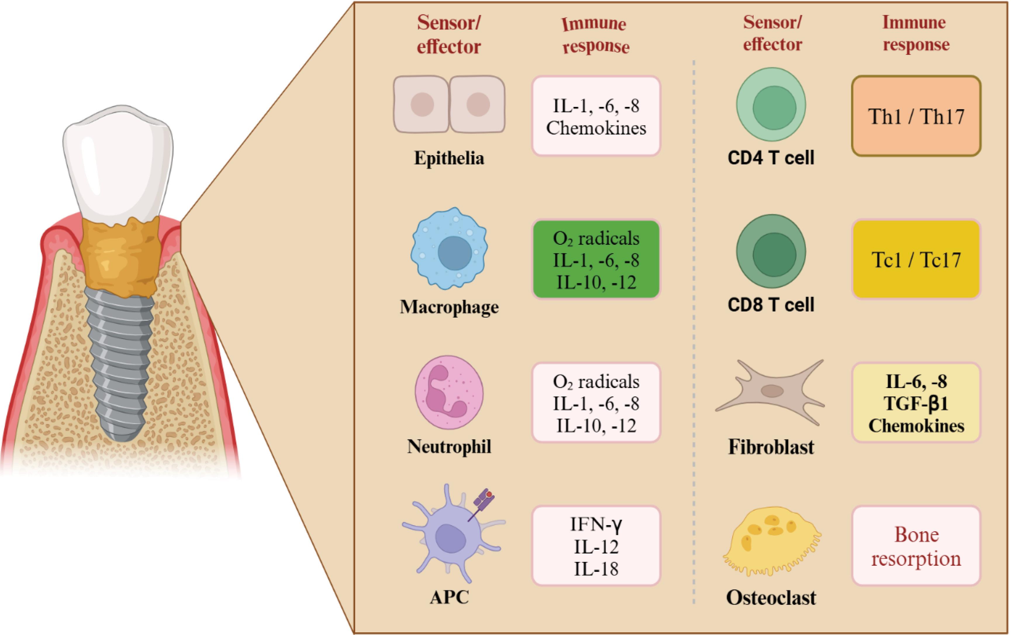
Fig. 1.
The diagram illustrates the immune response associated with peri-implantitis, highlighting the role of different sensor/effector cells and their corresponding immune mediators.
.
The diagram illustrates the immune response associated with peri-implantitis, highlighting the role of different sensor/effector cells and their corresponding immune mediators.
Ceramic based dental implants
Ceramic dental implants provide a better option of dental implants since they are biocompatible and contain no metals, they produce the best aesthetics as they are naturally coloured like teeth. However, contrary to conventional titanium implants, ceramic implants mainly made of yttria-stabilized zirconia (Y-TZP)-based are also beneficial to the patients with metal hypersensitivity or metal ion release concerns. Additional ceramic materials, including alumina (Al2O3), zirconia-toughened alumina (ZTA), HA, 3-tricalcium phosphate (3-TCP) and bioactive glass, have been identified as either a coating, a surface modification, or researchable but not as an implantation fixture on its own. Nanoparticles made of such materials are also used as ceramic nanoparticles on implant surfaces to boost the osseointegration, mechanical performance, and enhancement of long-term stability of restorations. Fig. 2 shows the experimental model employed to study the peri-implantitis formation and monitoring, which provides a biologically important model of evaluating the response of nano-engineered ceramic surfaces formed in inflamed peri-implant tissues.25,26
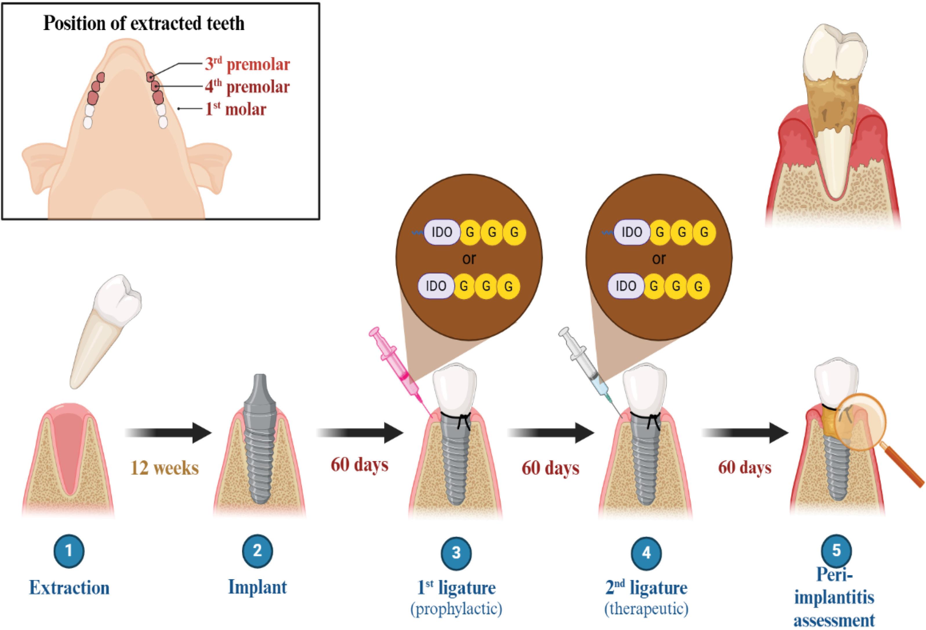
Fig. 2.
Experimental model of peri-implantitis development and assessment.
.
Experimental model of peri-implantitis development and assessment.
Polymer based dental implants
Polymers-based materials have notable advantages to the development of dental root and implants in comparison with other types of biomaterials such as customizable porosity, thermal and electrical neutrality, a high degree of biocompatibility, clinical ease of handling, and cost-effective manufacture. Being more elongated in their fractures, and having lower strength and lower elastic modulus, they are more likely to behave like soft tissues, alleviating stress shielding and enhancing patient comfort. Fig. 3 demonstrates the materials that are typically used in dental implants such as cobalt–chromium alloys, titanium and Ti6Al4V, and stainless steel and integration of PDMS-based microfluidic chips are used to study the interactions of implants and tissues.27 The dental implants manufactured using polymers are Polymethyl methacrylate (PMMA), Polytetrafluoroethylene (PTFE), polyamide and silicone. One of the latest materials is polyethers ether ketone (PEEK) that can boast of having elastic modulus (3.6 Gpa),28 comparable to bone and this is a major advantage over other conventional materials like titanium and zirconium.29
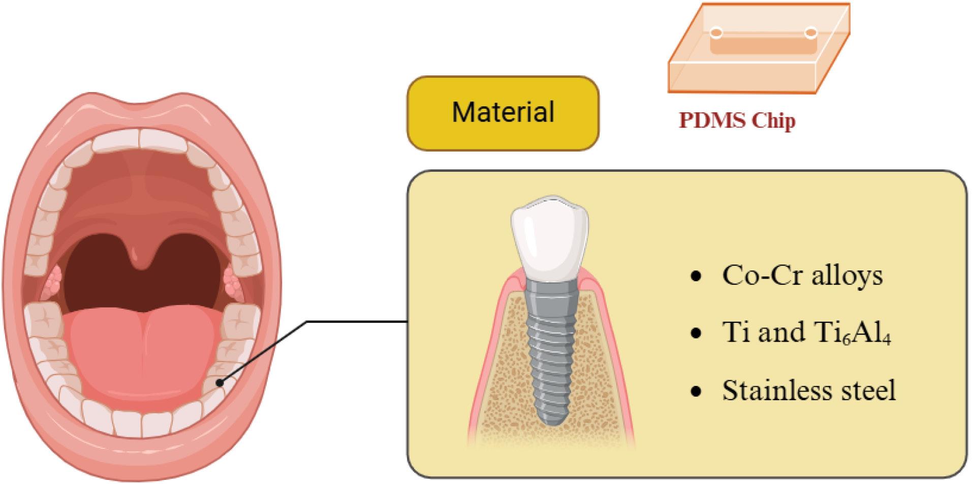
Fig. 3.
The materials commonly used for dental implants, including Co-Cr alloys, Ti and Ti₆Al₄, and stainless steel with PDMS chip.
.
The materials commonly used for dental implants, including Co-Cr alloys, Ti and Ti₆Al₄, and stainless steel with PDMS chip.
Nanoparticles
The nanoparticles have the ability to be used in a wide range of surface treatments of the dental implants including immunomodulation, osseointegration and soft-tissue integration, and antibiofouling. Different categories of nanoparticles which have been pursued to date in dental applications. These are metallic, ceramic and polymeric nanoparticles that have been used to improve the surface characteristic of implants, to give them antimicrobial properties or to allow them to deliver drugs locally. Their main features, modes of action, and the published results can be found in Table 1.30
Table 1.
Nanoparticle’s elements incorporated in dental implants
|
Prosthetic components
|
Construction
|
Key features
|
References
|
| Ti nanotubes |
Electrochemical
anodization |
Anti-inflammatory medications. |
31
|
| Silver nanoparticles |
Anodic spark
deposition |
Silver nanoparticles having antimicrobial properties, prevent bacterial contamination and provide biocompatible scaffolds |
32
|
| Gold nanoparticles |
Electro deposition
Sol-gel method |
Peri-implantitis disease. |
33
|
| Zinc nanoparticles |
Plasma electrolytic
oxidation |
Zinc nanoparticles have enhanced osteoblast proliferation and bone formation. |
34
|
| Copper nanoparticles |
Plasma electrolytic
Oxidation |
They can be used with a range of polymers and are stable and reasonably priced. They have been demonstrated to increase the bioactivity of the specified system and exhibit osteogenic, angiogenic, and persistent antimicrobial qualities. |
35
|
| Silica nanoparticles |
Hydrothermal
method |
Silica nanoparticles applied to tooth polishing, dental fillers, and hypersensitivity therapies. coating of silica applied to Ti-based implants to allow osseointegration and mainly serve as vessels for drug loading |
36
|
| Hydroxyapatite |
Electrophoretic
Deposition, |
Osteoconductivity, and good mechanical properties, improved stability at implants coated with Hydroxyapatite |
37
|
| Chitosan |
Microarc oxidized |
reducing the risk of infections |
38
|
| Carbon composites |
Meniscus-dragging
deposition |
Most promising materials due to their exceptional thermal, electrical, and mechanical characteristics. |
39
|
Silver (Ag)
Ag nanoparticles have also been explored widely in regard to dental applications.40 They have antimicrobial activity through two principal mechanisms direct and indirect. Silver ions attack and react in the direct mechanism with the bacterial cell wall and cytoplasmic membrane, resulting in oxidative stress and membrane destabilization, affecting organelle activity and cell growth and ultimately cell lysis.41 The indirect mechanism is associated with interference with DNA replication and generation of ROS that additionally lead to bacterial cell death.42
Gold
It is proved that there exist two different types of Au that have antibacterial properties. One of them is nanoporous gold (NPG), which has nanoscale open pores that have the potential of eliminating S. epidermidis and E. coli. The second type of nanoparticles is the gold nanoparticles (AuNPs). It is widely known that the antibacterial activity of AuNPs could be enhanced by their surface functionalization or conjugate preparation.
Zinc (Zn)
Zn is a vital trace element, which is present at all biological tissues and has antibacterial properties. Hu et al. used plasma electrolytic oxidation to impregnate TiO2 coating on titanium implants, which increased osteogenic activity and bactericidal effects.
Copper (Cu)
Copper nanoparticles provide an attractive choice of biomaterial use because they are rather cheap. According to Xia et al, titanium implants modified with copper nanoparticles had higher mechanical strength and better resistance to corrosion.43
Titanium (TiO₂) nanotubes
TiO₂ nanotubes have emerged as an effective platform for localized drug delivery due to their high surface area and drug-loading capacity. These nanostructures facilitate controlled and sustained release of therapeutic agents such as antibiotics and anti-inflammatory drugs.44 In patients with diabetes mellitus, implant success is often compromised due to hyperglycemia-induced oxidative stress, impaired osteoblast function, and delayed wound healing. TNT-modified implant surfaces can mitigate these challenges by enhancing protein adsorption, promoting osteoblast adhesion, and enabling localized drug delivery. Furthermore, multilayer coatings such as chitosan can be applied to regulate drug release kinetics, ensuring prolonged therapeutic effects and improved implant stability.45 Fig. 4 depicts the variety of nanoparticles applied in dental implant coating and surface modification, which gives a general idea of the existing methods of enhance the functionality and biological activity of implants.
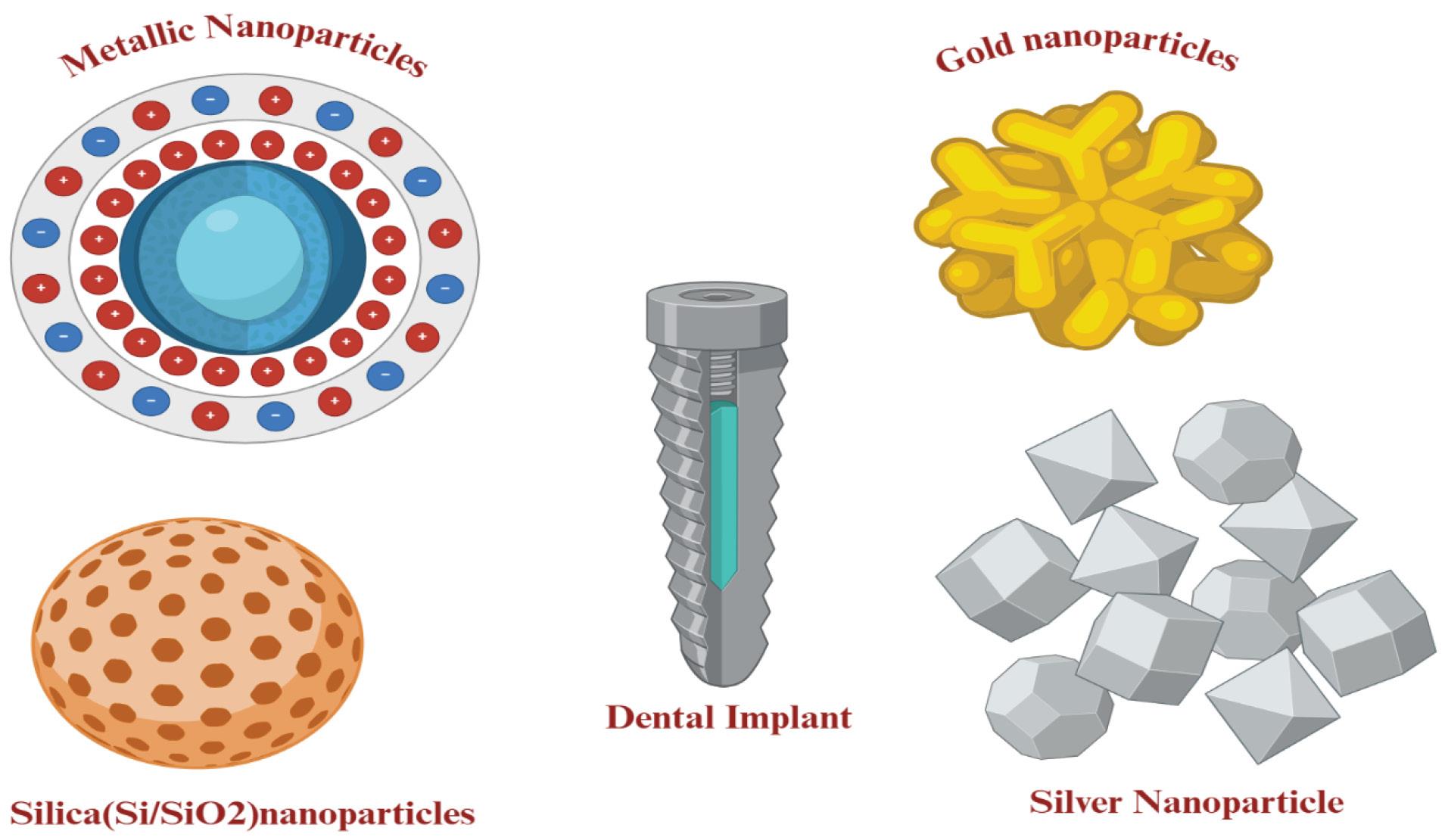
Fig. 4.
Various nanoparticles used in dental implant coatings and modifications.
.
Various nanoparticles used in dental implant coatings and modifications.
Silica (Si/SiO2) nanoparticles
Si nanoparticles have been applied in dental practice in the polishing of teeth, dental fillers and in hypersensitivity treatment. Silica nanoparticles are the inorganic nanomaterials that consist of silicon dioxide and they are either nonporous or mesoporous. Their biocompatibility is high, size and pore volume can be customized. Since the Si-OH groups may react with the biological fluids, it has been proved that the units are bioactive. Mesoporous silica nanoparticles are mainly used as drug loading vehicles. They may be applied together with the medicaments and other nanomaterials to stimulate the osteoblast differentiation and proliferation.46
Polymeric nanoparticles
Polymeric nanoparticleshave gained significant attention in dental implant applications due to their excellent biocompatibility, biodegradability, and ability to provide controlled drug release. Commonly used polymeric systems include poly(lactic-co-glycolic acid) (PLGA), chitosan, and polyethylene glycol (PEG)-based nanoparticles. These nanocarriers can encapsulate therapeutic agents such as antibiotics, anti-inflammatory drugs, and growth factors, enabling localized and sustained delivery at the implant site.47 PLGA-based nanoparticles are particularly advantageous due to their FDA-approved status and tunable degradation properties, allowing precise control over drug release kinetics. Chitosan nanoparticles exhibit inherent antimicrobial activity and mucoadhesive properties, which enhance their interaction with biological tissues. Similarly, PEGylated nanoparticles improve systemic stability and reduce immune recognition, thereby enhancing therapeutic efficiency.48 Compared to metallic nanoparticles, polymeric nanoparticles offer reduced cytotoxicity and improved safety profiles, although they may exhibit lower intrinsic antibacterial activity. Therefore, hybrid systems combining polymeric and metallic nanoparticles are increasingly being explored to achieve synergistic effects in improving osseointegration and preventing implant-associated infections.49
Hydroxyapatite
HA is a coating made of calcium phosphate, which is used on the surfaces of the implants, usually through the plasma spraying technique. It has a close chemical composition and structure to natural bone and this allows good interfacial bonding between the implant and the surrounding bone. The main advantages of HA coatings are high biocompatibility, osteoconductivity, and positive mechanical strength. Both clinical and experimental evidence have shown that primary stability of HA-coated implants has been improved and leads to a decrease in the time of healing and decrease in the overall treatment time. Regardless of all these benefits, traditional plasma-sprayed HA-based surfaces tended to produce a relatively thick (around 4050 μm) layer that had porous and non-uniform surface properties. These characteristics were correlated with such complications as marginal bone loss and, in certain instances, implant failure. In studies that utilized long-term outcomes to evaluate the success of these implants, they performed relatively lower.50 Moreover, first plasma-sprayed HA coatings had low adhesion performance in bone interfaces with implant, possible excretion of titanium particles into tissues, and poor interaction with antibiotics and other therapeutics. To address these shortcomings, a sophisticated fabrication approach was formulated and hence nano-hydroxyapatite (nano-HA) emerged. Nanocrystalline HA is similar to natural bone apatite crystals in size and form, which means it, is more bioactive on the surface and creates more implant reactivity. There are a number of methods by which Nano-HA can be produced with the most notable being electrodeposition and the wet chemical. These methods help to create very thin, uniform, and homogeneous surfaces, which act as bioactive scaffolds. These layers will enable the integration of other biological materials, such as antibiotics, growth factors and nanomaterials, such as graphene oxide and chitosan, to form multifunctional hybrid coating with better interfacial behaviour and enhanced biological incorporation.51
Carbon nanoparticles
Nanomaterials that are carbon based like graphene and graphene oxide have been explored to modify the surfaces of dental implants. Prepared by the physicochemical exfoliation of graphite. It has demonstrated that the implants, which are coated with graphene oxide, demonstrate enhanced osteogenic activity. Graphene oxide disclosed increased osteogenic differentiation, enhanced osteoblast activity and increased extracellular matrix deposition with no cytotoxic effect at all.52,53
Nanotechnologies in surface modification of titanium dental implants
The significance of nano-scale surface modification is because it has a great impact on the morphology of human tissues that do possess several nanostructured elements. These nanotubes and other micro and nanostructures are very instrumental in the control of cellular behaviour and initial bone formation surrounding the implant materials. Consequently, there has been an increased focus on fabricating integrated nano-topographies with superior and innovative technologies. These nanostructures promote osteogenic reaction and protein adsorption, which stimulate the osseointegration and improve the working of the implant (Fig. 5). Currently, some methods are used to produce nano-scale surfaces on TiO₂ implants.54 Besides new nanotechnological approaches, the traditional macro- and micro-scale techniques may also be adapted to obtain nano-level characteristics through a careful optimization of the process parameters. Grit blasting, acid etching and selective laser melting (SLA) techniques are often adopted and classified as nano-surface modification techniques when modified accordingly. Acid etching especially sandblasted large grit acid-etched (SLA) treatment was initially created to remove manufacturing contaminants and create roughness on an implant surface at micro- or nano-scales. Since the exact regulation of the topography of implants is needed, a commonly used clinical practice is the combination of sandblasting and acid etching. It has been shown that the SLA-treated implants lead to accelerated healing of the implantation process (osseointegration in one or two months) because of the micro- and nano-roughened surfaces. This physical-chemical approach to surface modification has been shown to be of great success in enhancing the surface of dental implants and its long-term outcomes have been a success both in the experimental and clinical fields. It should be noted, though, that surface properties of SLA could differ across the manufacturers of various types of implants, which makes them variable and comparatively challenging. Ti dental implants are surface engineered on a regular basis to reduce corrosion, increase implantation of the surface bone, and alter biocompatibility. In this respect, protective surface layer is formed to restrict oxidation and inhibit the release of metal ions in underlying layers. The strategies of surface modification can also strengthen the stability and passivation of the TiO2 layer, eventually leading to increased durability, corrosion resistance, and long-term success of implants.55 In patients with diabetes mellitus, implant success is often compromised due to impaired wound healing, chronic inflammation, and reduced osteoblastic activity caused by persistent hyperglycemia.56 These factors lead to delayed osseointegration and increased risk of implant failure. TNTs have emerged as a promising surface modification strategy to overcome these limitations by enhancing biological interactions at the implant interface. The unique nanostructured architecture of TNTs increases surface area, facilitating improved protein adsorption and promoting osteoblast adhesion and proliferation. TNTs serve as effective reservoirs for the localized delivery of therapeutic agents such as anti-inflammatory drugs, antibiotics, and growth factors. This controlled drug release capability is particularly beneficial in diabetic conditions, where infection risk and inflammatory responses are elevated. Furthermore, TNT-modified surfaces have been shown to modulate cellular signalling pathways, reduce oxidative stress, and enhance bone regeneration, thereby improving implant stability and success rates in compromised metabolic conditions. These properties make TNTs a valuable approach for improving clinical outcomes in diabetic patients.57
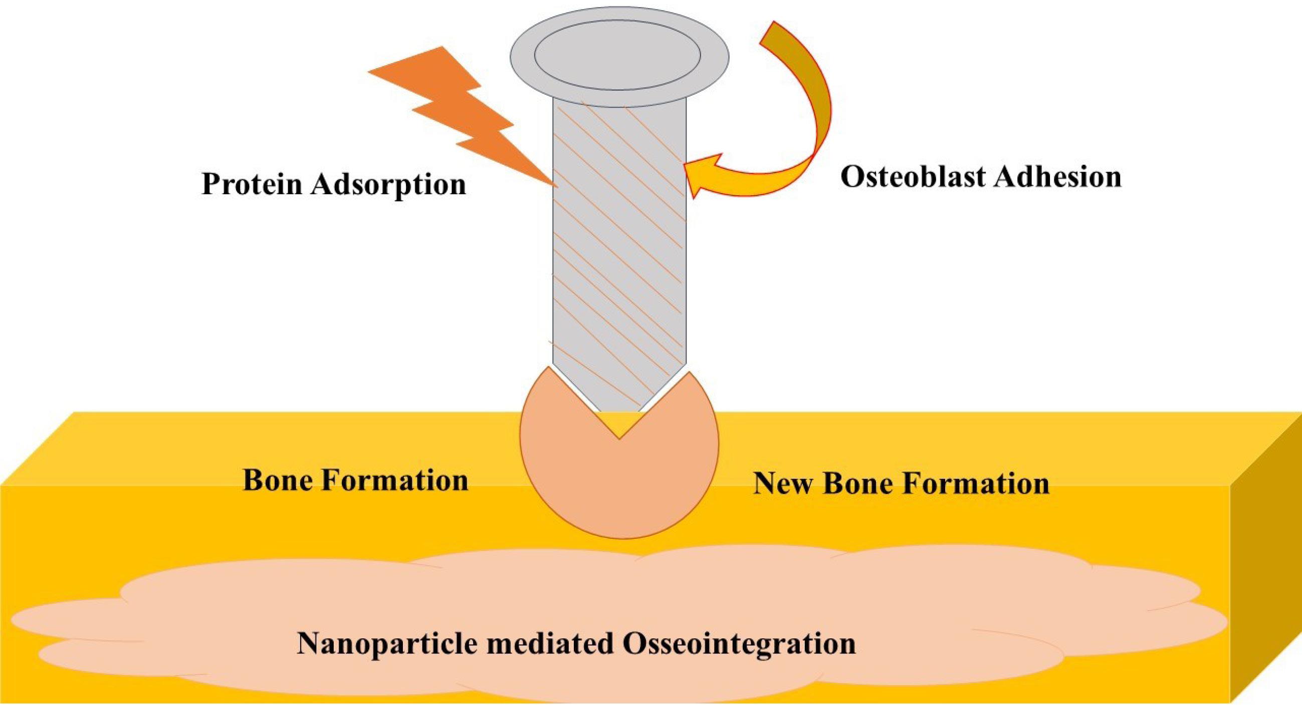
Fig. 5.
Schematic illustration of nanoparticle-mediated osseointegration, highlighting enhanced protein adsorption and cell adhesion, osteoblast adhesion, cellular proliferation, and improved bone–implant interface formation, ultimately leading to increased implant stability and long-term success.
.
Schematic illustration of nanoparticle-mediated osseointegration, highlighting enhanced protein adsorption and cell adhesion, osteoblast adhesion, cellular proliferation, and improved bone–implant interface formation, ultimately leading to increased implant stability and long-term success.
Techniques of inserting nanoparticles in dental implants
Nanoscale surface modification dental implants are common in the contemporary implant therapy. Nanotechnology has immensely contributed to the development of novel implant tissue interfaces that have improved the performance of the implants in its different surface engineering methods. These nano-modified surfaces enhance the adhesion of osteoblast, increase the rate of osseointegration and can offer the added antimicrobial or bioactivity which finally leads to the stability of the implants in the long term and clinical success.58
Nanoscale modification methods and coating of the implant surfaces
Nano-engineering of dental implants is made with high-level techniques. Those techniques are plasma treatment, which adjusts the energy of the surface to enhance cell adhesion; micro-machining and polishing/grinding, which generates fine surface texture; particle blasting and chemical etching, which introduces both micro- and nanotopographies; and electrochemical anodization, which forms controlled oxide layers with nanotopographies. Taken together, these methods are intended to improve on the conventional techniques of micro-roughness to enhance better results of osseointegration and implant life.59,60
Mechanical modification
Machining, blasting, polishing and attrition- are used to form nanoscale layers, which can increase the mechanical properties, such as hardness and wettability. Particles of Abrasive Blasting on the implant surface may enhance surface reactivity.61
Chemical modification
Surface chemistry modification on dental implants does not only modify their topography but enables incorporation of a certain chemical group, which makes them more bioactive and integrative of other tissues.62 Osteogenic potential of implants is improved as surface oxide layers ranging between several nanometers and micrometres are applied, whereas processes such as sol-gel processing and chemical vapour deposition are further used to increase their bioactivity.63
Physical modification
The coating of certain molecules or ions to the surface of the implants. Incident on Ti implants, thermal or plasma spraying including in the coating of hydroxyapatite, calcium silicate, alumina, zirconia and titania, to enhance their wear and corrosion resistance and bioactivity.63
Electrochemical modification
The formation of TiO2 layers of 10-40 nm is enhanced by the anodic oxidation.64
Biomolecule modification
The biological addition of dental implants with bioactive molecules, including collagen or peptides, has been revealed to increase bone-implant interactions.65
Differentiation of preclinical and clinical evidence
Current evidence supporting nanoparticle-enhanced dental implant systems is predominantly derived from preclinical investigations, and careful differentiation between in vitro, in vivo, and clinical findings is essential to avoid over interpretation. In vitro studies consistently demonstrate that nanoparticle-modified surfaces improve antibacterial activity, reduce biofilm formation, enhance osteoblast adhesion, and modulate inflammatory markers under controlled laboratory conditions. These experiments provide valuable mechanistic insights into ion release, ROS generation, protein adsorption dynamics, and cell–surface interactions.66 In vivo animal studies further suggest improved bone implant contact, enhanced new bone formation, reduced peri-implant inflammation, and improved mechanical stability in experimental models. However, while these findings are promising, they do not necessarily predict equivalent outcomes in humans due to biological variability, systemic influences, and long-term biomechanical factors. Clinical evidence remains comparatively limited. Only a restricted number of human studies have evaluated nano-engineered implant surfaces, and most report short-term outcomes with heterogeneous methodologies and follow-up periods.67 Long-term randomized controlled trials assessing survival rates, peri-implant bone loss, inflammatory parameters, and safety profiles are still scarce. From a regulatory standpoint, certain nano-roughened titanium surfaces have received approval as modifications of conventional implants; however, multifunctional nanoparticle systems particularly those involving drug-eluting platforms—face more complex regulatory pathways due to combined device–drug classification. Therefore, although preclinical data strongly support the biological potential of nanoparticle-based implant modifications, definitive conclusions regarding long-term clinical efficacy and safety require further robust human investigations and standardized evaluation protocols.68
Challenges and limitation
Although the development of implant has enabled the successful results of the clinical outcome to be enhanced, there are systemic and local ailments that may negatively affect the success of implants. The existence of systemic conditions as if uncontrolled diabetes, immunosuppression, recent myocardial infarction or stroke, and the use of glucocorticoids over a prolonged period may inhibit healing and osseointegration and may contraindicate the implant placement. Osteogenesis imperfect and osteoporosis are bone diseases that can impair the regeneration of alveolar bone and medications like corticosteroids or antiresorptive medications can contribute to a decreased quality of bone. The patient-related risk factors, including the presence of periodontitis in the history, have been found to lower the survival rates of implants and predispose people to the peri-implant disease. To overcome such issues, there is on-going research on titanium implant surfaces that are covered or altered with bactericidal nanoparticles, such as silver, copper and zinc, to prevent or control peri-implant disease. Although good results were seen in preclinical studies, clinical translation of nanoengineered titanium dental implants has been hard, since there are concerns of coating stability, long-term safety and regulatory approval. One of the greatest challenges in the use of nanoparticles in therapeutic applications is to maximize drug release profiles in order to: (i) assess cellular uptake and toxicity of released agents; (ii) attain long term, sustained, and controlled in vivo release; and (iii) measure potential toxicity of burst release or nanoparticle-derived metal ions. Important aspects affecting bioactivity, such as pore size and nanotopography have been established. Future studies will be directed towards the research of mechanotransduction of nanotubes on cellular systems, and the initial cell adhesion on surface of nano-engineered materials, to improve the osseointegration and implantation efficiencies.69-72 When a drug-releasing implant is placed, a number of cells rush to invade the position and often the nanotopography is immediately covered in cells and proteins, which may block the open pores. This could have an impact, considering the fact that medication release is based on a diffusion gradient, which is impeded in an insufficient perfusion within the bone microenvironment. The in-vitro and in silico simulation of these conditions may be difficult especially considering the fact that even in healthy individuals, surgical placement could result in damage. Consequently, the efficacy of the drug releasing implants has to be tested in the real injured tissue in vivo depending on the ex vivo identified therapeutic need.73 Two overriding concerns of using nano and biomaterials are safety and biocompatibility. Although the biocompatibility of these materials is well researched, there is not much information about their long-term effects and possible adverse response in the complex environment of the oral cavity. The sphere of nanotechnology and the use of biomaterials in the delivery of drugs into dental implants lacks standardized procedures and regulatory frameworks.74 The absence of generally accepted criteria may inhibit the smooth relation between research and clinical practice and the reproducibility of studies. Long-term clinical trials with large samples are not numerous, and their findings may not be able to fully explain the diversity of patients and their varying oral health issues. The stability of nanoparticle-engineered dental implants is a major factor that determines their clinical success since the stability of surface coating has a direct effect on the rate of osseointegration, mechanical performance, and resistance to peri-implant disease. Continuous loading of functionality, variation in pH and enzymatic degradation in the mouth cavity should not ruin nanoparticle’s coatings. Nanoparticles that are based on ceramics and oxides, e.g., TiO2 or ZrO2, generally show very high stability over time and have low solubility, whereas metallic nanoparticles, including silver or copper, need to be controlled in terms of their dose and release-profile because they can be dissolved over time. Biodegradable polymeric coatings have regulated routes of degradation but have to be designed to avoid the early loss of mechanical support.75 To translate the nanoparticle-engineered dental implants produced at the experimental research levels to the routine clinical practice, regulatory and clinical compliance is necessary. Any nano-modified implant systems should follow developed international standards, such as, ISO 10993 standards of biocompatibility, cytotoxicity, genotoxicity, and immunological evaluation, and ISO 14801 of mechanical stability under cyclic loading. Before clearance to the market (or CE marking) is given to a drug, regulatory bodies like the U.S. FDA or European Medicines Agency (EMA) demand detailed information on material safety, coating stability, ion-release profiles and possible adverse long-term effects of any drug. Nano-surface technologies have been clinically available (nano-sand-blasted, acid-etch (SLA)) and commercially (NanoTite-2) available that have shown regulatory approval under pre-clinical assessment, human clinical trials and post-market surveillance. It is thus important to establish strong regulatory channels and open reporting regulations in clinical domains so that advanced nanoparticle modified dental implants are deployed in a responsible manner.76
Future directions
Future development of nano-engineered dental implants is likely to involve the development of intelligent, bio-interactive system beyond passive drug release and structure where the dental implants are engineered to be biologically interactive. This mechanistic interaction of environmental signals with regulated therapeutic response is one of the greatest steps towards specificity in prevention of peri-implant disease. The recent advancements in biomaterials have been in the improvement of personalized drug delivery strategy, which has led to an improved clinical outcome with reduced side effects in the system. This is due to novel bioactive implant surfaces that exhibit osteopromotive behavior even compromised conditions (osteoporosis) or aging bone loss. Silica nanoparticles, titanium nanotubes and other nanocarriers have been used in the delivery of localized therapy, biological or gene therapy, where release is controlled by biopolymers to provide sustained treatment effect. In addition, the natural anti-bacterial effect of most metal nanoparticles offers a secondary protection against peri-implantitis, breaking down initial biofilm formation and reducing the bacteria load. It is also important to ensure corrosion resistance to avoid the ion leakage and inflammatory response in the peri-implant tissues. Although there is promise, existing drug-impregnated implant systems are prone to short term burst release that lasts a mere one to two months and hence the need to employ the long run, mechanistically regulated drug delivery models. The designs of the future need to be combined with stable nanotopographies and with smart release systems that will sustain therapeutic efficacy in the end. This is due to the fact that additive manufacturing provides novel options of custom, patient specific implants, and that growth factor, stem cells and regenerative agents delivery can be targeted to improve tissue regeneration and implant stability in the long term (Table 2).77-80
Table 2.
Overview of clinical evidence on nanomaterial-modified dental implants
|
Nanomaterial Used
|
Study Type
|
Key Findings
|
Limitations
|
| Silver nanoparticles |
Clinical trial |
Significant reduction in peri-implant bacterial load and inflammation |
Short follow-up duration81 |
| Titanium nanotubes |
Human study |
Enhanced osseointegration and improved implant stability |
Small sample size82 |
| Zinc oxide nanoparticles |
Clinical evaluation |
Improved antibacterial activity and reduced infection rates |
Limited long-term data83 |
| Gold nanoparticles |
Pilot study |
Promoted osteogenic activity and faster healing |
High cost and scalability issues84 |
| Polymeric nanoparticles (PLGA) |
Clinical study |
Controlled drug release and improved tissue response |
Complex fabrication process85 |
A comparative evaluation of different nanoparticle systems highlights significant variations in their biological performance and clinical applicability. Metallic nanoparticles such as silver, zinc oxide, and gold exhibit strong antibacterial properties primarily due to their ability to generate ROS, disrupt bacterial membranes, and inhibit biofilm formation. Among these, silver nanoparticles demonstrate superior antimicrobial efficacy; however, concerns regarding cytotoxicity at higher concentrations remain a challenge. Zinc oxide nanoparticles offer a balance between antibacterial activity and biocompatibility, making them suitable for implant surface modification. Gold nanoparticles, on the other hand, are known for their excellent biocompatibility and ability to promote osteogenic differentiation, although their antibacterial effects are relatively moderate and their high-cost limits large-scale application. In contrast, polymeric nanoparticles such as PLGA and chitosan provide controlled drug delivery with minimal toxicity, but lack inherent antimicrobial properties. Therefore, the selection of nanoparticle systems should be carefully optimized based on the desired balance between antibacterial efficacy, biocompatibility, and long-term safety. Importantly, nanoparticle-induced toxicity is dose-dependent, where lower concentrations promote beneficial cellular responses, while higher concentrations may induce oxidative stress, inflammation, and cytotoxic effects. This highlights the need for precise dose optimization and long-term safety evaluation to ensure successful clinical translation.
Conclusion
Nanostructured dental implants represent a transformative advancement in modern implantology by integrating structural and therapeutic functionalities at the nanoscale. The incorporation of nanoparticles, including metallic and polymeric systems, has demonstrated significant potential in enhancing osseointegration, improving antibacterial properties, and enabling localized drug delivery. These nanoscale modifications not only increase surface area and protein adsorption but also promote osteogenic activity and reduce the risk of peri-implant infections. Comparative analysis indicates that while metallic nanoparticles such as silver and zinc oxide provide strong antimicrobial effects, concerns related to cytotoxicity and long-term safety must be carefully addressed. In contrast, polymeric nanoparticles offer superior biocompatibility and controlled drug release, although their intrinsic antibacterial activity is limited. The development of hybrid nanoparticle systems may provide an optimal balance between efficacy and safety. Despite promising preclinical and early clinical outcomes, several challenges hinder the widespread clinical translation of nanoparticle-modified implants. These include issues related to dose optimization, long-term toxicity, large-scale manufacturing, cost-effectiveness, and regulatory approval. Addressing these challenges requires interdisciplinary research efforts and standardized evaluation protocols. Future perspectives in this field include the development of smart and multifunctional implant systems capable of responding to the local microenvironment, personalized treatment approaches, and advanced surface engineering techniques. With continued advancements, nanotechnology-based dental implants hold great promise for improving patient outcomes and revolutionizing the field of regenerative dentistry.
Review Highlights
-
Nanostructured dental implants enhance osseointegration by improving surface roughness, protein adsorption, and cellular adhesion.
-
This paper highlights nanostructured implants as dual-functional platforms combining structural integration with controlled therapeutic release.
Competing Interests
None to be declared.
Declaration of AI-assisted Tools in the Writing Procedure
The authors declare that AI assisted tools (such as QuillBot) were used solely for language refinement, grammar correction, and improvement of clarity and readability of the manuscript. The AI tools did not generate scientific content, interpret data, perform analyses, or influence the study design, results, or conclusions. All intellectual content, data interpretation, and scientific responsibility remain entirely with the authors.
Ethical Approval
Not applicable.
Acknowledgements
The author’s expresses his gratitude toward Hon. Chancellor, Bioorganic and Medicinal Chemistry Research Laboratory, Department of Pharmaceutical Sciences, Sam Higginbottom University of Agriculture, Technology and Sciences, Prayagraj, 211007, India.
References
- Ghodrati H, Goodarzi A, Golrokhian M, Fattahi F, Anzabi RM, Mohammadikhah M. A narrative review of recent developments in osseointegration and anti-corrosion of titanium dental implants with nano surface. Bone Rep 2025; 25:101846. doi: 10.1016/j.bonr.2025.101846 [Crossref] [ Google Scholar]
- Li Y, Han X, Ma Z. Research on the mechanical properties of PEEK material artificial bone implants fabricated by high-temperature air-assisted 3D printing. J Mech Behav Biomed Mater 2026; 173:107207. doi: 10.1016/j.jmbbm.2025.107207 [Crossref] [ Google Scholar]
- Sun F, Zhang J, Liu T, Yao H, Wang L, Meng H. A versatile microporous design toward toughened yet softened self-healing materials. Adv Mater 2024; 36:e2410650. doi: 10.1002/adma.202410650 [Crossref] [ Google Scholar]
- Frączek W, Kotela A, Kotela I, Grodzik M. Nanostructures in orthopedics: advancing diagnostics, targeted therapies, and tissue regeneration. Materials (Basel) 2024; 17:6162. doi: 10.3390/ma17246162 [Crossref] [ Google Scholar]
- Gallagher JE, Guarnizo-Herreño C, Kavanagh D, Makino Y, Mathur M, Mossey P. Global oral health: defining the research agenda in the public interest. J Dent Res 2026; 105:176-82. doi: 10.1177/00220345251372941 [Crossref] [ Google Scholar]
- Wang Y, Xu Y, Song J, Liu X, Liu S, Yang N. Tumor cell-targeting and tumor microenvironment-responsive nanoplatforms for the multimodal imaging-guided photodynamic/photothermal/chemodynamic treatment of cervical cancer. Int J Nanomedicine 2024; 19:5837-58. doi: 10.2147/ijn.S466042 [Crossref] [ Google Scholar]
- Hao X, Jiang B, Wu J, Xiang D, Xiong Z, Li C. Nanomaterials for bone metastasis. J Control Release 2024; 373:640-51. doi: 10.1016/j.jconrel.2024.07.067 [Crossref] [ Google Scholar]
- Luo J, Cui Y, Xu L, Zhang J, Chen J, Li X. Layered double hydroxides for regenerative nanomedicine and tissue engineering: recent advances and future perspectives. J Nanobiotechnology 2025; 23:370. doi: 10.1186/s12951-025-03448-1 [Crossref] [ Google Scholar]
- Wang S, Ma D, Yang M, Zhang Y, Wang S, Zhou W. Arsenic trioxide-based nanoparticles for enhanced chemotherapy by activating pyroptosis. Acta Pharm Sin B 2025; 15:6001-18. doi: 10.1016/j.apsb.2025.08.003 [Crossref] [ Google Scholar]
- Wang H, Tang C, Zhang H, Guo L, Zou C, Zhang S. A biocompatible tea polyphenol nanoplatform for efficient cytosolic delivery of protein therapeutics. J Control Release 2025; 388:114347. doi: 10.1016/j.jconrel.2025.114347 [Crossref] [ Google Scholar]
- Hu M, Chen H, Wang R, Zhang R, Yao J, Qiu X. Global, regional, and national burden of lip and oral cavity cancer and its attributable risk factors from 1990 to 2021: an analysis of the Global Burden of Disease study 2021. Int J Clin Pharm 2026; 48:93-106. doi: 10.1007/s11096-025-01961-9 [Crossref] [ Google Scholar]
- Anyikwa CL, Brennan PA, Ogwo CE. Why oral health deserves a seat at the global health table. J Oral Pathol Med 2026; 55:168-70. doi: 10.1111/jop.70065 [Crossref] [ Google Scholar]
-
Alias R, Abdullah SH, Fatah IYA, Pargi MN, Akhbar MF, Todoh M, et al. Introduction to the implant nanoscaled advanced materials. In: Behera A, Patra JK, eds. Advanced Nanomaterials in Biomedical Implants: Processing, Structures, Properties and, Applications. Elsevier; 2025. p. 3-28. doi: 10.1016/b978-0-443-27378-0.00001-5.
- Rani VV, Vinoth-Kumar L, Anitha VC, Manzoor K, Deepthy M, Shantikumar VN. Osteointegration of titanium implant is sensitive to specific nanostructure morphology. Acta Biomater 2012; 8:1976-89. doi: 10.1016/j.actbio.2012.01.021 [Crossref] [ Google Scholar]
- Losic D. Advancing of titanium medical implants by surface engineering: recent progress and challenges. Expert Opin Drug Deliv 2021; 18:1355-78. doi: 10.1080/17425247.2021.1928071 [Crossref] [ Google Scholar]
- Dohan Ehrenfest DM, Coelho PG, Kang BS, Sul YT, Albrektsson T. Classification of osseointegrated implant surfaces: materials, chemistry and topography. Trends Biotechnol 2010; 28:198-206. doi: 10.1016/j.tibtech.2009.12.003 [Crossref] [ Google Scholar]
- Jeevanandam J, Barhoum A, Chan YS, Dufresne A, Danquah MK. Review on nanoparticles and nanostructured materials: history, sources, toxicity and regulations. Beilstein J Nanotechnol 2018; 9:1050-74. doi: 10.3762/bjnano.9.98 [Crossref] [ Google Scholar]
- Gupta A, Dhanraj M, Sivagami G. Status of surface treatment in endosseous implant: a literary overview. Indian J Dent Res 2010; 21:433-8. doi: 10.4103/0970-9290.70805 [Crossref] [ Google Scholar]
- Cheng B, Niu Q, Cui Y, Jiang W, Zhao Y, Kong L. Effects of different hierarchical hybrid micro/nanostructure surfaces on implant osseointegration. Clin Implant Dent Relat Res 2017; 19:539-48. doi: 10.1111/cid.12471 [Crossref] [ Google Scholar]
- Thakral G, Thakral R, Sharma N, Seth J, Vashisht P. Nanosurface - the future of implants. J Clin Diagn Res 2014; 8:Ze07-10. doi: 10.7860/jcdr/2014/8764.4355 [Crossref] [ Google Scholar]
- Gaviria L, Salcido JP, Guda T, Ong JL. Current trends in dental implants. J Korean Assoc Oral Maxillofac Surg 2014; 40:50-60. doi: 10.5125/jkaoms.2014.40.2.50 [Crossref] [ Google Scholar]
- Mair LH. Wear in dentistry--current terminology. J Dent 1992; 20:140-4. doi: 10.1016/0300-5712(92)90125-v [Crossref] [ Google Scholar]
- Osman RB, Swain MV. A critical review of dental implant materials with an emphasis on titanium versus zirconia. Materials (Basel) 2015; 8:932-58. doi: 10.3390/ma8030932 [Crossref] [ Google Scholar]
- Saini M, Singh Y, Arora P, Arora V, Jain K. Implant biomaterials: a comprehensive review. World J Clin Cases 2015; 3:52-7. doi: 10.12998/wjcc.v3.i1.52 [Crossref] [ Google Scholar]
- Alkhawaldeh AK, Rheima AM, Kadhim MM, Abbas ZS, Al-Bayati AD, Talib Abed Z. Nanomaterials as transmitters of non-viral gene vectors: a review. Case Stud Chem Environ Eng 2023; 8:100372. doi: 10.1016/j.cscee.2023.100372 [Crossref] [ Google Scholar]
- Zheng W, Wu D, Zhang Y, Luo Y, Yang L, Xu X. Multifunctional modifications of polyetheretherketone implants for bone repair: a comprehensive review. Biomater Adv 2023; 154:213607. doi: 10.1016/j.bioadv.2023.213607 [Crossref] [ Google Scholar]
- Pye AD, Lockhart DE, Dawson MP, Murray CA, Smith AJ. A review of dental implants and infection. J Hosp Infect 2009; 72:104-10. doi: 10.1016/j.jhin.2009.02.010 [Crossref] [ Google Scholar]
- Edelmann A, Riedel L, Hellmann R. Realization of a dental framework by 3D printing in material cobalt-chromium with superior precision and fitting accuracy. Materials (Basel) 2020; 13:5390. doi: 10.3390/ma13235390 [Crossref] [ Google Scholar]
- Niinomi M. Mechanical properties of biomedical titanium alloys. Mater Sci Eng A 1998; 243:231-6. doi: 10.1016/s0921-5093(97)00806-x [Crossref] [ Google Scholar]
- Baltatu MS, Vizureanu P, Sandu AV, Florido-Suarez N, Saceleanu MV, Mirza-Rosca JC. New titanium alloys, promising materials for medical devices. Materials (Basel) 2021; 14:5934. doi: 10.3390/ma14205934 [Crossref] [ Google Scholar]
- Akbari Edgahi M, Naghib SM, Emamian A, Ramezanpour H, Haghiralsadat F, Tofighi D. A practical review over surface modification, nanopatterns, emerging materials, drug delivery systems, and their biophysiochemical properties for dental implants: recent progresses and advances. Nanotechnol Rev 2022; 11:637-79. doi: 10.1515/ntrev-2022-0037 [Crossref] [ Google Scholar]
- Luo R, Jiao Y, Zhang S, Wu J, Wu X, Lu K. Fabrication, properties and biological activity of a titanium surface modified with zinc via plasma electrolytic oxidation. Front Mater 2023; 10:1202110. doi: 10.3389/fmats.2023.1202110 [Crossref] [ Google Scholar]
- Pellosi DS, Paiva GS, Vital VG, Mendes AL, Santos NG, Kuriki FK. Harnessing copper nanoparticles for antimicrobial applications: advances and challenges. Antibiotics (Basel) 2025; 14:1170. doi: 10.3390/antibiotics14111170 [Crossref] [ Google Scholar]
-
Kawatra T, Gupta D, Gupta TK, Verma R. Role of nanomaterials in implant dentistry. In: Garg S, Chandra A, Sagadevan S, eds. Emerging Sustainable Nanomaterials for Biomedical Applications. Cham: Springer; 2024. p. 385-408. doi: 10.1007/978-3-031-63961-6_15.
- Asgari N, Rajabi M. Enhancement of mechanical properties of hydroxyapatite coating prepared by electrophoretic deposition method. Int J Appl Ceram Technol 2021; 18:147-53. doi: 10.1111/ijac.13638 [Crossref] [ Google Scholar]
- Zhou R, Zhou Y, Cheng J, Cao J, Li M, Yu H. Surface configuration of microarc oxidized Ti with regionally loaded chitosan hydrogel containing ciprofloxacin for improving biological performance. Mater Today Bio 2022; 16:100380. doi: 10.1016/j.mtbio.2022.100380 [Crossref] [ Google Scholar]
- Kang MS, Lee JH, Hong SW, Lee JH, Han DW. Nanocomposites for enhanced osseointegration of dental and orthopedic implants revisited: surface functionalization by carbon nanomaterial coatings. J Compos Sci 2021; 5:23. doi: 10.3390/jcs5010023 [Crossref] [ Google Scholar]
- Shah SA, Sohail M, Nakielski P, Rinoldi C, Zargarian SS, Kosik-Kozioł A. Integrating micro- and nanostructured platforms and biological drugs to enhance biomaterial-based bone regeneration strategies. Biomacromolecules 2025; 26:140-62. doi: 10.1021/acs.biomac.4c01133 [Crossref] [ Google Scholar]
- Wang Q, Huang Y, Qian Z. Nanostructured surface modification to bone implants for bone regeneration. J Biomed Nanotechnol 2018; 14:628-48. doi: 10.1166/jbn.2018.2516 [Crossref] [ Google Scholar]
- Priyadarsini S, Mukherjee S, Mishra M. Nanoparticles used in dentistry: a review. J Oral Biol Craniofac Res 2018; 8:58-67. doi: 10.1016/j.jobcr.2017.12.004 [Crossref] [ Google Scholar]
- Miyazawa N, Hakamada M, Mabuchi M. Antimicrobial mechanisms due to hyperpolarisation induced by nanoporous Au. Sci Rep 2018; 8:3870. doi: 10.1038/s41598-018-22261-5 [Crossref] [ Google Scholar]
- Rajchakit U, Sarojini V. Recent developments in antimicrobial-peptide-conjugated gold nanoparticles. Bioconjug Chem 2017; 28:2673-86. doi: 10.1021/acs.bioconjchem.7b00368 [Crossref] [ Google Scholar]
- Yuan JH, Chen Y, Zha HX, Song LJ, Li CY, Li JQ. Determination, characterization and cytotoxicity on HELF cells of ZnO nanoparticles. Colloids Surf B Biointerfaces 2010; 76:145-50. doi: 10.1016/j.colsurfb.2009.10.028 [Crossref] [ Google Scholar]
- Kulshrestha S, Khan S, Meena R, Singh BR, Khan AU. A graphene/zinc oxide nanocomposite film protects dental implant surfaces against cariogenic Streptococcus mutans. Biofouling 2014; 30:1281-94. doi: 10.1080/08927014.2014.983093 [Crossref] [ Google Scholar]
- Mascarenhas R, Hegde S, Manaktala N. Chitosan nanoparticle applications in dentistry: a sustainable biopolymer. Front Chem 2024; 12:1362482. doi: 10.3389/fchem.2024.1362482 [Crossref] [ Google Scholar]
- Stevanović MM, Qian K, Huang L, Vukomanović M. PLGA-based co-delivery nanoformulations: overview, strategies, and recent advances. Pharmaceutics 2025; 17:1613. doi: 10.3390/pharmaceutics17121613 [Crossref] [ Google Scholar]
- Mahmudi H, Adili-Aghdam MA, Shahpouri M, Jaymand M, Amoozgar Z, Jahanban-Esfahlan R. Tumor microenvironment penetrating chitosan nanoparticles for elimination of cancer relapse and minimal residual disease. Front Oncol 2022; 12:1054029. doi: 10.3389/fonc.2022.1054029 [Crossref] [ Google Scholar]
-
Gulati K, Ivanovski S. Dental implants modified with drug releasing titania nanotubes: therapeutic potential and developmental challenges. Expert Opin Drug Deliv2017. 14: 1009-24. doi: 10.1080/17425247.2017.1266332.
-
Overgaard S. Degradation of calcium phosphate coatings and bone substitutes. In: Di Silvio L, ed. Cellular Response to Biomaterials. Woodhead Publishing. 2009. p. 560-71. doi: 10.1533/9781845695477.3.560.
- Hrir H, Boudouma A, Layachi OA, Harrati A, Hsissou R, Khoumri E. An in-depth review of corrosion pathways in titanium dental and orthopedic implants and their biological interactions. Biomed Mater Devices 2026; 4:446-466. doi: 10.1007/s44174-025-00244-9 [Crossref] [ Google Scholar]
- Di Carlo R, Di Crescenzo A, Pilato S, Ventrella A, Piattelli A, Recinella L. Osteoblastic differentiation on graphene oxide-functionalized titanium surfaces: an in vitro study. Nanomaterials (Basel) 2020; 10:654. doi: 10.3390/nano10040654 [Crossref] [ Google Scholar]
- Gulati K, Hamlet SM, Ivanovski S. Tailoring the immuno-responsiveness of anodized nano-engineered titanium implants. J Mater Chem B 2018; 6:2677-89. doi: 10.1039/c8tb00450a [Crossref] [ Google Scholar]
- Sánchez-Salcedo S, García A, González-Jiménez A, Vallet-Regí M. Antibacterial effect of 3D printed mesoporous bioactive glass scaffolds doped with metallic silver nanoparticles. Acta Biomater 2023; 155:654-66. doi: 10.1016/j.actbio.2022.10.045 [Crossref] [ Google Scholar]
- Griger S, Sands I, Chen Y. Comparison between Janus-base nanotubes and carbon nanotubes: a review on synthesis, physicochemical properties, and applications. Int J Mol Sci 2022; 23:2640. doi: 10.3390/ijms23052640 [Crossref] [ Google Scholar]
- Paulose M, Shankar K, Yoriya S, Prakasam HE, Varghese OK, Mor GK. Anodic growth of highly ordered TiO2 nanotube arrays to 134 microm in length. J Phys Chem B 2006; 110:16179-84. doi: 10.1021/jp064020k [Crossref] [ Google Scholar]
- Ding Q, Zhang R, Zhang L, Sun Y, Xie Q. Effects of different microstructured surfaces on the osseointegration of CAD/CAM zirconia dental implants: an experimental study in rabbits. Int J Oral Maxillofac Implants 2020; 35:1113-21. doi: 10.11607/jomi.8207 [Crossref] [ Google Scholar]
- Dong H, Liu H, Zhou N, Li Q, Yang G, Chen L. Surface modified techniques and emerging functional coating of dental implants. Coatings 2020; 10:1012. doi: 10.3390/coatings10111012 [Crossref] [ Google Scholar]
- Stevanović M, Djošić M, Janković A, Kojić V, Stojanović J, Grujić S. The chitosan-based bioactive composite coating on titanium. J Mater Res Technol 2021; 15:4461-74. doi: 10.1016/j.jmrt.2021.10.072 [Crossref] [ Google Scholar]
- Diz P, Scully C, Sanz M. Dental implants in the medically compromised patient. J Dent 2013; 41:195-206. doi: 10.1016/j.jdent.2012.12.008 [Crossref] [ Google Scholar]
- Vissink A, Spijkervet F, Raghoebar GM. The medically compromised patient: are dental implants a feasible option?. Oral Dis 2018; 24:253-60. doi: 10.1111/odi.12762 [Crossref] [ Google Scholar]
- Gulati K, Chopra D, Kocak-Oztug NA, Verron E. Fit and forget: the future of dental implant therapy via nanotechnology. Adv Drug Deliv Rev 2023; 199:114900. doi: 10.1016/j.addr.2023.114900 [Crossref] [ Google Scholar]
- Hwang D, Wang HL. Medical contraindications to implant therapy: part I: absolute contraindications. Implant Dent 2006; 15:353-60. doi: 10.1097/01.id.0000247855.75691.03 [Crossref] [ Google Scholar]
- Wang MF, Yan T, Gao MC, Han CW, Yan ZQ, Gao YZ. A review of the advances in implant technology: accomplishments and challenges for the design of functionalized surface structures. Biomed Mater 2025; 20:032003. doi: 10.1088/1748-605X/adca7c [Crossref] [ Google Scholar]
- Hakim LK, Yari A, Nikparto N, Mehraban SH, Cheperli S, Asadi A. The current applications of nano and biomaterials in drug delivery of dental implant. BMC Oral Health 2024; 24:126. doi: 10.1186/s12903-024-03911-9 [Crossref] [ Google Scholar]
- Xing H, Wang X, Xiao G, Zhao Z, Zou S, Li M. Hierarchical assembly of nanostructured coating for siRNA-based dual therapy of bone regeneration and revascularization. Biomaterials 2020; 235:119784. doi: 10.1016/j.biomaterials.2020.119784 [Crossref] [ Google Scholar]
- Silva RC, Agrelli A, Andrade AN, Mendes-Marques CL, Arruda IR, Santos LR. Titanium dental implants: an overview of applied nanobiotechnology to improve biocompatibility and prevent infections. Materials (Basel) 2022; 15:3150. doi: 10.3390/ma15093150 [Crossref] [ Google Scholar]
- Choi SH, Cha JY, Joo UH, Hwang CJ. Surface changes of anodic oxidized orthodontic titanium miniscrew. Angle Orthod 2012; 82:522-8. doi: 10.2319/071311-448.1 [Crossref] [ Google Scholar]
- von Wilmowsky C, Bauer S, Lutz R, Meisel M, Neukam FW, Toyoshima T. In vivo evaluation of anodic TiO2 nanotubes: an experimental study in the pig. J Biomed Mater Res B Appl Biomater 2009; 89:165-71. doi: 10.1002/jbm.b.31201 [Crossref] [ Google Scholar]
- Cerverò-Varona A, Canciello A, Peserico A, Haidar Montes AA, Citeroni MR, Mauro A. Graphene oxide accelerates TGFβ-mediated epithelial-mesenchymal transition and stimulates pro-inflammatory immune response in amniotic epithelial cells. Mater Today Bio 2023; 22:100758. doi: 10.1016/j.mtbio.2023.100758 [Crossref] [ Google Scholar]
- Hossain N, Islam MA, Chowdhury MA, Alam A. Advances of nanoparticles employment in dental implant applications. Appl Surf Sci Adv 2022; 12:100341. doi: 10.1016/j.apsadv.2022.100341 [Crossref] [ Google Scholar]
- Chen L, Zhou X, He C. Mesoporous silica nanoparticles for tissue-engineering applications. Wiley Interdiscip Rev NanomedNanobiotechnol 2019; 11:e1573. doi: 10.1002/wnan.1573 [Crossref] [ Google Scholar]
- Gautam S, Bhatnagar D, Bansal D, Batra H, Goyal N. Recent advancements in nanomaterials for biomedical implants. Biomed Eng Adv 2022; 3:100029. doi: 10.1016/j.bea.2022.100029 [Crossref] [ Google Scholar]
- Cho JM, Hong N, Rhee Y, Park W, Oh KC, Seo Y. Clinical outcomes and bone marker changes in postmenopausal women with dental implants: a one-year prospective study. Int J Implant Dent 2025; 11:41. doi: 10.1186/s40729-025-00628-4 [Crossref] [ Google Scholar]
- Frankenberger T, Graw CL, Engel N, Gerber T, Frerich B, Dau M. Sustainable surface modification of polyetheretherketone (PEEK) implants by hydroxyapatite/silica coating-an in vivo animal study. Materials (Basel) 2021; 14:4589. doi: 10.3390/ma14164589 [Crossref] [ Google Scholar]
- Zhang Y, Gulati K, Li Z, Di P, Liu Y. Dental implant nano-engineering: advances, limitations and future directions. Nanomaterials (Basel) 2021; 11:2489. doi: 10.3390/nano11102489 [Crossref] [ Google Scholar]
- Humbert P, Kampleitner C, De Lima J, Brennan M, Lodoso-Torrecilla I, Sadowska JM. Phase composition of calcium phosphate materials affects bone formation by modulating osteoclastogenesis. Acta Biomater 2024; 176:417-31. doi: 10.1016/j.actbio.2024.01.022 [Crossref] [ Google Scholar]
- Dua B, Gupta R, Jamwal T, Ky M, Bhargava A. Advancements in dental implantology: from osseointegration to future directions. In: Advances in Sports Science and Technology. CRC Press. 2025. p. 736-40.
- Gao H, Jiang N, Niu Q, Mei S, Haugen HJ, Ma Q. Biocompatible nanostructured silver-incorporated implant surfaces show effective antibacterial, osteogenic, and anti-inflammatory effects in vitro and in rat model. Int J Nanomedicine 2023; 18:7359-78. doi: 10.2147/ijn.S435415 [Crossref] [ Google Scholar]
- Wang F, Li C, Zhang S, Liu H. Role of TiO2 nanotubes on the surface of implants in osseointegration in animal models: a systematic review and meta-analysis. J Prosthodont 2020; 29:501-10. doi: 10.1111/jopr.13163 [Crossref] [ Google Scholar]
- Zeidan NK, Enany NM, Mohamed GG, Marzouk ES. The antibacterial effect of silver, zinc-oxide and combination of silver/ zinc oxide nanoparticles coating of orthodontic brackets (an in vitro study). BMC Oral Health 2022; 22:230. doi: 10.1186/s12903-022-02263-6 [Crossref] [ Google Scholar]
- Qiao M, Tang W, Xu Z, Wu X, Huang W, Zhu Z. Gold nanoparticles: promising biomaterials for osteogenic/adipogenic regulation in bone repair. J Mater Chem B 2023; 11:2307-33. doi: 10.1039/d2tb02563a [Crossref] [ Google Scholar]
- Omidian H, Wilson RL. PLGA implants for controlled drug delivery and regenerative medicine: advances, challenges, and clinical potential. Pharmaceuticals (Basel) 2025; 18:631. doi: 10.3390/ph18050631 [Crossref] [ Google Scholar]
- Berger S, Berger M, Bantz C, Maskos M, Wagner E. Performance of nanoparticles for biomedical applications: the in vitro/in vivo discrepancy. Biophys Rev (Melville) 2022; 3:011303. doi: 10.1063/5.0073494 [Crossref] [ Google Scholar]
- Faiz N, Venugopal S, Sivasamy V. The effect of gold nanoparticle-coated dental implants on osseointegration - a systematic review. Indian J Dent Res 2024; 35:232-8. doi: 10.4103/ijdr.ijdr_761_23 [Crossref] [ Google Scholar]
- Ingrole RSJ, Shakya AK, Joshi G, Lee CH, Nesovic LD, Compans RW. Floss-based vaccination targets the gingival sulcus for mucosal and systemic immunization. Nat Biomed Eng 2026; 10:370-89. doi: 10.1038/s41551-025-01451-3 [Crossref] [ Google Scholar]